Course Articles
Acute Respiratory Distress Syndrome (ARDS) and What You Should Know
Tim Gilmore, RRT, RRT-ACCS, RRT-NPS, CPFT, AE-C, PhD
Editor’s note: This text-based course is an edited transcript of the webinar Acute Respiratory Distress Syndrome (ARDS) and What You Should Know, presented by Tim Gilmore, PhD, RRT, RRT-ACCS, RRT-NPS, CPFT, AE-C.
It is recommended that you download the course handout to supplement this text format.
Learning Outcomes
After this course, participants will be able to:
- Define and describe acute respiratory distress syndrome in alignment with current Berlin criteria
- Discuss classifications of ARDS
- Explain evidence-based mechanical ventilation strategies as part of ARDS treatment.
Acute Respiratory Distress Syndrome
The goal is for you to walk away with practical knowledge you can apply immediately in your bedside practice. Ultimately, our goal is to enhance patient care. To start, let's take a brief look at the historical perspective. The term "acute respiratory distress syndrome" implies a sudden onset and a respiratory-specific condition, which is accurate. "Syndrome" usually refers to a recognizable complex of symptoms rather than a distinct disease. When we talk about a syndrome, we are referring to a set of physical findings that indicate a specific condition, though the direct cause may not be fully understood. We understand ARDS better now, and we know that certain factors can predispose an individual to develop it. However, there is still no definitive cause or predictable factor that will absolutely lead to ARDS. it is somewhat predictable but also unpredictable. We will delve deeper into this as we progress.
The condition of ARDS was first reported in 1967 in a publication in The Lancet by the renowned Dr. David Ashbaugh from Ohio State University. He identified a dozen patients in a case series who had a particular presentation or pathology with some common characteristics. The respiratory distress syndrome in 12 patients was manifested by acute onset of tachypnea, hypoxemia, and loss of compliance after a variety of stimuli; the syndrome did not respond to usual and ordinary methods of respiratory therapy. Positive end-expiratory pressure was most helpful in combating atelectasis and hypoxemia (Asbaugh et al., 1967).
What was most interesting about the early observations of this so-called syndrome was the presence of persistent refractory hypoxia, often without direct lung involvement. In other words, patients developed persistent and pervasive respiratory distress regardless of whether a direct lung insult was initially suspected. Almost all these patients exhibited some degree of pulmonary edema on chest X-rays despite not being in left ventricular failure. They showed pulmonary edema symptoms without obvious cardiovascular compromise. Most of the early cases involved younger, generally healthy servicemen during the Vietnam War in the late 1960s.
ARDS Defined
ARDS had numerous descriptions and borderline formal definitions from the 1970s through the '80s and even into the early '90s, but there was very little agreement among key groups. It was not until 1994 that ARDS was formally defined by the American-European Consensus Conference. This led to a panel of experts convened in 2011 by the European Respiratory Society and the Society of Critical Care Medicine. They developed what is now known as the Berlin Definition, which has become the gold standard for defining ARDS. This Berlin Definition is likely the most familiar to everyone today.
They met in Berlin, which is why it is called the Berlin Definition. Starting from the early years of defining ARDS in the 1990s to the early 2000s, the PF ratio (the partial pressure of arterial oxygen compared to the inspiratory fraction of oxygen) and various other clinical indicators of significant respiratory impairment have long been part of the criteria to officially diagnose ARDS. However, it was not until the European Society of Intensive Care Medicine, with endorsement from the American Thoracic Society and the Society of Critical Care Medicine, convened an international expert panel in Berlin that the ARDS definition was revised. This is how the Berlin Definition was created.
The Berlin Definition changed everything. In fact, it is one of the most cited articles in critical care medicine. I will mention early on that the term "acute lung injury" (ALI) has come and gone, although the concept remains relevant. Clinically, acute lung injury is still a viable concept. However, we will focus solely on ARDS during today's lecture. Now, let's examine the original Berlin Definition that made such a significant impact. Compared to the 1994 AECC definition, the Berlin Definition has better predictive validity for mortality. The emphasis is on early identification and preventing avoidable deaths.
The original Berlin Definition in Figure 1., included three mutually exclusive categories of ARDS based on the degree of hypoxemia, all determined by evaluating the PF ratio (the partial pressure of arterial oxygen compared to the inspiratory fraction of oxygen). These categories are:
- Mild ARDS: PF ratio greater than 200 but less than or equal to 300
- Moderate ARDS: PF ratio less than or equal to 200 but greater than or equal to 100
- Severe ARDS: PF ratio less than or equal to 100
However, diagnosing ARDS involves more than just assessing the PF ratio.
Berlin Definition of Acute Respiratory Distress Syndrome
Timing
Within 1 week of a known clinical insult or new or worsening respiratory symptoms
Chest Imaging
Bilateral opacities - not fully explained by effusions, lobar/lung collapse, or nodules
Origin of Edema
Respiratory failure is not fully explained by cardiac failure or fluid overload. Need objective assessment (e.g., echocardiography) to exclude hydrostatic edema if no risk factor present
Oxygenation
Mild 200 mm Hg < Pa02/Fi02 ≤ 300 mm Hg with PEEP or CPAP ≥5 cm H20
Moderate 100 mm Hg < Pa02/Fi02 ≤ 200 mm Hg with PEEP ≥5 cm H20
Severe Pa02≥/Fi02 ≤ 100 mm Hg with PEEP ≥5 cm H20
Abbreviations: CPAP, continuous positive airway pressure; Fi02, the fraction of inspired oxygen; Pa02, partial pressure of arterial oxygen; PEEP, positive end-expiratory pressure.
Chest radiograph or computed tomography scan.
If the altitude is higher than 1000m, the correction factor should be calculated as follows: [Pa02/Fi02X (barometric pressure/760)].
This may be delivered noninvasively in the mild acute respiratory distress syndrome group.
JAMA, June 20, 2012—Vol 307, No. 23
Figure 1. Berlin Definition of ARDS.
As shown in Figure 1, timing is crucial. The term "acute" suggests that the onset occurs within a week or less. This rapid onset is significant. Additionally, a chest x-ray or CT scan is essential for diagnosis. These imaging techniques reveal opacities, also known as infiltrates, which must be present to confirm ARDS. Importantly, these thoracic infiltrates must originate from a non-cardiac source. Although heart failure can cause pulmonary edema, it does not classify as ARDS. The presence of true respiratory failure is assumed with ARDS. A key aspect of the classification process is the PF ratio, which categorizes the severity of the condition.
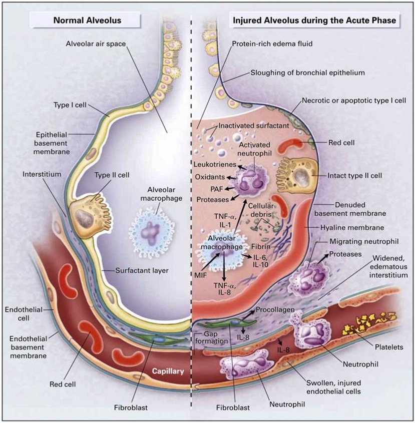
Figure 2. A diagram of the normal alveolus and injured alveolus during the acute ARDS phase (https://www.researchgate.net, CC BY-SA 4.0 via Wikimedia Commons).
Previously, the term "acute lung injury" (ALI) was used to describe a condition that could develop into ARDS. However, the ALI category is now obsolete. Today, we consider a patient to have ARDS if they meet specific clinical criteria. This is a clear, checkbox-based approach to diagnosis. ARDS, although somewhat unpredictable and sometimes discovered randomly, typically manifests after a significant infection or specific physical trauma. Importantly, this trauma does not necessarily need to directly involve the thorax. Figure 2, one of the more widely recognized images depicting ARDS, illustrates this well. On the far left of Figure 2, the key takeaway is that ARDS involves a severe inflammatory cascade. This cascade results in infiltrates and fluid accumulation in the lung acini, accompanied by a leaky pulmonary capillary bed. Platelets begin to aggregate, exacerbating the issues typically associated with an inflammatory response within the lung.
In Figure 2, you can see a comparison between normal alveoli on the left and injured alveoli on the right. The differences are striking. One critical aspect, which may not be clearly depicted in Figure 2, is the deactivation of surfactant. Normally, surfactant reduces surface tension in these tiny lung units. When it is washed out or deactivated, the lung becomes very stiff and non-compliant. Additionally, it is worth mentioning some of the common terminology associated with ARDS or ARDS-like conditions. These terms are often colloquial or clinical slang rather than standardized. However, our focus today is on the standard ARDS criteria.
ARDS stands for acute respiratory distress syndrome. As a side note, the term "baby lung" is often mentioned in the context of ARDS. "Baby lung" refers to the remaining healthy lung tissue that can still be ventilated when ARDS is present. Essentially, it is the functional lung tissue available for breathing. ARDS is considered a syndrome rather than a disease because there is no single known specific cause. it is not black and white. There are patterns and certain indications that a patient may be at risk for developing ARDS, but ultimately, it is a clinical syndrome diagnosed based on clinical features.
Here is a brief overview of those clinical features:
- Progressive dyspnea with increasing O2 requirements
- Alveolar infiltrates on chest x-ray & reduced SaO2 (SpO2) within 6-72h of inciting event
- Lab tests are nonspecific
Alveolar permeability presents a major challenge because it leads to intra-alveolar fluid accumulation, causing a ventilation-perfusion mismatch and right-to-left intrapulmonary shunting. This makes it extremely difficult to oxygenate these patients, who suffer from extremely low lung compliance. It is a vicious cycle: the stiffer the lung, the more creativity is required to ventilate. Otherwise, the cycle of alveolar infiltration continues.
In essence, ARDS can be summarized as sudden respiratory failure unrelated to heart failure or specific fluid overload. Patients present with pulmonary edema, but it is of non-cardiac origin. While there are other aspects to discuss, such as pulmonary capillary wedge pressure monitoring, today, we will focus on the essential items you need to know. Now, back to Figure 2. We know that ARDS pathophysiology is complex. Although it primarily affects the lungs, there is a significant risk of multi-organ involvement. In fact, much of the mortality associated with ARDS is due to multi-organ dysfunction syndrome (MODS). However, that is a topic for another day.
Before we move on, let's focus not only on the alveolar involvement depicted on the right side of Figure 2 but also on the intravascular and extravascular activities, also known as the interstitial processes. Notice the inflammatory mediators, such as interleukin, that are pervasive in both the alveoli and the capillaries. Due to increased capillary permeability, there is a massive fluid shift from inside the capillaries toward the interstitial space and into the gas exchange units. These patients are extremely difficult to oxygenate. Does this sound familiar to something We have encountered en masse over the last few years? The COVID-19 condition often presents similarly, making it challenging to differentiate between the pulmonary presentations of ARDS and COVID-19.
Epidemiology
When discussing any disease, disorder, or syndrome, it is helpful to quickly outline the epidemiology. Here is what we know. A well-done article by Hendrickson et al. (2021), published in the American Journal of Respiratory and Critical Care Medicine, reviewed 50 years of research and publications on ARDS. This makes it a fairly comprehensive source. What is mildly disturbing is the admission from the Canadian group of researchers, despite their bold statement that "No other intensive care syndrome has been as extensively studied as ARDS."
Many basic epidemiologic questions remain unsolved. In other words, the incidence, distribution, control methods, and overall contributing factors of this syndrome are not easily identified. We can define ARDS and occasionally recognize it, but piecing it all together remains challenging. The researchers cite a lack of standardization, and the exact onset of ARDS is not always easy to pinpoint. Even though our understanding of ARDS pathophysiology has certainly improved over the past five decades of published data, gaps remain. Several articles, not just this one, have referred to this extensive timeframe of available information.
Here is what we know: while pharmacologic interventions, such as corticosteroids and diuretics, have their place, the primary target of ARDS management should be the avoidance of ventilator-induced lung injury. We have a lot to consider, including noninvasive ventilation, high-flow nasal cannula, ECMO, and even extracorporeal CO2 removal, which we perform at my local facility. Before diving into these topics, it is important to quickly discuss the famous Lung Safe study by Bellani et al. (2016). Lung Safe stands for "Large Observational Study to Understand the Global Impact of Severe Acute Respiratory Failure." This international, multi-center prospective cohort study included some well-known researchers and investigated patients undergoing invasive or non-invasive ventilation over four consecutive weeks nearly a decade ago.
The study included patients from 459 ICUs across 50 countries on five continents, resulting in a large sample size of just over 29,000 ICU admissions. Remarkably, over 10% of these patients met the criteria for ARDS. This highlights the prevalence of ARDS in ICU settings. If you work in an ICU, you will inevitably encounter ARDS. However, the study also revealed that we are not particularly good at recognizing or treating ARDS, which, unfortunately, remains associated with a high mortality rate. The landscape changed dramatically around 2020 with the onset of COVID-19. It is now clear that differentiating between ARDS and COVID-19 is challenging, as their pulmonary presentations, chest X-rays, lab results, and overall clinical manifestations are often indistinguishable.
Severe cases of COVID-19 are arguably more difficult to treat and require more creative approaches to both ventilator and overall clinical management. Nevertheless, ARDS criteria remain the same. The treatment of the respiratory side of things does not differ dramatically between COVID-19-induced ARDS and classic ARDS. There are over 60 known etiologies of ARDS. Hendrickson et al. (2019) listed some of the most common precursors to the development of this syndrome. If you work at a facility with high trauma cases or a large ICU patient census, you will encounter ARDS frequently. If you have worked in a hospital setting for any significant amount of time, you have likely seen most of these common precursors to ARDS: ARDS in various clinical settings.
- Sepsis
- Aspiration pneumonitis
- Infectious pneumonia (including mycobacterial, viral, fungal, parasitic)
- Severe trauma and/or multiple fractures
- Pulmonary contusion
- Burns and smoke inhalation
- Transfusion-related acute lung injury and massive transfusions
- HSCT
- Pancreatitis
- Inhalation injures other than smoke (e.g., near drowning, gases)
- Thoracic surgery (eg, post-cardiopulmonary bypass or other major surgery
- Drugs (chemotherapeutic agents, amiodarone, radiation)
Understanding these common precursors helps in recognizing and managing. If you work at a trauma center, you are likely to encounter ARDS more frequently than the average clinician. However, ARDS can develop in almost any patient, regardless of age. The official diagnosis is often unclear. There is a great variety of presentations, and no definitive diagnostic test exists to conclusively diagnose ARDS. While we can consider markers like C-reactive protein, no lab value consistently and sensitively alerts us to ARDS. The chest X-ray remains a key feature in the overall assessment of ARDS. At a minimum, a chest X-ray is required, though a CT scan would be ideal. Fluid specifically infiltrates the alveoli without any indication of cardiac failure or typical fluid overload. ARDS is a pulmonary condition, and although it is classified as a syndrome, it manifests as a pulmonary disease.
Stages of ARDS
- Early Exudative Stage (Early)
- Fibroproliferative Stage (Mid)
- Fibrotic Stage (Later)
As with any lung condition, ARDS typically follows an acute phase. While ARDS is characterized by rapid onset and unpredictability, it can be described in stages. These stages help in understanding the progression of the syndrome despite its acute nature. In the early stage, also known as DAD (diffuse alveolar damage), this occurs within the first 10 days of the process. This stage is not dissimilar to various other acute lung conditions. ARDS can also involve a prolonged recovery process. The histological hallmark of this early exudative stage is diffuse alveolar damage, characterized by edema and cellular infiltration, with cells migrating to the site of gas exchange. These cells gather in areas where they did not originate and should not reside.
The next stage, occurring beyond the first 10 days, is known as the fibroproliferative stage. In this stage, pulmonary edema resolves, but cellular infiltration and multiplication continue. Essentially, while the lung fluid decreases, cellular debris continues to accumulate. It is almost like leftover trash in the pulmonary system. The latter stage of ARDS is known as the fibrotic stage. Not all ARDS survivors necessarily progress to this stage, but many do. This stage is characterized by the deterioration of normal lung architecture and the presence of fibrosis or scarring. The extent of scarring varies, with some individuals experiencing minimal scarring while others suffer from severe scarring, requiring home oxygen for the rest of their lives, even after a short period of ARDS in the ICU. According to Dr. Yoshikawa, the subacute phase is a proliferative or organizing phase of ARDS In this phase, polyp-like spindle-shaped fibroblasts infiltrate the alveoli, further complicating the lung's recovery. Of course, anytime the distance between the free surface and the diffusible layer of the alveoli thickens, some degree of refractory hypoxemia is likely to develop. The red arrow in the image points to those spindle-shaped fibroblasts.
ARDS Mortality
To be clear, there is no magic potion or perfect combination of treatments for ARDS. Despite advances in technology and knowledge, the mortality rate remains extremely high. Maca et al. (2017) conducted a systematic review published in the Respiratory Care Journal, categorizing mortality rates by type, such as at 28 days, one month, 60 days, three months, and one year. From nearly 1,100 studies initially evaluated, about 177 papers were included.
Their conclusion was that reported mortality rates are highly variable among institutions, often biased by numerous factors. Without offering an exact number with confidence, we recognize that ARDS-related mortality rates remain high, typically ranging from 30% to 45%, depending on various factors. The highest mortality rate, unsurprisingly, occurs in the ICU. Patients who are critically ill enough to be admitted to the ICU are subject to the most intensive monitoring and diagnostic evaluation. An interesting study by Hendrickson et al. (2020) specifically identified adult patients hospitalized at a single tertiary care center over approximately one year. These patients had acute hypoxic respiratory failure, defined by a PF ratio of less than or equal to 300.
These patients met ARDS criteria while receiving invasive mechanical ventilation for at least 12 hours and ultimately died during hospitalization. The study focused on those who experienced mortality during their hospital stay. ARDS was verified by multiple physicians using the Berlin definition. Ketcham et al. (2020) studied under 400 patients and found that sepsis, pulmonary dysfunction, and neurologic dysfunction were the most commonly identified causes of death. Notably, 70% of those who died exhibited multi-organ failure at the time of death. This underscores the severe impact of ARDS.
Now, let's return to the broader medical community. Although there is rarely complete consensus worldwide, let's review the most updated Berlin criteria for ARDS. We will then quickly move into classification and conclude with some evidence-based ventilator strategies. The overarching summary is that ARDS has a rapid onset, typically less than a week. Chest X-rays will show bilateral infiltrates. Respiratory failure is present and is relatively unexplained by cardiac failure, indicating that the pulmonary edema is of non-cardiac origin. The PF ratio is decreased to 300 or below; normally, this number should be well above 400. In the oxygenation section, it is assumed that all patients are on a minimum PEEP of at least five centimeters of water pressure. This encapsulates ARDS in a nutshell. These criteria allow the ordering provider to code a patient with an ARDS diagnosis.
ARDS Redefined
Very recently, Villar et al. (2023) published a paper in Critical Care suggesting that we should further define or standardize the assessment of ARDS to include both pulmonary and extrapulmonary involvement. They argue that the PF ratio is insufficiently sensitive to guide treatment selection. I cannot disagree with that. They also include a compelling case study of a septic patient with a urinary tract infection. This patient was initially placed on a high-flow nasal cannula, then transitioned to invasive mechanical ventilation, but eventually improved and was discharged after a few days. The key question in their article was whether this patient truly had ARDS.
They contend that their patient initially met the ARDS criteria but quickly fell off after a short period of mechanical ventilation. According to the latest Berlin definition, which includes the PF ratio, their patient had ARDS. However, they question whether the patient truly had ARDS after showing slight improvement in mechanical ventilation. This points to the PF ratio not being the most sensitive indicator of true respiratory status.
Today’s topic is not meant to stir up controversy; we are simply examining the evidence. Villar et al. recommend using new data sets from 2010 and beyond, utilizing more current data. They also suggest that the PF ratio be calculated under more specific standard conditions, such as at predetermined FIO2 and PEEP levels. This approach aims to provide a more accurate assessment of respiratory status in ARDS patients. In other words, leveling the playing field with standardized conditions is not a bad idea. While theoretical concepts do not always easily translate into bedside practice, this one seems feasible. Let's dive into it.
We Are Sure It Is ARDS, Now What?
Let me reiterate that there is no magic potion or absolutely effective treatment for ARDS. We will focus on one of the most important aspects of therapeutic intervention for ARDS, which is mechanical ventilation. ARDS results in respiratory failure with complex oxygenation needs. Sometimes, a less-is-more approach may be acceptable in respiratory failure, but what about with ARDS? I hope to address this moving forward. Multi-organ failure is an unfortunate reality in the wake of ARDS. While not always, it occurs frequently. How do we avoid it? For starters, we seek to ventilate the lungs conservatively and protect the lung tissue as much as possible. A minimalist approach may be best.
Regardless of the journal, it is well established that ARDS and organ failure often go hand in hand, affecting more than just the lungs. Let's look at some strategies to manage this:
- Judicious Ventilation: Employing lung-protective ventilation strategies to minimize further lung injury.
- Minimalist Approach: Using the least amount of intervention necessary to support the patient.
- Organ Protection: Implementing measures to prevent or mitigate multi-organ failure.
By focusing on these aspects, we aim to improve outcomes for ARDS patients and reduce the incidence of multi-organ failure.
Mechanical Ventilation in ARDS
Many recommendations have been made over the years regarding the mechanical ventilation of ARDS patients. Let's focus on the evidence. A team from Vanderbilt (Fuller et al., 2015) published a review in Chest titled "Mechanical Ventilation in ARDS: A State-of-the-Art Review." This article, published over 15 years ago, remains one of the most cited articles of its time. They evaluated only high levels of evidence, specifically randomized control trials, including some of the most renowned trials of the day. In summary, a low tidal volume strategy with the maintenance of plateau pressure below 30 centimeters of water pressure, when possible, is the only method of positive pressure ventilation shown to date that improves survival.
They recommended several useful rescue therapies for severe hypoxemia, including high PEEP, often referred to as "Super PEEP" in some articles, alveolar recruitment maneuvers, and prone positioning. Those of you who regularly work in an ICU have likely seen these techniques integrated over the years for both ARDS and non-ARDS patients when managing severe hypoxemia refractory to usual care.
Do these recommendations, which are approaching 20 years old, still hold today? Let's take a closer look. Before we dive into more recommendations, it is important to note that this is a multi-center clinical trial (Fuller et al., 2015) conducted in collaboration among four academic medical centers, specifically their emergency departments.
Institution-specific influences and other potentially unaccounted-for confounding variables are always at play. Unfortunately, their conclusions may align with what we see at any given time in our emergency departments. Lung-protective ventilation is infrequently used in patients receiving mechanical ventilation in the ED, regardless of ARDS status. It is a concerning finding, especially since this study was conducted among four academic medical centers. We in academic medicine are supposed to be at the cutting edge and evidence-based, right? Yet, this is a reality many of us can relate to. The study also emphasized that the progression to ARDS is very common after admission to the emergency department. The title of the publication says it all.
Spontaneous Breathing
As I mentioned earlier, extensive research on ARDS spans over 50 years. To be clear, a publication by Yoshida et al. (2017) focused on whether ARDS patients, particularly those with severe cases, should be allowed to breathe spontaneously while on positive pressure ventilation. The group presents accumulating evidence that spontaneous breathing may cause or worsen acute lung injury, especially if the breathing is vigorous. We have all seen this type of patient.
In the end, the net impact of allowing ARDS patients to spontaneously breathe while on mechanical ventilation depends on the severity of the lung injury or the ARDS condition. The authors recommend ventilator strategies that either allow spontaneous breathing or use muscle paralysis during mechanical ventilation, depending on the stage and severity of ARDS.
A recent publication by Pelosi et al. (2021) in Critical Care outlines a personalized mechanical ventilation approach. This ongoing recommendation includes a low tidal volume strategy, with volumes as low as four milliliters per kilogram of predicted body weight being acceptable in some patients. There is also a focus on driving pressure, known as Delta P. Additionally, the use of an esophageal balloon to monitor pulmonary pressures, specifically pleural pressures, has well-established merit.
However, most experts argue that there are many barriers to acquiring accurate results and properly interpreting them. Even when the measurements are reliable, their fidelity can sometimes be questionable. While a personalized mechanical ventilation approach is theoretically ideal, implementing it in practice poses significant challenges. How do we truly attain this level of individualized care?
In 2021, a group of Italian physician researchers published what they called the "10 Golden Rules for Mechanically Ventilating ARDS Patients" (Battaglini, 2021). While not groundbreaking, it offers some valuable perspectives. First and foremost, an accurate diagnosis is essential before treating any patient. Classifying the severity of ARDS is also important. Key recommendations include maintaining conservative tidal volumes and minimal plateau pressures, as well as monitoring driving pressures (ΔP). PEEP is crucial, along with recruitment maneuvers and other strategies we have mentioned. Consider prone positioning for patients with refractory hypoxemia and extreme V/Q mismatching, as changing their position can allow gravity to help improve blood flow to the dependent regions of the lung.
- Maintaining conservative tidal volumes: Aim for low tidal volumes to prevent further lung injury.
- Minimal plateau pressures: Keep plateau pressures as low as possible.
- Monitoring driving pressures (Delta P): Focus on maintaining safe driving pressures.
- PEEP: Use appropriate positive end-expiratory pressure to keep the alveoli open.
- Recruitment maneuvers: Implement strategies to reopen collapsed alveoli.
Consider prone positioning for patients with refractory hypoxemia and extreme V/Q mismatching. Changing their position can allow gravity to aid in blood flow to dependent regions of the lung, which can be beneficial. These guidelines reinforce the importance of precise, patient-specific management strategies to optimize outcomes for ARDS patients.
- Classify severity
- Monitor and maintain Vt, Pplateau, ΔP
- Implement/exploit PEEP
- Utilize recruitment maneuvers
- In certain cases, implement paralyzation
- Decrease MV support when possible
- Use prone positioning
- Consider non-traditional rescue therapies as available (i.e., artificial lung, iNO)
- Consider ECMO
- Wean PPV when possible
ARDS-Related Complications
- Barotrauma
- Nosocomial infection (VAP/VAE)
- Delirium
- Thrombi
- GI bleeding
- Sleep disturbances
- Poor nutrition
- Multiorgan failure
If your institution has it, ECMO is a possibility, wean as soon as you can from mechanical ventilation. Almost every breath on positive pressure mechanical ventilation is antiphysiologic. As a side note, there is a relatively short yet well-known list of common mechanical ventilation-related complications encountered by ARDS patients. Many of these pathologies are also present outside the ARDS condition, but they are more commonly seen in ARDS patients, especially those experiencing mechanical ventilation and multi-organ failure. If you have worked with ARDS patients, you have likely seen these complications repeatedly. They almost develop into a pattern, with ARDS-related complications manifesting consistently.
Ventilate This!
Variables of PPV
- Flow
- Pressure
- Volume
- Time
This is a friendly reminder that we are dealing with extremely delicate lung tissue. When a surfactant is suddenly deactivated by the influx of fluid into the gas exchange spaces, you must be creative in avoiding underinflation and the risk of atelectrauma while also being cautious of hyperinflation and volume trauma. Studies suggest that both repeated cyclic opening and closing of lung units, as well as excessive driving pressure, contribute to the already significant pulmonary and systemic inflammation. Therefore, it is crucial to stabilize the lung unit and ventilate minimally.
When considering full inflation, bordering on hyperinflation, versus slight under-inflation, flirting with borderline atelectasis, the sweet spot lies somewhere in the middle. , how do you ventilate fluid-filled, stiff lungs? What is the takeaway? Given the multiplicity of modes, fine-tuned boutique settings, and proprietary options available to some but not to others, what can we do about mechanical ventilation? Clinicians vary in their openness to new methods. Some are evidence-based, while others rely on empirical experience. We have all encountered this variability. As a respiratory therapist, I understand the barriers, but there often comes a point when traditional options are exhausted, and oxygenation remains difficult. In those moments, even the most conservative caregivers become open to trying whatever may work.
I want to leave you with some evidence-based considerations. Ventilating many alveoli with various filling pressures and times, interconnected through the pores of Kohn and alveolar ducts, is a complex task. However, we can simplify it by focusing on the three main variables we can manipulate in real-time mechanical ventilation: flow, pressure, and volume. These variables are all plotted over time.
Flow and Volume
A ventilator is essentially a pressurized flow generator. Oversimplified, it is akin to pouring water into a bag; at its core, it is all fluid dynamics. The process of positive pressure ventilation today is almost identical to its original method, much like auscultation has not fundamentally changed over the years. Understanding these basics helps us navigate the complexities of mechanical ventilation more effectively.
Old Process, New Method
Thanks to technological advancements, the method has indeed evolved. For example, the original stethoscope involved simply placing your head on a patient to listen. The first formal stethoscope, invented in 1816 by French physician Rene Laennec, looks quite different from the modern stethoscope we use today. Similarly, with mechanical ventilation, we are still taking compressed gas and forcing flow under high velocity into a delicate system. While the basic process has not changed much over the decades, our methodologies certainly have.
Equation of Motion
- PVENT + PMUSC = (E x V) + (R x V)
- Pvent – pressure generated by ventilator > PEEP
- Pmusc – pressure generated by inspiratory muscles
- E – respiratory system elastance
- V – Δ lung volume above FRC
- R – respiratory system resistance
- V - flow
Consider Newton's equation of motion without getting too deep into the math. Think of acceleration as the derivative of velocity and velocity as the derivative of position. This understanding of dynamics helps us appreciate the intricate control we now have over mechanical ventilation settings.
Factors to “Overcome”
- Airways resistance (Raw)
- Lung Compliance (CL)
- Elasticity
- Thoracic Wall
In positive pressure ventilation, we must overcome the lung's elastic and resistive forces. The lung resists stretching, especially when the diaphragm is not contracting, and we are controlling each breath. Therefore, the ventilator and the respiratory musculature must work in harmony, particularly when a patient makes spontaneous efforts to encourage adequate ventilation and maintain synchrony.
As the term implies, these are challenges we must address while ventilating patients and each case is unique. According to Poiseuille's law, flow is indirectly proportional to the length of the tube and the viscosity of the gas. This means flow will decrease with a decrease in driving pressure or a decrease in tube radius. Understanding these principles helps us fine-tune our approach to mechanical ventilation for each patient.
Time Constant (T)
- 1 TC = CL x Raw
- How quickly pressure equilibrates between the circuit and alveoli
- 3-to-5-time constants required for complete inspiration/expiration
What is interesting is that even a small change in the cross-sectional area of an airway can dramatically alter the flow received into the lung due to its exponential relationship. Mathematically, if a patient's airway radius decreases by just 16%, the flow rate through that tube is cut in half. This sensitivity underscores the importance of maintaining airway patency.
Now, let’s talk about time constants. A time constant is the time required to fill a particular lung region to 63% of its potential capacity. Understanding this helps in managing ventilation, as different lung regions can have varying time constants, affecting how quickly they fill and empty. The goal is to target three to five-time constants for complete filling. In a normal lung, surface tension is not constant due to a mixture of phospholipids and proteins, known as surfactants, floating atop the alveolar epithelium. This surfactant helps gas flow from larger to smaller alveoli, producing homogeneity in alveolar volume and stabilizing the lung.
In ARDS, however, surfactant is deactivated by the mass infiltration of lung fluid, disrupting this balance. As a result, you can encounter very high airway resistance and, more critically, low compliance. When compliance decreases, as it does in ARDS, the time constant increases because it is a product of airway resistance and compliance. This is why an increase in inspiratory time is sometimes necessary to adequately recruit and oxygenate the lungs. Theoretically, if you allow enough time, even the most noncompliant lung can accept volume within reason. By extending the time, you enable a more equal distribution of gas, allowing the lung to accommodate the necessary volume despite its reduced compliance.
What About Ventilator Mode... Volume-Targeted or Pressure-Tageted?
This question is undoubtedly debated worldwide, let's examine some data. At face value, you could argue either side if you truly know how to manipulate the ventilator. However, many clinicians assume that pressure-targeted modes are more lung-protective, but let’s see what the evidence says. A systematic review and meta-analysis were conducted by Rittayam et al. (2015) to determine whether pressure-control continuous mandatory ventilation (PCCMV) or pressure-control inverse ratio ventilation (PCIRV) has demonstrated advantages over volume-controlled CMV. The Cochrane tool for risk of bias was used to assess methodological quality.The researchers conducted their due diligence and introduced physiological criteria as quality indicators for selecting studies. They identified 34 studies that met the inclusion criteria and compared PCCMV and PCIRV to volume-control CMV. Their results indicated no significant differences in compliance or gas exchange, even with inverse ratio ventilation or when using pressure control with prolonged inspiratory time. They calculated the oxygen index and, after analyzing several patients, observed a poor effect of pressure-controlled inverse ratio ventilation on the oxygenation index in a large portion of patients. Additionally, there was no difference between the modes in terms of hemodynamics, work of breathing, or clinical outcomes. This suggests that the assumed superiority of pressure-targeted modes for lung protection may not hold true, as both modes showed comparable results across various parameters. Simply some things to think about.
Another systematic review by Chacko et al. (2015) evaluated acute lung injury in ARDS, analyzing over 60 years of data from studies conducted between 1950 and 2014. Their conclusions, which you can find in the article, indicate that currently available data from randomized controlled trials are insufficient to confirm or refute whether pressure-controlled or volume-controlled ventilation offers any advantage for patients with acute respiratory failure or ARDS. This suggests that while we may have some insights, the definitive answer remains elusive.
In 2000, the landmark ARDSNet study, a famous randomized controlled trial, demonstrated an approximate 9% absolute mortality reduction using a strategy of low tidal volume. This strategy has been well established since then, with a recommendation of 6 ml/kg of predicted body weight and a limitation of plateau pressure to less than 30 cm H2O as a starting point. This approach is considered ideal, though it represents just the minimum standard for managing ARDS patients.
However, current research suggests that driving pressure might be more crucial than simply focusing on tidal volume and plateau pressure. Driving pressure is the difference between peak inspiratory pressure and plateau pressure, or in pressure-control ventilation, the difference between inspiratory pressure and PEEP. This metric reflects the movement of lung tissue from rest to full inflation.
Gayat et al.'s (2018) article, related to the well-cited FROG study (French and European Outcome Registry study), evaluated 1,570 patients after discharge from the ICU. The term "ICU survivors" highlights that the ICU is indeed a dangerous environment, where patients are often more critically ill than average. The study underscores the importance of considering driving pressure as a significant factor in the management and outcomes of ICU patients, suggesting a need for a more nuanced approach to ventilation strategies beyond traditional metrics.
Ventilator Mismanagement Sign
ICU patients are already at higher risk for mortality upon admission. However, it has also been established that ICU survivors have decreased long-term survival compared to the general population, meaning they often die earlier, even after surviving an ICU stay. This highlights the need for careful management and consultation with pulmonary critical care specialists or respiratory therapists before placing a patient on a ventilator to avoid potential mismanagement.
This situation is sometimes referred to as the "ventilator mismanagement sign," which can occur right after intubation and the initiation of positive pressure ventilation. Always begin conservatively if possible. Where is the so-called sweet spot? The idea is to aim for a balance, finding a middle ground in ventilation strategies. This approach promotes optimal outcomes by avoiding the extremes of under- or over-ventilation and mitigating potential complications.
If you examine a microscopic view of alveoli under hyperinflation, you will see atelectasis and appreciate the dramatic differences between various levels of expansion. These alveoli are delicate, compressible, contractible, and recruitable lung units closely associated with adjacent capillaries that can also become recruited, dilated, constricted, collapsed, or compressed. Pulmonary blood flow is affected in real time as the alveoli expand and contract.
Finding the sweet spot of ideal inflation has been a long-standing concept. This so-called safe window of ventilation aims to avoid lung collapse at the end of expiration while preventing overfilling and overdistension during inspiration. The goal is to maintain this balance, ensuring optimal gas exchange without causing additional injury to the lung tissue.
Safe Window
To ensure this safe window, start with conservative tidal volumes. Typically, this means using 4 to 6 ml/kg of predicted body weight, occasionally up to 7 ml/kg, but preferably on the lower side to minimize lung injury. However, be mindful of hypercarbia, which can develop if you under-ventilate a patient. Permissive hypercapnia might be an option in some cases.
Monitor ventilating pressures carefully, whether you are using volume-targeting or pressure-targeting modes. Evaluate all the available scalars. Modern ventilators often provide a volume-pressure scalar, flow-volume loop, or other compliance curves, which can be evaluated quickly without adjusting settings or stimulating the patient. These tools help ensure you are within the safe window, balancing adequate ventilation and lung protection.
To evaluate a pressure-volume curve in its pure form, the patient should be in a flow-controlled mode with a very slow and prolonged inspiratory time. However, modern technology allows us to approximate these curves more easily. Evaluating lung mechanics in ARDS can be challenging, especially when determining the best PEEP. To generate an accurate pressure-volume curve, the patient must not make spontaneous efforts, and controlled flow delivery is typically achieved in a volume-targeting mode. However, the concept is more important than the specific method.
The goal is to identify two critical points:
- The pressure level at which the lung begins to rapidly fill with volume.
- The pressure level at which the lung begins to collapse on expiration.
By setting the PEEP just above the pressure level where the lung begins to collapse on expiration, you can prevent lung collapse, ensuring a more stable and effective ventilation strategy. To prevent ventilator-induced lung injury, we want to avoid the cyclic closing and reopening of alveoli with each breath. Since their lungs are already injured if they have ARDS, we should strive to minimize additional pulmonary inflammation whenever possible.
Here is a clinical tip: with the ordering provider's approval and supervision, you can occasionally step to the bedside and slightly increase the PEEP while observing the patient's chest rise. If you see an improvement in chest rise, an increase in oxygen saturation, and a sudden increase in expiratory volume (especially if you are in pressure control mode), you have likely recruited additional lung tissue.
There are other strategies to consider as well:
- Using recruitment maneuvers to open collapsed alveoli
- Adjusting inspiratory and expiratory times to optimize ventilation
- Monitoring for signs of overdistension to avoid further lung injury
By carefully adjusting these parameters, you can better manage ARDS patients and improve their respiratory outcomes. You can auscultate while slightly increasing the PEEP, allowing the patient to take a few breaths. If you notice an increase in aeration and air movement, it is likely that you have successfully recruited additional lung tissue. Sometimes, it requires a bit more creativity, such as adjusting inspiratory times and other settings, but this is a good starting point. In mechanical ventilation, we should be particularly concerned with the concepts of stress and strain. These terms, crucial in material science and engineering, are equally important in healthcare and medicine.
- Stress refers to the force applied to a material per unit area
- Strain is the deformation or change in the shape of the material resulting from the applied force
In the context of mechanical ventilation, stress corresponds to the pressure applied to the lung tissues, while strain represents the resulting changes in lung volume. Understanding and managing these concepts is essential to prevent ventilator-induced lung injury and optimize patient outcomes.
Generally Speaking…
- End-inspiratory plateau pressure and Vt/IBW ratio are inadequate surrogates to qualify and/or quantify end-inspiratory stress and strain Gattinoni et al., 2012)
- Large static strains are less harmful than large dynamic strains (Protti et al., 2013)
- An optimal “protective breath” describes one of minimal dynamic strain and energy/power load (Nieman et al., 2016)
- Lung protection > diaphragm protection (Goligher et al., 2020)
Within an alveolus, you can continue to increase pressure, applying stress without seeing any change in shape or recruitment. Only when you achieve a change in shape, through recruitment and increased volume in a particular lung unit, does the lung unit react to the applied force. Some of the most well-known names in clinical research support these concepts. Let's review the key points:
- Lower Tidal Volumes: Using lower tidal volumes is generally better for reducing lung injury.
- Lowest Possible Pressures: Maintaining the lowest pressures possible is crucial to minimize stress and strain on the lung tissues.
- Sustained Inflation: Sustained inflation (such as Super PEEP) is preferable to cyclic closing and reopening of alveoli. This approach reduces the risk of ventilator-induced lung injury.
This concept of sustained inflation aligns closely with the principles of high-frequency oscillatory ventilation (HFOV), which aims to maintain alveolar recruitment and minimize cyclic atelectasis and overdistension. Understanding and applying these principles can help optimize ventilation strategies and improve outcomes for patients with ARDS and other forms of acute lung injury.
The most gentle delivery possible, as tolerated by the patient, is ideal. However, this is often easier said than done. Recently, there has been much debate about whether to allow spontaneous breathing during the more severe stages of ARDS. The consensus seems to be shifting towards prioritizing lung protection over maintaining diaphragm activity, at least in the short term during the most severe phase of ARDS.
If a patient is asynchronous in deteriorating and unable to oxygenate due to issues like clamping down, then removing spontaneous efforts from the equation may be necessary. This approach can help ensure more consistent and controlled ventilation, potentially improving oxygenation and reducing the risk of further lung injury. Ultimately, the goal is to balance lung protection with overall patient stability, adapting strategies as the patient's condition evolves.
Summary
- Lung protective ventilation is a must
- Lower tidal volumes
- Application of optimal (and adequate) PEEP may require higher than traditional settings
- Target low plateau pressure (and low driving pressure)
- Use recruitment maneuvers cautiously
- Consider prone positioning
- Seek to maintain ventilator synchrony – may require neuromuscular blockade
- ***Don’t disregard potential non-traditional intervention as an effective option: NIV, HFOV, APRV, ECMO (Liaqat et al., 2022)
Here are some key points to take away:
- Lung Protective Ventilation: This is essential and begins with using conservative tidal volumes. Even in pressure-control or pressure-targeted modes of ventilation, it is crucial to monitor tidal volumes alongside plateau pressure.
- Adequate PEEP: Finding the optimal PEEP is vital. This "sweet spot" varies for each patient and is dynamic, meaning the PEEP setting at the start of a shift may not be appropriate later on. Regularly reassess and adjust PEEP as needed throughout the day.
- Monitoring and Adaptation: Continuous monitoring and adaptability are key. Pay attention to how the patient's condition evolves and be ready to adjust ventilation settings accordingly to maintain lung protection and optimize oxygenation.
- Balancing Lung Protection and Diaphragm Function: While lung protection is a priority, especially in the severe stages of ARDS, sometimes it may be necessary to eliminate spontaneous breathing efforts if they cause significant asynchrony and impede oxygenation.
By adhering to these principles, you can help minimize ventilator-induced lung injury and improve patient outcomes.
- Recruitment Maneuvers:
- Use Cautiously: Recruitment maneuvers can be beneficial but should be implemented cautiously.
- Open Lung Technique: This technique, which involves applying extremely high pressure for a short period to recruit alveoli, has been around for several years. However, it is less promoted in recent literature, with a preference for simpler recruitment strategies based on scalars and clinical monitoring.
- Prone Positioning:
- Team Effort: Prone positioning requires coordination and buy-in from the ordering provider.
- Quick Transition: When done correctly, it only takes a few moments to move a patient from supine (facing up) to prone (facing down). This position can significantly improve oxygenation and lung mechanics.
By integrating these strategies, you can enhance ventilation management, reduce lung injury, and improve patient outcomes.:
- Driving Pressure: Focus on maintaining a safe driving pressure to minimize lung injury.
- Recruitment Maneuvers: Implement these with caution and base them on clinical monitoring.
- Prone Positioning: Consider this effective strategy, especially for patients with severe ARDS, as it can significantly improve outcomes.
Monitoring for skin breakdown, lines, and other potential issues is crucial when implementing prone positioning, but the benefits can be significant. I recently saw a patient with ARDS who had reached the end of conventional ventilator options, and their oxygenation status improved dramatically upon being placed in the prone position.
- Key Points to Consider:
- Prone Positioning:
- Monitor for Skin Breakdown: Regularly check for skin breakdown, dislodged lines, and other complications.
- Significant Impact: Prone positioning can rapidly improve oxygenation status in patients with severe ARDS.
- Maintaining Synchrony:
- Neuromuscular Blockade is sometimes necessary to eliminate spontaneous breathing efforts and ensure ventilator synchrony. There is supporting literature for this approach.
- Alternative Ventilation Strategies:
- Non-Invasive Ventilation (NIV): Useful in certain scenarios, particularly if the primary issue is oxygenation rather than ventilation.
- High-Frequency Airway Pressure Release Ventilation (HFAPRV): Another option for specific cases.
- ECMO: Considered for severe cases where other interventions fail.
- High-Flow Nasal Cannula (HFNC): Can be effective for oxygenation problems without significant ventilation issues.
- Prone Positioning:
This is indeed a broad but vital topic in respiratory care and clinical medicine. By employing these strategies thoughtfully, we can improve patient outcomes and manage complex cases of ARDS and other respiratory conditions more effectively.
Questions and Answers
Do you find that a high-flow nasal cannula is helpful early on in the weaning process from the ventilator?A high-flow nasal cannula (HFNC) can be very helpful, and I can tell you why. There are two trains of thought here. With HFNC, you can deliver a massive amount of flow with high FIO2s. This can be particularly beneficial in cases of refractory hypoxemia where lung recruitment is not an immediate priority, and the patient is simply in a distressed state. HFNC allows the patient to calm down and may decrease their oxygen consumption, giving you time to perform a better assessment. Not every patient will respond to HFNC, and once they deteriorate and become truly refractory, they will need positive pressure ventilation. However, if you have the option at your institution, trying HFNC early is worthwhile unless you know the patient is in a more severe ARDS condition. With limited information, it is best to try oxygen first. I always recommend trying high levels of oxygen flow before resorting to mechanical ventilation unless you have a chest X-ray and a good clinical history indicating otherwise.
What additional metrics would you find valuable at the bedside to quantify inspiratory stress and strain? For example, if you're closely monitoring driving pressure or dynamic strain based on compliance, what parameters seem easiest to capture and interpret at the bedside?
I wish I had a definitive list, too; it would probably have me giving talks around the world. However, over the years, I have noticed a few key insights beyond what We have discussed today. When you look at the compliance curve or the volume-pressure scalar, you can identify what used to be called the "bird's beak" or the point of overdistention. This is where you see a flattening of the curve after a progressive, almost linear increase in pressure and volume. Suddenly, with each unit increase in pressure, there is very little to no change in volume. This indicates you're overstretching the lung tissue, and it is crucial to avoid this. Not all ventilators provide scalar data with enough detail for precise assessment, but you can still make informed decisions. As you increase the PEEP or other pressure settings, watch for a point where the return volume increases and then suddenly stops. Continuing to increase pressure beyond this point is likely not beneficial. Sometimes, after recruiting lung tissue, you can slightly reduce the pressure while maintaining lung recruitment. Other times, it might not be as straightforward. This is where your assessment skills are essential. Monitor chest rise, the patient’s degree of comfort, the absence of grimacing (if they looked distressed before), oxygen saturation, blood pressure, and heart rate. Simple indicators like these can help you determine if the patient is responding positively. When making changes, observe if they are improving or heading in the right direction. The patient’s response can often guide you better than any scalar or curve alone.
When you're looking at sustained plateau pressures over 30 despite the usual interventions, what other considerations come into play?
One of the first things that come to mind for refractory hypoxemia is permissive hypercapnia. If we are really concerned about their oxygenation level, reducing O2 consumption is key. For instance, using a cooling blanket or even packing the patient in ice in extreme scenarios can help lower their metabolic rate. Administering a paralytic can also reduce O2 consumption in hypermetabolic patients, decreasing the lung's demand for oxygen. Additionally, proning the patient or maintaining sustained pressure for longer periods can help. This technique essentially splints the lung tissue open, promoting more equal distribution of gases and allowing oxygen molecules to diffuse more efficiently across the alveolar membrane. Monitoring fluid status is another critical aspect. Many patients are underhydrated, and ensuring they are adequately hydrated can improve their oxygenation status. In some cases, making the patient hyperdynamic can be beneficial, provided their cardiovascular system is not compromised. Potential pulmonary edema can be managed with diuretics later if necessary. Anti-inflammatories, particularly corticosteroids, are also frequently discussed in the literature. While they are not a cure-all, they can help manage inflammation, although they may not have an immediate impact on oxygenation status. By integrating these strategies—reducing oxygen consumption, optimizing gas distribution, ensuring proper hydration, and managing inflammation—clinicians can better address refractory hypoxemia and improve patient outcomes in severe respiratory distress.
References
ARDS Definition Task Force; Ranieri, V. M., Rubenfeld, G. D., Thompson, B. T., Ferguson, N. D., Caldwell, E., Fan, E., Camporota, L., & Slutsky, A. S. (2012). Acute respiratory distress syndrome: The Berlin Definition. JAMA, 307(23), 2526-2533.
Ashbaugh, D., Boyd Bigelow, D., Petty, T., & Levine, B. (1967). Acute respiratory distress in adults. The Lancet, 290(7511), 319-323.
Battaglini, D., Sottano, M., Ball, L., Robba, C., Rocco, P. R. M., & Pelosi, P. (2021). Ten golden rules for individualized mechanical ventilation in acute respiratory distress syndrome. Journal of Intensive Medicine, 1(1), 42-51.
Bellani, G., Laffey, J. G., Pham, T., Fan, E., Brochard, L., Esteban, A., Gattinoni, L., van Haren, F., Larsson, A., McAuley, D. F., Ranieri, M., Rubenfeld, G., Thompson, B. T., Wrigge, H., Slutsky, A. S., & Pesenti, A.; LUNG SAFE Investigators; ESICM Trials Group. (2016). Epidemiology, patterns of care, and mortality for patients with acute respiratory distress syndrome in intensive care units in 50 countries. JAMA, 315(8), 788-800. doi: 10.1001/jama.2016.0291. Erratum in: JAMA, 316(3), 350.
Fuller, B. M., Mohr, N. M., Miller, C. N., Deitchman, A. R., Levine, B. J., Castagno, N., Hassebroek, E. C., Dhedhi, A., Scott-Wittenborn, N., Grace, E., Lehew, C., & Kollef, M. H. (2015). Mechanical ventilation and ARDS in the ED: A multicenter, observational, prospective, cross-sectional study. Chest, 148(2), 365-374.
Gattinoni, L., Carlesso, E., & Caironi, P. (2012). Stress and strain within the lung. Current Opinion in Critical Care, 18(1), 42-47.
Goligher, E. C., Dres, M., Patel, B. K., Sahetya, S. K., Beitler, J. R., Telias, I., Yoshida, T., Vaporidi, K., Grieco, D. L., Schepens, T., Grasselli, G., Spadaro, S., Dianti, J., Amato, M., Bellani, G., Demoule, A., Fan, E., Ferguson, N. D., Georgopoulos, D., Guérin, C., Khemani, R. G., Laghi, F., Mercat, A., Mojoli, F., Ottenheijm, C. A. C., Jaber, S., Heunks, L., Mancebo, J., Mauri, T., Pesenti, A., & Brochard, L. (2020). Lung- and diaphragm-protective ventilation. American Journal of Respiratory and Critical Care Medicine, 202(7), 950-961.
Hendrickson, K. W., Peltan, I. D., & Brown, S. M. (2021). The epidemiology of acute respiratory distress syndrome before and after Coronavirus Disease 2019. Critical Care Clinics, 37(4), 703-716.
Ketcham, S. W., Sedhai, Y. R., Miller, H. C., Bolig, T. C., Ludwig, A., Co, I., Claar, D., McSparron, J. I., Prescott, H. C., & Sjoding, M. W. (2020). Causes and characteristics of death in patients with acute hypoxemic respiratory failure and acute respiratory distress syndrome: A retrospective cohort study. Critical Care, 24(1), 391.
Liaqat, A., Mason, M., Foster, B. J., Kulkarni, S., Barlas, A., Farooq, A. M., Patak, P., Liaqat, H., Basso, R. G., & Zaman, M. S. (2022). Evidence-based mechanical ventilatory strategies in ARDS. Journal of Clinical Medicine, 11(2), 319.
Máca, J., Jor, O., Holub, M., Sklienka, P., Burša, F., Burda, M., Janout, V., & Ševčík, P. (2017). Past and present ARDS mortality rates: A systematic review. Respiratory Care, 62(1), 113-122.
Matthay, M. A., & Zimmerman, G. A. (2005). American Journal of Respiratory Cell and Molecular Biology, 33, 319-327.
McGurk, K., Riveros, T., Johnson, N., & Dyer, S. (2020). A primer on proning in the emergency department. Journal of the American College of Emergency Physicians Open, 1(6), 1703-1708. doi: 10.1002/emp2.12175.
Nieman, G. F., Satalin, J., Andrews, P., Habashi, N. M., & Gatto, L. A. (2016). Lung stress, strain, and energy load: Engineering concepts to understand the mechanism of ventilator-induced lung injury (VILI). Intensive Care Medicine Experimental, 4(1), 16.
Pelosi, P., Ball, L., Barbas, C. S. V., Bellomo, R., Burns, K. E. A., Einav, S., Gattinoni, L., Laffey, J. G., Marini, J. J., Myatra, S. N., Schultz, M. J., Teboul, J. L., & Rocco, P. R. M. (2021). Personalized mechanical ventilation in acute respiratory distress syndrome. Critical Care, 25(1), 250.
Pham, T., & Rubenfeld, G. D. (2017). Fifty years of research in ARDS. The epidemiology of acute respiratory distress syndrome: A 50th birthday review. American Journal of Respiratory and Critical Care Medicine, 195(7), 860-870.
Protti, A., Andreis, D. T., Monti, M., Santini, A., Sparacino, C. C., Langer, T., Votta, E., Gatti, S., Lombardi, L., Leopardi, O., Masson, S., Cressoni, M., & Gattinoni, L. (2013). Lung stress and strain during mechanical ventilation: Any difference between statics and dynamics? Critical Care Medicine, 41(4), 1046-1055.
Villar, J., Szakmany, T., Grasselli, G., & Camporota, L. (2023). Redefining ARDS: A paradigm shift. Critical Care, 27(1), 416.
Citation
Gilmore, Tim (2023). Acute respiratory distress syndrome (ARDS) and what you should know. Continued.com - Respiratory Therapy, Article 228. Available at www.continued.com/respiratory-therapy
Jun 17, 2024
1 CE HourRespiratory Management of Patients With Neuromuscular Weakness Series: Review Recommendations and Best Practice
Duane Reed, RRT, RCP, EdD
Editor’s note: This text-based course is an edited transcript of the webinar, Respiratory Management of Patients With Neuromuscular Weakness Series: Review Recommendations Best Practice, presented by Dr. Duane Reed, EdD RRT, RCP. Any errors in transcription or editing are the responsibility of Continued.com and not the course presenter. It is recommended to download the course handout to supplement this text format.
Learning Outcomes
After this course, participants will be able to:
- Identify neuromuscular diseases that affect the respiratory system.
- Identify evidence-based recommendations for patients with neuromuscular diseases.
- Discuss best practice management for patients with neuromuscular diseases.
Overview of Neuromuscular Diseases (NMD)
Many forms of neuromuscular disease exist:
- Amyotrophic Lateral Sclerosis (ALS)
- Charcot Marie Tooth Disease (CMT)
- Multiple Sclerosis (MS)+
- Muscular Dystrophy
- Myasthenia Gravis
- Myopathy
- Myositis
- Guillain Barre Syndrome (not a disease, but when the immune system attacks the neuro system)
The foundation for our discussion comes from a panel discussion centered around 128 studies presented in the Journal of Chest. This panel has yielded 15 graded recommendations and a good practice and consensus-based statement. Our primary aim is to uncover evidence-based best practices for effectively managing the respiratory aspects of neuromuscular diseases.
Neuromuscular diseases manifest when the nerves governing movement are negatively impacted. This brings our attention to the nervous system. The resulting muscle weakness, a notable complication stemming from neuromuscular diseases, significantly affects the respiratory system. This connection becomes evident when considering the role of the diaphragm, a pivotal component of the respiratory system. The diaphragm relies on nerve impulses for stimulation. Thus, any damage to the nerves jeopardizes the function of the respiratory system. This is where the link between nerve damage and respiratory system vulnerability emerges. Respiratory failure is a prevalent complication closely linked to neuromuscular diseases.
This serves as a pivotal reason for us to delve into a discussion about various neuromuscular diseases. When nerve functionality is compromised, complexities can emerge, especially concerning respiratory function. This can lead to disruptions in the processes necessary for breathing and lung function. While you might be familiar with some of these conditions, others may be less known, yet all are intrinsically tied to the risk of respiratory failure. Understanding neuromuscular diseases' influence on the respiratory system is crucial because they often lead to respiratory complications due to their impact on nerves and muscles involved in breathing.
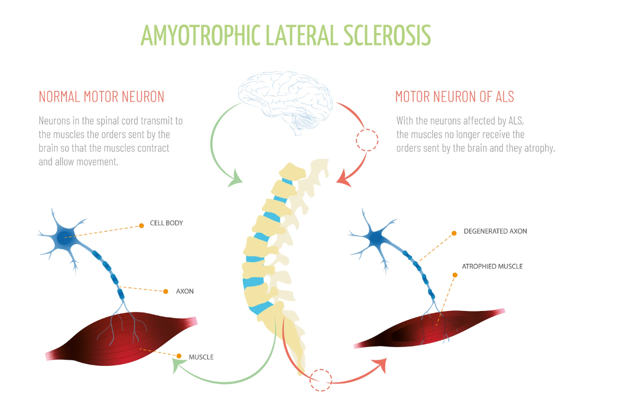
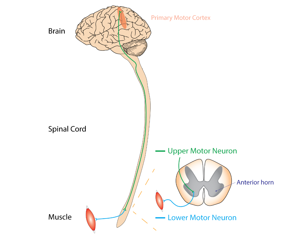
Figure 1. Diagram illustrating motor neuron of ALS (Continued Getty Images).
Many forms of neuromuscular diseases exist, with some being more familiar to us than others. One well-known example is ALS or amyotrophic lateral sclerosis in Figure 1. This disease primarily affects the nervous system, leading to a gradual weakening of the muscles. You may recognize it by its former name, Lou Gehrig's disease. It leads to muscle cell degeneration, resulting in a decline in muscle functionality and overall mobility.
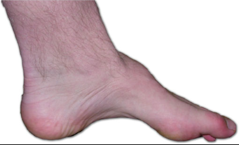
Figure 2. The foot of a person with Charcot-Marie-Tooth. (Benefros at English Wikipedia, CC BY-SA 3.0 via Wikimedia Commons).
Another example is Charcot-Marie-Tooth disease, commonly referred to as CMT. This condition is essentially a hereditary disorder that primarily induces nerve damage in the arms and legs seen in Figure 2. CMT disease leads to reduced muscle size and strength, accompanied by potential sensory loss, muscle contractions, and challenges in walking. Commonly observed are foot deformities like hammertoes and high arches. While symptoms often initiate in the lower extremities, progression to the hands and arms can occur over time. The manifestations of CMT disease generally emerge during adolescence or early adulthood, although they can also manifest in midlife.
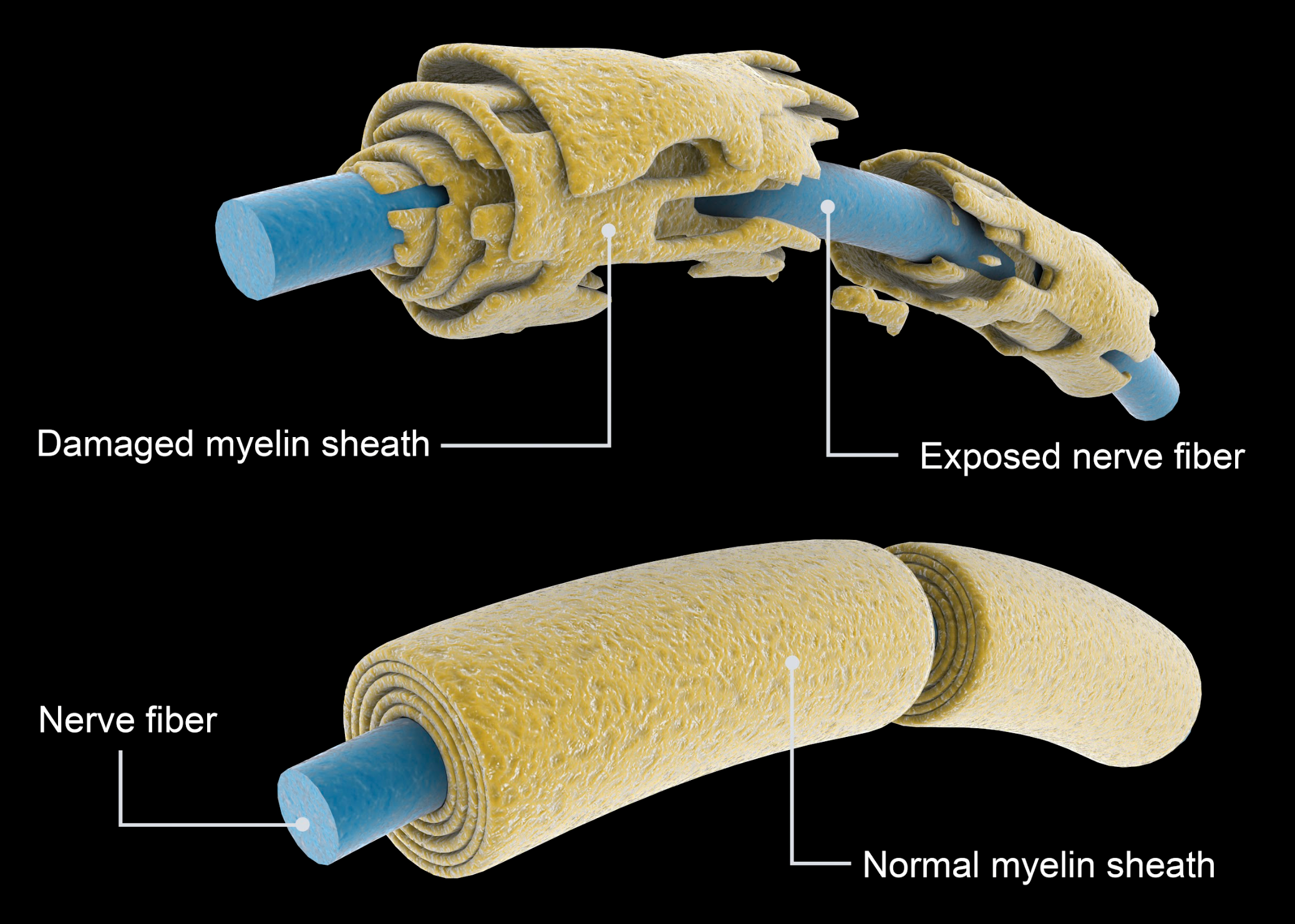
Figure 3. Damage myeline sheath of Multiple Sclerosis. (Continued Getty Images).
Multiple sclerosis in Figure 3, often referred to as MS, is a widely recognized condition. It is a potentially disabling disease affecting the brain, spinal cord, and central nervous system (CNS). In this disease, the immune system erroneously targets the protective sheath, known as myelin, covering nerve fibers. This leads to various issues, as the degradation of myelin disrupts the proper functioning of nerves, resulting in communication problems within the body. This condition can cause various symptoms and challenges, and we will provide an illustrative image to convey its effects better.
Muscular dystrophy is a widely recognized condition known for inducing muscle-related issues. This disorder also intricately affects the surrounding protective sheath of nerves. Over time, these issues inevitably extend their reach to the respiratory system, raising a pivotal concern. Delving into the context of ALS, a compelling image illustrates the contrast between a healthy motor neuron and one that is emblematic of ALS. These distinctions become apparent in the cell bodies and axons, both crucial for maintaining optimal nerve function. These transformations culminate in muscle atrophy, setting apart ALS-affected motor neurons from their healthy counterparts within the spinal cord. ALS is unquestionably a profoundly debilitating condition.
Moreover, we have previously explored Charcot-Marie-Tooth disease. A comparison between unaffected and affected nerves visually portrays the scarring of the myelin sheath, a distinctive feature of this hereditary disorder. The damage predominantly manifests in the arms and legs, impairing nerve and myelin sheath. This disease can significantly impair affected individuals, and its impact may even extend to lung function. It is important to note that while these diseases, such as multiple sclerosis, exhibit distinct origins, they share commonalities in terms of their repercussions.
Regarding multiple sclerosis, a parallel arises concerning the damage inflicted upon the myelin sheath. A visualization on the right effectively underscores the compromised state of the myelin, underscoring its pivotal role in safeguarding nerves. This assumes particular significance when considering nerves stemming from the cervical spine, which extend to the diaphragm. Any compromise in these nerves disrupts the necessary transmission for stimulating impulses, posing a considerable challenge to respiratory function. This emphasizes the significance of delving into neuromuscular diseases, given their substantial influence on the respiratory system, particularly within the realm of inherited disorders.
Among the array of disorders highlighted, myasthenia gravis emerges as a notable case. This condition typically initiates in the brain and progresses downward, impacting muscles and culminating in rapid fatigue. This importance cannot be understated, as it targets voluntary muscle groups, including those implicated in respiration. Conversely, while Guillain-Barre syndrome is classified as a syndrome rather than a disease, its effects on muscle function are noteworthy. Additionally, it is essential to distinguish myositis, characterized by inflammation of muscles, as a rare disorder. Both Guillain-Barre syndrome and myositis present substantial challenges.
All these diseases collectively manipulate nerve function. Once nerve integrity is compromised, the essential nerve impulses the diaphragm relies upon to generate pressure changes are disrupted. This underscores the potential complexities that can arise when nerve functionality is compromised, particularly concerning respiratory function.
Advancing Research on Respiratory Management of Patients with Neuromuscular Weakness
An expert panel conducted a systematic review focusing on the respiratory management of neuromuscular diseases (NMD). They employed the Grading of Recommendations, Assessment, Development, and Evaluations approach to assess the certainty of evidence, formulate recommendations, and assign grades. The consensus was reached through a modified Delphi technique.
Drawing insights from 128 studies, the panel established 15 graded recommendations, one good practice statement, and one consensus-based statement. It is important to note that the evidence supporting best practices for NMD respiratory management is limited, primarily relying on observational data from amyotrophic lateral sclerosis.
The guidelines offered an understanding of areas such as mouthpiece ventilation, the transition to home mechanical ventilation, salivary secretion management, and airway clearance therapies. It's emphasized that the pathologic features of NMD encompass a wide spectrum of disorders, each with varying rates of lung function decline. In clinical practice, the role of clinicians extends beyond medical evaluation to shared decision-making with patients and families. This involves respecting patient preferences and treatment goals, considering the quality of life, and judiciously utilizing available resources in decision-making processes.
Panel Recommendation 1: Use and Timing of Pulmonary Function Test (PFT)
In the context of patients with neuromuscular diseases at risk of respiratory complications, experts advise incorporating the timing of Pulmonary Function Testing (PFT) as a valuable tool for management. The panel strongly recommends the integration of PFT into the management process. These recommendations aim to serve as a basis for developing protocols and guidelines.
Panel Suggestions:
The panel proposes PFT as a cost-effective intervention. They recommend spirometry, focusing on FVC (forced vital capacity) and SVC (slow vital capacity), along with MIP (maximum inspiratory pressure), MEP (maximum expiratory pressure), and SNIP (nasal inspiratory pressure) measurements for patients with neuromuscular conditions. Additionally, peak cough flow should be considered. FVC and SVC techniques should be employed to assess respiratory health comprehensively. A slower version like SVC is crucial for patients with COPD who may experience prolonged air trapping. Evaluating respiratory reserve and muscle strength involves measuring MIP and SNIP. These parameters are indispensable when managing neuromuscular patients, expanding our toolkit. Panel recommendation two emphasizes utilizing PFTs and understanding their optimal timing.
Panel Recommendation 2: Use and Timing of PFT for Patients with NMD at Risk of Respiratory Failure
For patients with neuromuscular disease prone to respiratory complications. The panel advises conducting PFTs at least every six months, as deemed suitable. This recurring six-month interval aligns with the recommended frequency.
Panel Suggestions:
Employing a consensus-based approach, the panel arrived at a six-month testing interval. While recognizing that the progression rates vary among NMDs, particularly evident in cases like ALS, where significant respiratory parameter changes could manifest in 3 to 6 months, the panel acknowledged that for more stable or slowly progressing conditions like Duchenne muscular dystrophy, PFTs could be conducted at less frequent intervals, such as every 12 months. The panel proposes considering one or more of the following assessments within this timeframe. While not mandatory to perform all, options include measuring vital capacity, slow vital capacity, MIP, MEP, SNIP, and cough.
Panel Recommendation 3: Screening for Respiratory Failure and Sleep-Related Breathing Disorders
Let's delve into recommendation three, which revolves around sleep-related disorders in the context of neuromuscular diseases. When working with patients with symptomatic patients with NMD who have normal PFT, the panel proposes employing overnight oximetry, often referred to as ONO. Additionally, the suggestion involves utilizing polysomnography to evaluate the necessity for non-invasive ventilation. This step relates to the consideration of sleep studies as well.
Panel Suggestions:
The panel highlights polysomnography as a valuable tool for determining non-invasive ventilation's appropriateness. Clinicians, consider polysomnography to assess whether noninvasive ventilation (NIV) is clinically indicated. This particular ventilation approach includes methods like BiPap.
Panel Recommendation 4: Noninvasive Ventilation (NIV)
Moving on to recommendation four primarily centers around patients with neuromuscular diseases experiencing chronic respiratory failure. This scenario necessitates a closer look at non-invasive ventilation strategies. When discussing chronic respiratory failure, it is important to note that this often pertains to COPD patients.
Panel Suggestions:
The panel's recommendation of employing non-invasive ventilation aligns logically with this context. However, it is worth highlighting that while clinical indications for non-invasive ventilation are present, they can vary based on factors such as the specific neuromuscular disease, patient age, and the pace of disease progression. In essence, these multifaceted variables influence the suitability of non-invasive ventilation.
The evidence was primarily derived from adults with ALS, given its prevalence as the most common NMD. However, its application should be approached thoughtfully when considering younger children, adolescents, and adults with non-ALS diagnoses. The panel further suggests that its applicability remains subject to these diverse factors even with established indications for non-invasive ventilation. The panel highlights key indicators, such as a decrease in forced vital capacity (FVC) to less than 80% of predicted accompanied by symptoms or an FVC below 50% of predicted symptoms. Similarly, a SNIP or MIP measurement indicative of a reserve of -40 centimeters of water can signify the need for non-invasive ventilation.
The guidelines have transitioned from recommendations and marked a significant development in 2023. Specifically, if a patient's FVC falls below 80% with symptoms or below 50% without symptoms, the situation warrants the implementation of non-invasive ventilation.
Panel Recommendation 5: Sleep-related Breathing Disorders
Panel recommendation number five pertains to the intersection of neuromuscular diseases and sleep-related breathing disorders. The panel highlights the utilization of non-invasive ventilation as a treatment approach for individuals with neuromuscular diseases facing sleep-related breathing disorders.
Panel Suggestions:
Based on limited available data, transcutaneous and end-tidal CO2 measurements exhibited potential in identifying hypoventilation and initiating as well as managing NIV. Home-based overnight capnography emerged as a practical option and criterion for initiating respiratory support. Furthermore, the panel advises referencing the criteria outlined by the American Academy of Sleep Medicine (AASM). Familiarity with these criteria is valuable, and considering a sleep course might be beneficial if you are not already acquainted. The AASM criteria pertain to sleep disorder-related hypoventilation in adults. It is also important to note that the European Respiratory Society (ERS) offers its own guidelines and recommendations tailored specifically for pediatric patients. While the panel endorses the use of non-invasive ventilation, they emphasize the significance of aligning with the appropriate guidelines for comprehensive and effective treatment.
Panel Recommendation 6: Respiratory Parameters for NIV
When it comes to determining the parameters for non-invasive ventilation in patients with neuromuscular diseases, the panel suggests employing diagnostic tests such as FVC, MIP/MEP, ONO, or evidence of sleep-disordered breathing or hypoventilation on polysomnography to predict the timing of NIV initiation. Additionally, the panel proposes using parameters observed on the polygraph to predict the appropriate timing for initiating non-invasive ventilation in sleep-related breathing disorders or hyperventilation cases. Polysomnography is not deemed obligatory for adult NMD patients under NIV management if PFT or ONO criteria validate its use. In situations where concerns linger regarding whether PFT and clinical assessments adequately address potential complications like hypoventilation, sleep testing can prove beneficial.
Panel Suggestions:
The panel emphasizes that initiating non-invasive ventilation does not necessarily require polysomnography for adults. Instead, relying on the criteria established through pulmonary function tests (PFT) alone can be sufficient for guiding the decision. Notably, ONO presents distinct advantages of being cost-effective and easy to repeat. ONO serves dual roles: evaluating nocturnal desaturation to levels below 88% for a 5-minute interval, potentially qualifying NMD patients for NIV devoid of OSA indications, and monitoring NIV effectiveness by ensuring oxygen saturations exceed 90% during over 90% of the NIV recording. This shift in focus minimizes the necessity for polysomnography in the initiation process.
Panel Recommendation 7: Individualizing NIV Treatment
Recommendation number seven addresses the treatment approach for patients with neuromuscular disease necessitating non-invasive ventilation. The panel highlights the importance of tailoring the non-invasive ventilation treatment to align with individual ventilation goals, as It is not a one-size-fits-all solution.
Panel Suggestions:
The panel recommends adjusting variables like mode selection, inspiratory time, and inspiratory/expiratory pressures. While no specific mode is favored over others, It is crucial to select a mode and then fine-tune parameters like inspiratory time and pressures to meet the patient's unique ventilation objectives. This adaptation process varies on an individual basis, taking into account the broader hospital context, yet each patient's distinct needs must be prioritized when making these determinations.
The panel also suggests that implementing a backup respiratory rate could enhance patient-ventilator synchrony and improve gas exchange. It is important to note that patients with bulbar impairment might face challenges in tolerating non-invasive ventilation. Furthermore, the panel advocates for ongoing assessment encompassing factors such as sleep quality, digital data analysis, leak management, oximetry, and capnography. This comprehensive approach ensures an effective treatment strategy and encompasses the multifaceted aspects of patient care.
Figure 4. Diagram of bulbar neuron impairment (Rcchang16, CC BY-SA 4.0 via Wikimedia Commons).
The topic of bulbar impairment is a crucial consideration in non-invasive ventilation. Bulbar impairment in Figure 4, often referred to as bulbar palsy, results from conditions affecting the lower cranial nerves, typically the seventh and eighth nerves. This condition can lead to speech deficits due to paralysis affecting these nerves.
In the accompanying visual in Figure 4, you can observe the damage to the lower motor neuron, which stems from the primary motor cortex of the brain. This damage can significantly impact speech functions. It is important to understand that non-invasive ventilation might not be a suitable solution when we refer to bulbar impairment due to the nature of the impairment itself. Certain populations, particularly individuals with bulbar dysfunction, may face challenges in performing PFTs. Additionally, daytime PFTs can serve as a valuable indicator of sleep-related hypoventilation, exhibiting a correlation with durations of oxygen saturation ≤ 90% and arterial blood gas Pco2 levels ≥ 45 mm Hg.
Diagnosing bulbar impairment is the realm of medical professionals, but It is essential for us to comprehend that non-invasive ventilation would not effectively address this condition. This understanding is pivotal for making informed decisions about the appropriate course of action in such cases.
Panel Recommendation 8: Bulbar Function
Recommendation number eight addresses the use of non-invasive ventilation in patients with preserved bulbar function who are affected by neuromuscular diseases. In such cases, the panel advises considering the utilization of mouthpiece ventilation (MPV) during daytime hours, serving as an adjunct to the traditional nocturnal mask ventilation.
Panel Suggestions:
While mouthpiece ventilation has been employed to delay the need for mechanical ventilation in neuromuscular diseases, it is important to acknowledge that its effectiveness could be limited by bulbar symptoms in some cases. If non-invasive ventilation is necessary, opting for mouthpiece ventilation is recommended. Various designs exist for mouthpiece ventilation, which is distinct from conventional ventilation due to the absence of intubation. Patients are fitted with a mouthpiece instead. This method is endorsed as a viable approach in cases where preserved bulbar function exists.
Panel Recommendation 9: Mechanical Ventilation
In cases where patients with neuromuscular diseases experience non-responsive non-invasive ventilation and their bulbar function deteriorates, leading to issues like aspiration, inadequate cough, and declining lung function, adopting home mechanical ventilation via tracheostomy becomes a viable alternative. The panel strongly recommends initiating mechanical ventilation as early as feasible, alongside careful consideration of care objectives. This move is particularly pertinent if non-invasive ventilation proves ineffective.
Panel Suggestions:
The positive impacts of MPV remain uncertain, yet they might encompass postponing or circumventing tracheostomy, enhancing speech, augmenting cough efficacy, and refining the synchronization of breathing and swallowing. Given the limited evidence, the equilibrium between risks and advantages likely tips in favor of the intervention, although the certainty of evidence remains very low. Even when confronted with worsening bulbar function, aspiration, ineffective cough, and declining lung function, the panel suggests transitioning to regular mechanical ventilation. The key is to ensure appropriate ventilation and care despite these challenges.
Furthermore, the panel advocates for optimizing secretion management and airway clearance, tailoring these approaches according to patient preferences, treatment objectives, quality of life factors, and available resources. This comprehensive strategy assists in making informed decisions regarding patient care. This emphasis on personalized care accounts for various facets and aligns with the overall care approach.
Panel Recommendation 10: Sialorrhea Management
Moving on to panel recommendation number ten, which addresses sialorrhea in patients with neuromuscular diseases. In such cases, the panel suggests commencing with a therapeutic trial of an anticholinergic medication as the initial treatment option. This approach serves as the primary line of therapy for managing sialorrhea in these patients. Sialorrhea is a prevalent issue in NMD, especially notable in ALS, and can elicit significant distress, leading to a diminished quality of life. It also amplifies the risk of aspiration and pneumonia due to challenges in swallowing, safeguarding the airway, and fostering effective coughing. Consequently, the panel strongly recommends initiating treatment with a trial of anticholinergic agents, which offer affordability and accessibility.
Panel Suggestion:
The panel recommends starting with a trial of oral anticholinergic medication. We are familiar with anticholinergic medicines like DuoNeb and Atrovent, which block cholinergic responses. This action not only addresses their intended effects but also has a secondary effect of reducing secretions. This dual function is important to grasp when considering their use for conditions like sialorrhea.
Sialorrhea
Figure 5. Diagram of salivary glands. (Blausen Medical WikiJournal of Medicine, CC BY 3.0).
Sialorrhea, also known as drooling, is characterized by the presence of excessive saliva beyond the lip margin seen in Figure 5. This condition can manifest due to various neurologic disorders, including ALS, cerebral palsy, Parkinson's disease, and medication side effects. In children, cerebral palsy is the most frequent cause of sialorrhea. Anticholinergic medications can be beneficial for managing excessive drooling in children as they offer a drying effect. Beyond its role in this context, anticholinergics are known to block acetylcholine, which can affect bodily functions, including heart rate regulation.
In the accompanying image (Figure 5), you can observe the various affected areas when someone experiences sialorrhea. The buildup of excessive secretions can pose significant challenges. While anticholinergic drugs are a valuable option, they aren't the sole approach available for addressing this issue.
Panel Recommendation 11: Botulinum toxin (BT) Therapy for Salivary Glands
For patients with NMB and sialorrhea who have an inadequate response or are intolerant of the side effects of anticholinergic therapy, the panel suggests using Botulinum toxin (BT) or Botox.
Panel Suggestion:
A potential alternative for individuals facing challenges with anticholinergic therapy due to side effects is Botulinum Toxin (BT) treatment targeted at the salivary glands. Some individuals find it difficult to tolerate anticholinergic agents because they can impact heart rate regulation, accelerating the heart rate by blocking the slower processes. In cases where anticholinergic therapy is not suitable, the panel also suggests considering the use of Botox injections for the salivary glands. This option stands as a valid panel recommendation for addressing sialorrhea.
Panel Recommendation 12: Salivary Gland Radiation
Panel recommendation number 12 addresses the same concern with sialorrhea. It pertains to patients with neuromuscular disease and sialorrhea who face an inadequate response to or cannot tolerate the side effects of anticholinergic therapy. Alongside the option of using Botox injections, the panel recommends considering salivary gland radiation as an alternative. This treatment approach is more comprehensive and specific to the disease context.
Neuromuscular diseases can present significant challenges, particularly when accompanied by uncontrollable drooling due to sialorrhea. In such cases, exploring advanced options like salivary gland radiation underscores the gravity of the issue and the need for tailored interventions to enhance the patient's quality of life.
Panel Suggestion:
The panel suggests using salivary gland radiation as an option when anticholinergic therapy or Botox injections do not yield the desired results. This approach serves as an additional avenue for managing sialorrhea in patients with neuromuscular disease, particularly when other interventions prove ineffective. The aim is to provide a comprehensive range of options to address this challenging condition and improve patients' well-being.
Panel Recommendation 13: Glossopharyngeal Breathing for Lung Volume Recruitment (LVR) and Airway Clearance
For patients with NMB and hypoventilation, the panel recommends Glossopharyngeal Breathing (GPB) for lung volume recruitment (LVR). This technique is preferred due to its low cost and the patient's ability to perform it independently. The technique requires minimal training or assistance.
Panel Suggestion:
For patients experiencing hypoventilation, an approach to consider is glossopharyngeal breathing (GPB) for lung volume recruitment. This technique aims to counter the development of atelectasis by recruiting alveoli. In essence, GPB is a positive pressure breathing technique suggested by the panel to aid failing respiratory muscles, keeping in mind the context of muscle weakness.
The glottis plays a key role in this method, trapping air in the lungs while a gulp of air is drawn in. While numerous breathing techniques exist, GPB emerges as a valuable option highlighted by the panel for its potential to enhance lung function and counter the challenges associated with muscle weakness and hypoventilation.
Glossopharyngeal Breathing (GPB)
Glossopharyngeal breathing offers a distinct approach to enhance voice and cough effectiveness. This technique focuses on trapping air within the lungs through a stepwise process. Beginning in a relaxed position, the patient sequentially employs the glottis to retain air while taking a gulp of air, followed by a passive exhalation. This method effectively maintains lung expansion and function by harnessing the glottis to hold air, ensuring optimal lung capacity and assisting in voice and cough improvement. This recommendation presents an alternative breathing technique to optimize lung function and address issues like muscle weakness.
Panel Recommendation 14: Reduced Cough
Panel recommendation number 14 addresses patients with neuromuscular diseases who experience diminished cough. Recognizing the critical significance of effective coughing, the panel suggests the adoption of manually assisted cough techniques independently or added to other modalities such as LVR. These techniques encompass various approaches to aid patients in achieving a productive cough.
Panel Suggestion:
The panel recommends employing these cough techniques independently or integrating them with other modalities, like lung volume recruitment (LVR). Essentially, any effective cough technique that can be utilized is encouraged. The panel emphasizes the value of attempting various assisted cough techniques, including those previously employed, to enhance the patient's ability to clear their airways effectively.
Panel Recommendation 15: Reduced LVR
Panel recommendation number 15 pertains to patients with neuromuscular disease experiencing diminished lung volume recruitment (LVR) and cough capabilities. In such cases, the panel advises the utilization of standard LVR techniques employing a handheld resuscitation bag or a mouthpiece.
Panel Suggestion:
The technique itself is cost-effective, yet it does require training and assistance. Notably, manual-assisted coughing proves more effective when combined with volume recruitment or expiratory cough assist. The essence of this technique lies in utilizing a handheld resuscitation bag or a mouthpiece to facilitate coughing. However, it's essential to provide patient training for proficient execution. This recommendation emphasizes a comprehensive approach to enhance lung function and cough effectiveness.
Panel Recommendation 16: Reduced Cough Effectiveness
For patients with neuromuscular disease experiencing compromised cough effectiveness that is not responsive to alternative techniques, the panel recommends integrating a regular mechanical insufflation-exsufflation (MI-E) cough assist device. You might be familiar with these devices, which aid in clearing secretions.
Panel Suggestion:
The panel's suggestion to employ MI-E devices underscores their importance in addressing excessive secretions and the associated risks posed by muscle weakness. The accumulation of excess secretions can lead to serious issues, including atelectasis. Consequently, the recommendation urges the use of cough assist devices like MI-E to mitigate these concerns. While various types of devices are available, the one shown here represents the approach. Assisting individuals with coughing is vital for maintaining respiratory health, whether they have neuromuscular diseases or not. However, providing assistance becomes even more critical for those with neuromuscular conditions due to the challenges they face. Handheld devices play a significant role in aiding individuals in effectively clearing their airways and managing secretions. The goal is to ensure optimal lung function and minimize the potential complications associated with muscle weakness and compromised cough effectiveness.
Panel Recommendation 17: Secretions
Panel recommendation number 17 focuses on secretion management, introducing various devices. For patients experiencing challenges in clearing secretions due to neuromuscular diseases, the panel suggests the utilization of High-Frequency Chest Wall Oscillation (HFCWO), commonly known as vest therapy, for secretion mobilization. In addition, HFCWO is suggested to be combined with airway clearance therapies such as cough assistance or LVR.
Panel Suggestion:
This approach is recommended and can be combined with other airway clearance devices like the cough assist or lung volume recruitment (LVR). These familiar devices, commonly used in our practice, gain enhanced significance in the context of neuromuscular diseases. Notably, the panel recommends using the vest as part of a comprehensive strategy to ensure effective coughing and secretion management. The primary objective is to prevent potential complications that may arise when patients struggle to clear their secretions effectively.
The panel has devised a flowchart (click on the link to access or refer to the course handout) for initiating non-invasive ventilation (NIV) in patients with NMD who exhibit respiratory failure symptoms. Beginning at the top left with the neuromuscular disease, the flowchart outlines a structured process. This includes recommendations for performing PFTs every six months. If criteria are met, progression to the next phase occurs. In instances where criteria are not met, scheduling a follow-up PFT in six months is suggested. Monitoring for current symptoms is essential. If the criteria are met, NIV initiation can be considered. Additionally, ABG and sleep study results are considered, demonstrated in the flowchart.
This flowchart effectively breaks down the discussed criteria, including forced vital capacity thresholds and symptoms, providing a visual representation that aids in understanding and retention. Consider utilizing this informative slide as a resource to reinforce the concepts discussed in our conversation. While not mandatory, this new research offers valuable insights into managing patients with neuromuscular diseases.
Clinical Scenarios
Question 1:
When caring for a patient with an NMD, the panel suggested that a PFT be done at least how often?
A. Once a year
B. Every 3 months
C. Every 6 months
D. PFTs are not recommended for NMD patientsRationale:
For patients with neuromuscular diseases prone to respiratory failure, the recommendation is to schedule evaluations at least every six months, aligned with the progression of the particular neuromuscular condition. When discussing Pulmonary Function Tests (PFT), it is crucial to focus on Forced Vital Capacity, Slow Vital Capacity, as well as assessing respiratory muscle strength through Maximum Inspiratory Pressure (MIP) and Maximum Expiratory Pressure (MEP). Clear comprehension of these elements is essential as they form an integral aspect of the treatment protocol for patients afflicted by neuromuscular disorders.
Question 2:
For patients with systemic NMD who have a normal PFT and overnight oximetry (ONO), what is suggested by the panel to assess whether noninvasive ventilation (NIV) is needed?
A. Polysomnography
B. Chest X-Ray
C. ECG
D. EEGRationale:
In the case of patients affected by systemic neuromuscular diseases who exhibit normal overnight oximetry results but require assessment for the necessity of non-invasive ventilation, the recommended approach by the panel involves polysomnography. Polysomnography serves as a diagnostic tool to determine the suitability of non-invasive ventilation for systemic patients with normal Pulmonary Function Test (PFT) results. Therefore, when patients present with normal overnight oximetry data, the appropriate course of action involves conducting a polysomnography, commonly referred to as a sleep study, to ascertain the potential requirement for non-invasive ventilation.
Question 3:
For the treatment of patients with NMD and sleep-related breathing disorders, the panel suggests which of the following?
- Tracheostomy
- Bronchodilator therapy
- PFT
- Noninvasive Ventilation (NIV)
Rationale:
When addressing the treatment approach for patients with neuromuscular diseases who exhibit sleep-related disorders, and considering options such as tracheostomy, bronchodilator therapy, Pulmonary Function Tests (PFT), or non-invasive ventilation, the recommended course of action is to opt for non-invasive ventilation.
This decision aligns with the criteria established by the American Academy of Sleep Medicine (AASM) for adults and the European Respiratory Society's guidelines for pediatric patients experiencing sleep-related breathing hypoventilation. As per these guidelines, once patients meet the specified criteria during evaluations, it becomes appropriate to initiate non-invasive ventilation. It is noteworthy that the decision to employ non-invasive ventilation applies to both adult and pediatric patients, depending on which set of guidelines is applicable to the specific case.
Question 4:
For patients with NMD and sialorrhea, the panel suggests the use of which treatment?
- Sympathomimetic bronchodilator medication
- Anticholinergic medications
- Steroidal medications
- Nonsteroidal anti-inflammatory drug
Rationale:
In the scenario where a patient with a neuromuscular disorder is experiencing issues with excessive drooling and impaired fluid control, the recommended solution is the utilization of anticholinergic medication. The panel suggests an initial trial of a cost-effective oral anticholinergic medication as a suitable intervention.
When considering anticholinergic drugs, examples such as Atrovent and Spiriva are relevant. These medications work to address the symptoms associated with excessive drooling and inadequate fluid control caused by neuromuscular disease.
Question 5:
After evaluating patients with NMD and difficulties with secretion clearance, the panel recommends using which for secretion mobilization?
- Suctioning with a large catheter
- High-Frequency Chest Wall Oscillation (HFCWO)
- Incentive spirometer
- Tracheostomy
Rationale:
To facilitate the movement and clearance of secretions, a strategy often employed is secretion mobilization. The term "mobilization" signifies movement in this context. An effective solution, as recommended by the panel, is the utilization of high-frequency chest wall oscillation, commonly known as the vest. This approach has been suggested by the panel for enhancing secretion mobilization.
Furthermore, it is worth noting that incorporating vests, like the high-frequency chest wall oscillation device, can be enhanced by combining them with other airway clearance devices. This combined approach aligns with the panel's suggestions for optimizing the effectiveness of secretion mobilization techniques.
Summary
Our discussion has revolved around neuromuscular diseases and their respiratory management. The panel's recommendations have been formed based on an amalgamation of 128 studies. These recommendations are categorized as graded and consensus-based statements.
These guidelines underscore the challenges posed by limited research, largely reliant on observational data from ALS patients. Our primary focus has been on pulmonary function assessment. Low-cost interventions have been highlighted, particularly forced vital capacity (FVC), slow vital capacity, maximum inspiratory and expiratory pressures, including SNIP (nasal inspiratory pressure), and peak cough flow. These parameters should be considered in patients with neuromuscular diseases, bearing in mind clinical practice variances.
Furthermore, the panel endorsed the utilization of pulmonary function tests (PFTs) at least every six months, based on the specific neuromuscular disease progression. The essentials include FVC, MIP, and SNIP. For those with normal PFTs but sleep-related disorders, polysomnography is recommended to assess the need for non-invasive ventilation.
In the context of chronic respiratory failure in neuromuscular disease patients, non-invasive ventilation is advocated. The criteria include FVC dropping below 80% of predicted with symptoms or below 50% without symptoms, as well as SNIP/MIP falling below 40 cmH2O or the presence of hypercapnia.
Polysomnography aids in predicting the timing of non-invasive ventilation. In specific cases, PFT criteria alone suffice. Non-invasive treatment aligns with hospital goals, emphasizing an individualized approach. For patients with bulbar impairment, toleration of non-invasive ventilation can be challenging. Mouthpiece ventilation is considered, along with nocturnal mechanical ventilation for those with preserved bulbar function. Management includes anticholinergic medication or Botox for sialorrhea. In severe cases, salivary gland radiation might be necessary.
For lung recruitment, glossopharyngeal breathing can be explored. Reduced cough capacity necessitates assisted cough techniques, potentially utilizing insufflation-exsufflation devices. Secretion mobilization is vital, with options including vests and PEP devices to aid in preventing complications from impaired cough reflexes. This comprehensive approach, from PFTs to non-invasive ventilation, underscores the commitment to aiding patients facing neuromuscular diseases.
References
Berry, R. B., Chediak, A., Brown, L. K., Finder, J., Gozal, D., Iber, C., Kushida, C. A., Morgenthaler, T., Rowley, J. A., Davidson-Ward, S. L., & NPPV Titration Task Force of the American Academy of Sleep Medicine (2010). Best clinical practices for the sleep center adjustment of noninvasive positive pressure ventilation (NPPV) in stable chronic alveolar hypoventilation syndromes. Journal of clinical sleep medicine: JCSM : official publication of the American Academy of Sleep Medicine, 6(5), 491–509.
Khan, A., Frazer-Green, L., Amin, R., Wolfe, L., Faulkner, G., Casey, K., Sharma, G., Selim, B., Zielinski, D., Aboussouan, L. S., McKim, D., & Gay, P. (2023). Respiratory Management of Patients With Neuromuscular Weakness: An American College of Chest Physicians Clinical Practice Guideline and Expert Panel Report. Chest, S0012- 3692(23)00353-7. Advanced online publication.
Silva, I. S., Pedrosa, R., Azevedo, I. G., Forbes, A. M., Fregonezi, G. A., Dourado Junior, M. E., Lima, S. R., & Ferreira, G. M. (2019). Respiratory muscle training in children and adults with neuromuscular disease. The Cochrane database of systematic reviews, 9(9), CD011711.
Warren, V.C. (2002). Glossopharyngeal and neck accessory muscle breathing in a young adult with C2 complete tetraplegia resulting in ventilator dependency. Physical
therapy, 82 6, 590-600.Citation
Reed, D. (2023). Respiratory management of patients with neuromuscular weakness series: review recommendations best practice. Continued.com - Respiratory Therapy, Article 192. Available at www.continued.com/respiratory-therapy
Aug 15, 2023
1 CE HourAutogenic Drainage: Technique for Airway Clearance
Duane Reed, RRT, RCP, EdD
Editor’s note: This text-based course is an edited transcript of the webinar, Autogenic Drainage: Technique for Airway Clearance, presented by Duane Reed, EdD, RRT, RCP.
It is recommended that you download the course handout to supplement this text format.
Learning Outcomes
After this course, participants will be able to:- Explain the indications for performing autogenic drainage
- Describe the benefits of implementing autogenic drainage
- Identify the procedure for set-up and troubleshooting autogenic drainage
Introduction
We will discuss autogenic drainage techniques for airway clearance management. We will demonstrate how respiratory therapists teach patients the autogenic drainage technique. One thing you need to know about it is it can take several days to learn. It is a difficult subject to teach someone, but once they learn it, it can help them be self-sufficient when trying to remove secretions. Another thing, the patient should know how to do the diaphragmatic breathing technique because it is similar and one of the things that will help the patient to understand what autogenic drainage is all about. The technique requires training, concentration, and effort from the individual. We will include instruction because we will teach the patient how to do this for themselves. We will talk about exactly what autogenic drainage will do and how it is an adequate technique for patients to move some of the secretions without buying expensive devices. In the hospital, we use positive expiratory pressure (PEP) devices and vests, but autogenic drainage is something that a patient can do at home. Once we can get this technique down, we can have the patient learn how to move the secretions without any devices. That is a good thing that I like about autogenic drainage.
What is Autogenic Drainage (AD)?
Autogenic drainage is an airway clearance technique that Jean Chevaillier developed in 1967. Autogenic drainage means "self-drainage" by definition. It is characterized by breath control and self-drainage. The different breathing frequencies are used to mobilize secretions from the lower to the upper airway. It is all about how the patient will control his breathing. The patient focuses on adjusting the rate, how fast the patient breathes, the depth of how they breathe, and the location. We are teaching the patient how fast to breathe, how deep to breathe, and where the location of the movement of the lung volume during respiration.
We are teaching the patient to do this technique to move their secretions from the lower airway until it gets to the upper airway. It sounds like a lot, but once you do it a few times, the patient gets it down. It is not as hard as it looks, but the first time you ever do it might be challenging. That is why I wanted to do this presentation. If you have more expensive secretion-moving devices the patient can use, that is totally different. This is for the patient to learn how to move secretions independently, which I think is a big deal if you can teach them how to do it. We know what will happen once the secretions get to the upper airway when the coughing mechanism occurs, and we can have them cough up the sputum. When we get to the coughing section, we will discuss the huff maneuver and the huff cough. This is also going to be a part of the autogenic drainage.
What is the Autogenic Drainage Principle?
Autogenic drainage is based on multiple principles in which the patient is able to achieve airway clearance independently. Autogenic drainage consists of controlled expiratory and inspiratory reserve volume breathing that is practiced at different levels of lung volume. In this, the subject self-adjusts the force or velocity of the expiratory airflow at different levels of inspiration in order to reach the maximum possible airflow generated in the bronchi without resulting in airway collapses during coughing. The respiratory therapist will teach the patient. Then the patient will be able to achieve airway clearance independently. Remember, it is adapted to their own pathology and pulmonary function. It might be a good idea to have a pulmonary function test on hand to understand the disease pathology that the patient has.
Indications for Autogenic Drainage
- Treat patients with retained secretions
- Treat patients with asthma
- Treat patients with cystic fibrosis
In the past mainly, this was done for cystic fibrosis patients, but as we go through the presentation you will see that it is not only for those patient populations. In the 1980s, the autogenic drainage technique was used throughout Europe for the treatment of patients with asthma who had retention of secretions in the chest and difficulty in clearing the secretions. Who will use autogenic drainage? Anyone with retained secretions is eligible to do autogenic drainage. Retained secretions will decrease flow and lung volume, causing atelectasis and other complications. As we mentioned, it was originally used to treat patients with cystic fibrosis and asthma, but it is not the only thing we can use it for. Anybody with retained secretions that you think will benefit from autogenic drainage, I would try it.
Benefits of Using Autogenic Drainage
- No equipment needed
- Can perform airway clearance as an independent procedure
- Less coughing effort is needed for sputum production
In the past mainly, this was done for cystic fibrosis patients, but it is not only for those patient populations. In the 1980s, the autogenic drainage technique was used throughout Europe for the treatment of patients with asthma who had retention of secretions in the chest and difficulty in clearing the secretions. You do not have to look for equipment, nor is it required. PEP devices, acapella, and vest equipment are not needed. We teach a technique that has been lost in many of the therapy of today because there is much equipment out there. The patient can perform airway clearance as an independent procedure. We know that many patients, if they are weaker, have trouble with their cough, and they are not able to cough effectively as you would think. With this procedure, it requires less coughing because you are going to move the secretions with the technique, and in order to get it to the areas or "zones" that you need to get to, you will not have to cough as effortfully. It is almost like the PEP device where you can do the huff maneuver that would cause you to cough as well. We will talk about it when we get to that section.
Disadvantages of using Autogenic Drainage
- Patient needs to be over the age of eight
- Technique can be difficult to teach
- Patients need the cognitive ability to understand the physiology behind the methods you are teaching
There are always disadvantages to anything, even though we are not presenting or using a device. One is age specific. The patient needs to be over the age of eight years old. If you are going to consider using autogenic drainage on patients, including patients with cystic fibrosis, we want to make sure that the patient should be at least eight or over the age of eight years old. Yes, the technique can be difficult to teach. As we get through this presentation we hope to make it a little easier for you to understand. It can be difficult to teach, and you might have to do it multiple times. For example, you may have to practice for up to three or four days sometimes before the patient is able to grasp it. It depends on the patient and their condition. The patient will need some cognitive ability to understand the physiology behind the methods you are teaching. If they have some cognitive impairments and cannot follow directions, they might not be a good candidate for autogenic drainage. So not a lot of disadvantages to something that you can teach someone to do.
Autogenic Drainage Rationale
Autogenic drainage aims to reach the highest possible expiratory airflow in different generations of the bronchi simultaneously with an active but not forced expiration. We are going to say that again. Autogenic drainage aims to reach the highest possible expiratory flow rate. Breathing out in different generations of the bronchi. It can be from the large bronchi all the way down to the terminal bronchi in three phases, starting in the smaller areas and working our way up. Chevaillier previously described these three phases as ‘unstick,’ ‘collect,’ and ‘evacuate.’ Nowadays, these phases are not looked at as separate, yet rather that they blend into one another.
The different levels of the lungs we call zones. Autogenic drainage is a technique that requires commitment from the individual. Autogenic drainage is a controlled technique of breathing that uses different depths and speeds of exhaled breath to move mucus up the airways resulting in a spontaneous or voluntary cough. It can be used without help but requires training, concentration, and effort. The rationale for the technique is the generation of shearing forces induced by airflow. The speed of the expiratory flow may mobilize secretions by shearing them from the bronchial walls and transporting them to the central airways. The main objective is to transport secretions to the upper airways by using the principle of shearing and flow. Autogenic drainage aims to move the secretions to a different area.
We are going to start low, at the lower generations of the lungs, and we are going to work up by using this technique. We talk about the different areas of the lungs, which are called drainage zones. In Figure 1, you have three drainage zones. We will use our technique to move secretions from zone one, your smaller peripheral airways, which are deeper into your lungs. Then from zone two, we call that your middle airway. Finally, zone three is going to be your large central airways. Our goal is to use the zones from one to three, moving those secretions from the peripheral airways, middle lobes, and then to your larger central airways in order to be able to cough up the secretions. When we talk to the patients about that, they sometimes do not understand the technique.
In zone one, we are going to try to unstick the mucus. The smaller airways are where your zone one is, and it does not take much to decrease the radius in that area. It is where your mucus gets stuck, and we have to unstick them with autogenic drainage therapy. When we move to zone two, we can start getting ready to collect the mucus. When we go to zone three, that is what we call excavating or coughing it up at that point.
Figure 1. Illustration of lung drainage zones.
What Is the Goal of Autogenic Drainage?
Figure 2. The 3 phases of autogenic drainage (Image source: physiopedia.com).
- Stage 1: Low lung breaths to mobilize secretions from the peripheral airways
- Stage 2: Medium (tidal) volume breaths to collect mucus from the middle airways
- Stage 3: Large volume breaths enabling expectoration from the central airways
Now we are talking about the secretions systematically transported from the peripheral airway to the more central airways by breathing at lower lung volumes. From the expiratory reserve volume (ERV) to progressively higher lung volumes in the inspiratory reserve volume (IRV). We call these stages (Figure 2). The expiratory reserve volume is below your tidal volume. We will start low and then go higher to your inspiratory reserve volume. Your inspiratory reserve volume sits on top of your tidal volume, and it is closer to stage three, where we are trying to cough it all out. It helps to know a little about your pulmonary function tests (PFT), which, if you are a respiratory therapist, you understand what ERV and IRV are. If you look at the graph in Figure 2, you see Stage one, Stage two, and Stage three. Stage one is where the low-volume breaths mobilize secretions from the peripheral airways. Stage two is the medium tidal volume breath to collect mucus from the middle airways. Stage three, the large volume breaths would enable expectoration or coughing up from the central airway. If you take a snapshot of your lungs or do a PFT simultaneously performing autogenic drainage it would look similar to Figure 2. Starting low, remember the ERV is below the tidal volume, then you would go up to your inspiratory reserve, above the tidal volume. If you can visualize how your lungs are moving secretions from the lower to the upper part of your airways, then you can cough it out. A lot of times, when we look at the PFT graphs, we do not think about it simplistically enough.
Autogenic Drainage Frequency and Length of the Sessions
The autogenic procedure sounds more difficult than it is, but I have to admit you have to do this a few times before you get the hang of it. Do not let it discourage you when you are trying, and your patient does not feel the mucus, or you do not see it but feel it. If you keep doing the procedure, it is very successful but takes some work. Once a patient understands how a procedure works and are asked to do the procedure themselves, they usually will continue to do the procedure.
- The length and frequency of a session is dependent on the disease severity, knowledge of the technique, quantity of secretions, etc.
- The goal is to clear the most secretions possible by the last of the treatment so that previously obstructed areas of the lung re-expand.
- The more retained secretions, the more frequency times and/or sessions per day are recommended
Stage 1
Phase 1: Moving the mucus from the small airways (unsticking)
- Start with breathing control: air in, air out
- Relax
- Take in a deep breath and blow all of the air out of your lungs until you feel like the lungs are empty of air
- Once the lungs are empty take in a small breath of air through your nose.
- Hold the breath for three seconds then exhale all of the air out
- Repeat this two to three times (cff.org)
Stage one is located in the lower peripheral airways, and in this stage, it is basically used to what we call "unstick secretions." First, the patient breathes in as much air out of their lungs as they can. Second, they take a small breath, feeling their stomach, and breathe at the bottom of the chest using their hands. If you demonstrate it to yourself, you can see what the patient is doing when the patient is trying to unstick their secretions. You breathe as much air out of your lung and then start taking smaller breaths while holding the bottom of your stomach and abdomen area. During Stage one, you may hear, or they may hear secretions, and it starts to crackle. At this stage, we are in the lower peripheral airway. The patient resists any desire to cough during this stage.
By using slow deep air movement, the patient will loosen the peripheral secretions by breathing at low lung volumes. You start taking big, deep breaths, which will probably force you to cough. Even though you might hear secretions start to crackle, this is not the time for the patient to cough, and you must tell them that. As we go through the procedure, secretions are not ready to come up because they are still deep in the airways, but it is a good technique to loosen them up. We are "unsticking" the secretions. All we are trying to do is to unstick the airways during this stage.
Stage 2
Phase 2: Moving mucus from the small airways to the medium-sized airways (collecting)
- Take in a normal sized breath
- Hold the breath for three seconds
- With some gentle force exhale all of the air out of your lungs. This may make you feel like you want to cough but hold the cough to the very end
- Repeat three times (cff.org)
Stage two is used to collect secretions as the crackles of the secretions start to get louder, and why you want the patient to resist coughing in stage one. When you start to hear a lot of crackles during this time. As the crackles get louder, you tell the patient to take medium breaths. As the secretions move upward we can breathe in a little more, but we will continue the small breaths out. You have to remember that when you are teaching the patient how to do autogenic drainage, remind them not to exhale forcefully. You tell the patient to continue to take the small breaths out but to have them breathe in more.
Once you finish stage two, you need to repeat it. Do not jump to stage three. You will repeat stage two for at least three breaths. Continue collecting secretions to the central airways by breathing at low to middle lung volumes. Remember when you were in stage one, you were breathing only low lung volumes, but now you are increasing from low to middle lung volumes, and the breaths are slow mid-range air movements. We are not to the point where we are ready to start telling the patient to cough, but we are getting there, and the patient can feel the secretions in the mid-central area of the lungs. You have to make sure when teaching it will take some time to learn. The breathing pattern is going to be big when it comes to being successful in teaching someone autogenic drainage.
Stage 3
Phase 3: Moving mucus from the medium-sized airways to the large airways to be coughed out (evacuation)
- Take in a deep breath
- Hold for three seconds
- With gentle force, exhale all of the air out of your lungs
- Repeat three times
- Do not cough until after the third breath
- Each phase should take two to three minutes to complete. Completing all three phases (one cycle) should take about six to nine minutes to complete
- Repeat the cycle until you have cleared your lungs as much as possible, which should take between 20 and 45 minutes (cff.org)
We have moved the secretions up midway, and now we are ready for stage three to excavate the secretions. Stage three is used to move the secretions when the crackles are louder. The patient will take long, slow, full breaths into maximum inspiration while continuing to take small breaths out. Remember, the expiratory phase is going to remain small. The patient will repeat stage three for at least three breaths. The patient will expel secretions from the central airways by breathing at mid to high lung volumes (shallow air movements). This is the stage where you will start to get rid of the secretions. Here we can have the patient do the huff cough that can cause the patient to want to cough up as well. The patient inhales a deep breath which is to be followed by the huff cough maneuver. The patient should never huff cough to the point of exhaustion.
The huff cough is a gentle way of coughing, which speeds up airflow while you keep the throat open. To save energy and oxygen, you must practice and master the method of controlled huff cough.
- Begin in a sitting position with your chin slightly upward
- Use your diaphragm (stomach muscle) for breathing in slowly
- Hold your breath for 2 to 3 seconds
- Force the breath out your mouth in one quick burst of air
- Make sure the back of your throat is kept open
- Perform 2 or 3 huff breaths
- Cough when you feel the mucus collected in your breathing tubes
- Repeat the huffs until you feel you have cleared mucus or you become tired
The patient will repeat stage three for at least three breaths. Then you allow the patient to expel the secretion, and the velocity of the force or the expiratory flow will help.
- Stage 1 - low volume breaths to mobilize secretions from peripheral airways
- Stage 2 - medium (tidal volume) breaths to collect mucus from the middle airways
- Stage 3 - large volume breaths enabling evacuation from central airways
Stage one, you are in the peripheral airways, and you tell the patient to have low-volume breaths to mobilize secretions. When trying to "unstick," remember the term unstick the secretion. Then stage two is when the patient can breathe in more. In this phase, you want them to exhale with not a lot of great force. In stage two, when they can breathe in medium breaths, we are "collecting the mucus" from the middle airways. When the patient gets ready to cough up, we want him to be able to cough up by having those secretions up go up to the middle and loosen those up in the middle and have that collected there. We can get ready to bring it up to the larger airway, which is stage three. The large volume of breaths, when they take deep breaths on inspiration, enables them to eliminate the secretions from the central airways. Those are the stages that you are going to have to go through, and a lot of people when who get ready to do autogenic drainage may not understand the concept behind it. If you do not do it correctly, it will not work.
Autogenic Drainage
Now the actual procedure. When the doctor writes an order for autogenic drainage, what are you going to do? The first thing you need to do is choose a breathing-stimulating position like sitting or reclining. The patient can be lying down or sitting up, and they should be relaxed with the next neck slightly extended. It is better, I think if they are sitting upward, but they do not have to be. Make sure they are sitting straight up, as shown in Figure 3.
Figure 3. Illustration of sitting position.
First, have the patient clear their throat by blowing their nose and using the huff maneuver. Remember the huff maneuver that you may have seen in positive expiratory pressure devices, and there are different brands. We are getting the same result without any particular thing or device. Huffing is a technique that helps move mucus from the lungs. It involves taking a breath in, holding it, and actively exhaling. Breathing in and holding it enables air to get behind the mucus and separates it from the lung wall so it can be coughed out. Huffing is not as forceful as a cough, but it can work better and be less tiring.
Huff coughing technique:
- Sit up straight with chin tilted slightly up and mouth open
- Take a slow deep breath to fill lungs about three-quarters full
- Hold breath for two or three seconds
- Exhale forcefully, but slowly, in a continuous exhalation to move mucus from the smaller to the larger airways
- Repeat this maneuver two to three times and then follow with one strong cough to clear mucus from the larger airways (cff.org)
When breathing in, slowly have the patient breathe in through the nose to keep the upper airways open. Place hands on the diaphragm and /or abdomen if possible. Second, have the patient inhale a large breath and hold it momentarily. The patient will then breathe all the way out for as long as possible (this is low lung volume). Then the patient will take a small to normal breath in and pause. They will then hold their breath for about 3 seconds.
When breathing out, the patient will breathe out through the mouth to keep the airways open. At the low lung level, the patient will breathe using their abdominal muscles to squeeze all the air out. If you can depict or use your abdominal muscles to squeeze all the air out, that is what you will tell a patient to do. You will hear mucus when this is done correctly. You can have the patient feel the mucus vibrating. During this procedure, the patient may better understand if you let them feel. High frequencies mean that the mucus is in the small airways. Low frequencies mean that the mucus is in the large airways. Repeat the cycle and perform a huff cough to remove mucus. An example video demonstrating autogenic drainage can be viewed by clicking here.
Summary
What I want to do now is summarize what we talked about because we did talk about a lot of stuff and procedures. It is not a hard procedure, and it is not a long procedure. It is one of those procedures that you have to do correctly. We are trying to teach the patient how to move his secretions—bottom line. We talked about some indications for autogenic drainage, mainly patients that have retained secretions. Then we talked about patients who are asthmatics who also can benefit from autogenic drainage. Originally this technique was designed for cystic fibrosis patients over eight.
Some of the benefits include no equipment. The patient does not have to buy anything. All you have to do is teach them how to do it. They can use it for their self-care, which is a good tool to have as well. The hardest thing I had to do was resist, get them to resist the cough when it wasn't time. The autogenic sessions can last up to 30 minutes or longer. You continue until the patient has cleared all the secretion. If the patient cannot continue the procedure, do not wear them out, they have to stop. Do not think you are just going to go in there for a few minutes and your autogenic drainage procedure is done. No, it will take a while, and it might take some days before you say this patient has it. The patient can do this at home. You might want to explain this to the family, as it is always good to do that. When describing the technique to them, do not be in a rush.
Your main job when you working with autogenic drainage is coaching. If you ever wanted to be a coach, this will be one of those procedures where that is what you will be doing. Challenges can arise if you have some patients who may not understand and have low mental capacity, you might have difficulty trying to teach them autogenic drainage. They might be better off buying or getting devices to help them move secretions. I wanted to make sure that you are familiar with the procedure, and if you get a chance, you can try it because it does not cost anything. You want the patient can help themselves. Home care and rehab that is what it is all about having a patient become sufficient. When they call you, and they need you, you have to go and do what you need to do. The first goal of home care is to help the patient become independent. The autogenic procedure allows the patient to become independent and move their secretion independently without help.
Poll and Audience Questions & Answers
Poll 1: During phase 1 of autogenic drainage, the RT instructs the patient to breathe:
A. At low lung volumes
B. At regular tidal volumes
C. Out using large-volume breaths
D. Normally using short breaths and holding every 3 secondsWe have results from our first poll question, 88% chose answer A (at low lung volumes). We had a few that chose answer D (normally using short breaths and holding every three seconds). Let's take a look and see what the correct answer was. The correct answer is A. Remember that it is phase one. The patient takes a deep breath in and blows all his air out, and once he blows all his air out, that is at the low lung volume area. Then the patient can inhale some smaller breaths.
Poll 2: The RT explains to the patient that the reason for using autogenic drainage is to move secretions from:
A. From the larger airways to the smaller airways
B. From the smaller airways to the larger airways
C. To the midsize airways to larger airways only
D. From the large airways, onlyWe will give everyone a chance to answer the question, and then we will return and look at the poll results. From the audience, 18% chose Answer A (from the larger airways to the smaller airways), and 82% chose answer B (from the smaller airways to the larger airways). The correct answer is B (from the smaller airways to the larger airways). Most of you got that one, which is excellent. We do have a couple of questions that were in the question and
[Audience]: Is there a difference between autogenic drainage and ACBT?
ACB is an active cycle breathing technique. There is really not too much difference between those two. Mainly the terminology has changed a little bit, but both are basically trying to allow the patient to manage their secretions.
[Audience]: Can this technique be used for tracheostomy patients with thicker secretions who may tolerate capping for longer durations?
Yes, this can be used for anyone with retained secretions, whether they have a tracheostomy.
References
Bokov, P., Gerardin, M., Brialix, G., Da Costa Noble, E., Juif, R., Foucher, A. V., Le Clainche, L., Houdouin, V., Mauroy, B., & Delclaux, C. (2022). Beneficial short-term effect of autogenic drainage on peripheral resistance in childhood cystic fibrosis disease. BMC pulmonary medicine, 22(1), 241.
McCormack, P., Burnham, P., & Southern, K. W. (2017). Autogenic drainage for airway clearance in cystic fibrosis. The Cochrane database of systematic reviews, 10(10), CD009595.
Burnham, P., Stanford, G., & Stewart, R. (2021). Autogenic drainage for airway clearance in cystic fibrosis. The Cochrane database of systematic reviews, 12(12), CD009595.
Citation
Reed, D. (2022). Autogenic Drainage: Technique for Airway Clearance. Continued.com - Respiratory Therapy, Article 176. Available at www.continued.com/respiratory-therapy
Mar 10, 2023
1 CE HourCystic Fibrosis: A Comprehensive Overview
Prigi Varghese, MSN, APRN, CPNP-PC
Editor’s note: This text-based course is an edited transcript of the webinar, Cystic Fibrosis: A Comprehensive Review, presented by Prigi Varghese, RN, PNP.
Learning Outcomes
After this course, participants will be able to:
- Recognize the basic pathophysiology of Cystic Fibrosis
- Describe how to screen and diagnosis CF
- Describe how CF affects the body systems
- Identify treatments used in the management of CF
- Discuss the multidisciplinary approach to CF care
Introduction
I have been with the Cystic Fibrosis Center in Dallas for the past seven years and very excited to share an overview of Cystic Fibrosis. Respiratory therapists, please know that you guys are instrumental to the CF community. Thank you for the work that you do.
What Is Cystic Fibrosis?
Cystic Fibrosis is a progressive autosomal recessive genetic disorder that affects multiple body systems. The mutations or errors in the Cystic Fibrosis transmembrane conductance regulator gene (CFTR) disrupts the sodium chloride ion exchange within the cells (Figure 1), causing a buildup of thick mucus in the lungs, pancreas, and other organs.
Figure 1. Illustration of the CFTR gene (Source: Continued licensed from Getty Images).
CFTR:
Cystic Fibrosis Transmembrane Conductance RegulatorThe cystic fibrosis transmembrane conductance regulator (CFTR) gene located on chromosome number seven, contain codes from each parent to create the CFTR chloride protein channel within the cells. If the genes are without any mutations, the CFTR protein, as you can see in this diagram (Figure 2), can transport chloride in and out of the cells without any disruption. The CFTR gene was identified in 1989 and is responsible for encoding the CFTR protein. the CFTR protein regulates the chloride ion content of epithelial cells that line nasal cavities, lungs, and stomach to name a few. These cells secrete fluids such as sweat, mucus, and tears.
Figure 2. Diagram of CFTR gene (Source: Primal Images).
When there is a defect with the CFTR gene (Figure 3), as in the case of Cystic Fibrosis, the CFTR protein is not working correctly and is unable to help move chloride, a component of salt, to the surface. The mucus on the cell surface becomes very thick and sticky.
Figure 3. Illustration of the CFTR gene mutation (Source: Primal Images).
CFTR Mutation Class
There are over 2000 Cystic Fibrosis mutations that have been identified worldwide. Therefore, the scientists have grouped these mutations into five classes by the problems that they cause and the production of the CFTR protein. Class one is the protein production mutations, which interfere with the production of the CFTR protein. Class two is the protein processing mutations, and these mutations cause an amino acid to be deleted or incorrect amino acid to be added. Therefore, the CFTR protein cannot form its correct shape and function properly. Class three is the gating mutations. The CFTR protein is generally shaped like a tunnel or channel with a gate. The cell can open the gate when chloride needs to flow through the channel. Otherwise, the gate stays closed. Gating mutations lock the gate in the closed position so that chloride cannot get through at all. Class four is the conduction mutations, and these mutations change the shape of the inside of the tunnel so that chloride cannot move through as quickly as it should. Finally, class number five is the insufficient protein mutations that cause a reduction in the amount of normal CFTR protein at the cell surface, no matter what class the mutation is in at the end of the day. They all lead to disruption of sodium and chloride exchange in the cells. Sodium-ion plus chloride ion make up salt, and we know in the body, water follows salt.
The Gene Factor
The top diagram (Figure 4) shows a normal cell that does not have any defective CFTR genes. The exchange between sodium and chloride is balanced, and therefore the water can enter and exit quickly from the cell. In the bottom diagram (as shown in Figure 4), the CFTR gene is not there, or it is defective. This disrupts the sodium chloride exchange in the cell, causes the water to accumulate inside the cell and not disperse the cell's surface to help keep the mucus out. How are genes passed along? The diagram in Figure 4 shows mom and dad, and both of them are carriers for CF, meaning they each have one Cystic Fibrosis mutation. If both are carriers, then with each pregnancy, they have a 25% chance of having a child that has Cystic Fibrosis, a 25% chance of having a child without Cystic Fibrosis and not a carrier, and a 50% chance of having a child that is a CF carrier.
Figure 4. Diagram of the Cystic Fibrosis gene factor (Source: Autorecessive.svg CC BY 3.0 via Wikimedia Commons).
Cystic Fibrosis Stats
Here are a few current statistics on CF from the CF Foundation. The CF Foundation sets forth the evidence-based guidelines for every aspect of care, and all CF centers must adhere to these guidelines, or we can lose our accreditation and funding for our centers. There are over 100 CF centers across the nation, more than 30,000 people living in the United States currently have a diagnosis of Cystic Fibrosis, and worldwide there are over 70,000 people. Approximately a thousand new cases of CF are diagnosed every year. More than 75% of people with Cystic Fibrosis are diagnosed by two due to newborn screening. Right now, more than half the CF population is age 18 or older. Meaning our patients are living longer.
Race Distribution
Years ago, Cystic Fibrosis was thought to be a European or Caucasian disease. However, we see Cystic Fibrosis in all races and ethnicities. One in approximately 3,500 white newborns is diagnosed with CF, while one in 17,000 African-American babies and one in 31,000 Asian-American newborns. Recently, patients of Hispanic origin with Cystic Fibrosis are the largest growing minority representing about 8.5% of patients in the United States with Cystic Fibrosis.
SCREENING
Let's look at different types of screening for Cystic Fibrosis.
Prenatal Screening
CF can be screened prenatally. The American College of Obstetricians and Gynecologists recommended CFTR 32 mutation panel to screen expecting couples for CS carrier status. Especially if there was a family history on either side, the screen is more sensitive among the Caucasian population at about 78%. Sensitivity is lower in other races because they could have a mutation not listed on that 32 mutation panel.
Newborn Screen For Cystic Fibrosis
The most common and widely used is the newborn screen. A newborn screen is a simple heel stick done at birth. And then one to two weeks repeated at the pediatrician's office—the newborn screen checks over 53 genetic conditions. Cystic Fibrosis was added to this list as recently as 2009 in the state of Texas. This means that kids born before 2009 were not screened at birth for CF. Those affected are now being picked up on clinical signs and symptoms by their pediatricians or other specialty doctors like pulmonologists, allergists, GI doctors, and ENT doctors.
Specific to CF, the newborn screen checks for a chemical called immunoreactive trypsinogen (IRT). IRT is made by the pancreas and helps with digestion. Everyone has small amounts in their body, but IRT can be high or very elevated in infants with Cystic Fibrosis. If the IRT is high, the second step is to look for changes in the gene that causes CF. Newborn screen checks for the most common mutations in each state. It can vary between states. In Texas, our newborn screen only has 60 of the most common mutations, but as mentioned before, there are over 2,000 mutations documented. This is why we advise pediatricians and families to obtain a sweat test.
How Is CF Diagnosed?
With increased diversity in all of our states, there is a high chance a patient could have a mutation that is not on the newborn screen mutation panel. But not all elevated IRT means the baby has CF. Premature birth and or traumatic delivery can stress out the pancreas, causing it to release a high amount of IRT into the bloodstream. This is why in Texas, we wait for the second newborn screen. If the IRT is also elevated, then we recommend getting a comprehensive genetic screen.
Sweat Test
Sweat chloride testing is the gold standard for the diagnosis of Cystic Fibrosis (Figure 5). The sweat test needs to be done in a CF Foundation accredited center, where personnel is trained to obtain a sweat test. It provides a conclusive CF diagnosis by measuring the concentration of salt in a person's sweat. The simple, painless test is the most reliable way to diagnose Cystic Fibrosis. Infants do not sweat much. Sweat glands have to be stimulated by using an electrical current, along with a chemical called pilocarpine. We usually test on two sides. Often it is both forearms. After the stimulation, a plastic coil device the size of a quarter is placed and covered with gauze to collect the sweat. We advise families to keep babies hydrated and wrapped in a blanket to obtain enough sweat for the test.
After the sweat test is completed, how do you interpret the results? Well, 60 is the magic number. Always think 60 in Cystic Fibrosis. Sweats are measured in milliequivalent per liter units. Anything over 60 is a positive indication of Cystic Fibrosis. Sweat tests between 31 and 59 are intermediate and will need to be repeated. If the repeat sweat is also intermediate, we get a CFTR gene test, either with blood or a buccal swab. Any sweat tests below 30 are consistent with normal findings. As with any test, there is a chance for false positives and false negatives. Some conditions that can cause false positives include hypothyroidism, Addison's disease, and malnutrition. I have seen many malnourished patients with elevated sweats, and once their malnutrition is corrected, their sweat will normalize.
We can also get false negatives if the patient has edema, if the sample was diluted, or if they have milder CF mutations. There are patients with two Cystic Fibrosis mutations of milder severity that can have normal sweat tests. In which case, we follow them annually or every six months to see if they ever develop any signs and symptoms of Cystic Fibrosis.
Figure 5. Sweat Test (Attribution: Prigi Varghese).
Multi-Disciplinary Model
Let's look at the multidisciplinary care model. Multidisciplinary care is when professionals from various disciplines work together to deliver comprehensive care that addresses as many of the patient's needs as possible. As a patient's condition changes over time, the team's composition may change to reflect the changing clinical and psychosocial needs of the patient. This care model was set forth as a standard by the Cystic Fibrosis Foundation. Since Cystic Fibrosis is a complex disease, it requires a team approach. The foundation requires each center to have a core team of nurses, dieticians, physicians, social workers, respiratory therapists, and program coordinators. The rest of the disciplines are recommended but not required. This includes a physical therapist, psychologist, research coordinator, and pharmacist. In keeping with this model, it is not unusual for our team members to go in together to see a patient if they are coming in for a clinic visit.
When I see patients, I go in with the respiratory therapist, the dietician, and sometimes even the GI doctor. Visits go by faster, and patients are appreciative of this team collaboration. This team of various disciplines is also charged with meeting before patient visits to discuss a comprehensive care plan for each patient. At our center, we meet as a team once a week to discuss every patient coming in the next week. This way, each team member knows about any concerns the family has, any abnormal labs, or any changes that will need to occur as part of their care. As part of CF Foundation guidelines, patients over one year of age must have quarterly visits.
Babies are required to have monthly visits until they are six months of age. And then every two months till they turn one. All visits, labs, CF cultures, plans of care are also entered into our center's Cystic Fibrosis registry. This is an extensive registry of CF patients, which includes information on genetic mutations, Clinical features like lung function, CF cultures, height, and weight percentiles, CF medications, respiratory cultures, and any CF-related complications, such as diabetes. The CF registry provides the basis for public reporting of CF Center outcomes, which are provided on the CF Foundation website and are available to families and patients. The registry data is reviewed annually by our whole team, and it helps us to see where we need to make improvements in our care and guides us in our quality improvement projects. The CF Foundation website is www.cff.org.
Mucus, Mucus, Everywhere!
CF affects many parts of the body, not just the lungs. We will look at the central systems affected. This is a great visual of what all CF entails (Figure 6). Sinus problems, salty sweat, trouble breathing, and large hard gallstones, abnormal pancreas function, trouble digesting food, and fatty bowel movements. We will start at the top of the body and work our way down.
Figure 6. Illustration of body parts affected by CF (Source: Blausen.com staff. Medical gallery of Blausen Medical 2014).
ENT-Sinus Disease
Many CF patients have sinus disease. Normal sinus drainage is when the mucus is thin and efficiently secreted. In CF, the mucus in the sinuses is very thick and sticky. This leads to inflammation and more mucus production (Figure 7). Eventually, the accumulation of bacteria leads to an infection. We treat sinus infections with oral antibiotics, nasal saline sprays, steroids, sinus rinses, and sometimes referrals to the ear, nose, and throat doctors for surgical intervention. Sinuses are often colonized with similar bacteria, as in the lungs. We have had a few older patients be referred to our center by ear ENT doctors because of Pseudomonas in their sinus cavities. That is not normal. When those patients were sweat tested, they were positive for CF.
Figure 7. Illustration of sinusitis (Source: Continued licensed from Getty Images).
Nasal Polyps
The other ENT issue that most CF patients have is called nasal polyps (Figure 8). Nasal polyps are soft, painless non-cancerous growth on the lining of the nasal passages or sinuses. They occur due to chronic inflammation due to infection and seasonal allergies. They hang down like teardrops or grapes and are often not bothersome to the patients, but some can be very large and block the nasal passages. It can lead to breathing problems, a loss of sense of smell, and frequent infections. Treatment for nasal polyps is daily use of nasal steroids, sinus rinses, and if bad enough, referral again to the ENT doctor for surgical intervention.
Figure 8. Nasal polyps (Source: MathieuMD via Wikimedia Commons).
NON-CF LUNG
- THIN, RUNNY mucus coats the surface of the airways -produced by the cells lining the breathing tubes
- Tiny hairs called cilia on the surface of the breathing tubes sweep the mucus and foreign particles upward into the larger air passages and then up to the throat where they can be swallowed/coughed up
As this course is geared towards respiratory therapists, the respiratory system is one of the leading systems affected by CF. Mucus is essential in the lungs, where it traps germs and pollutants that we inhale. Cilia, tiny hairs on the outside of the cells, propel the mucus out of the lungs and into the throat, where mucus can be swallowed or coughed out.
CF Lung
- THICK & STICKY mucus makes it hard for cilia to sweep germs & particles out of the lungs leading to persistent lung infections/PNA
- Chronic cough, chronic inflammation leading to airway damage, hemoptysis, Pneumothorax, clubbing, hypoxia, bronchiectasis, mucus plugging
However, in a Cystic Fibrosis lung, the mucus is what? Thick and sticky. And therefore it is very hard for cilia to sweep up the germs and particles from the lungs. As a result, the mucus clogs the airways making it difficult to breathe. Because the mucus in people with CF is also abnormal in other ways, it is less able to kill germs than the mucus in healthy individuals, thus leading to a fertile breeding ground for lung infections, inflammation, and damage. Other symptoms of CF in the respiratory system include hemoptysis, pneumothorax, clubbing of digits, hypoxia, mucus plugging, and bronchiectasis.
I wanted to share a video from the CF Foundation. The video explains what happens when the Cystic Fibrosis transmembrane conductance regulator channel causes this thick, sticky buildup of mucus.
CF Pathogens
Let's look at different CF pathogens or bacteria. Pseudomonas are among the most common bacteria found in people with CF. More than 60% of adults with Cystic Fibrosis have cultured Pseudomonas. It is not a question of if they will get Pseudomonas, but rather when will they get Pseudomonas. When Pseudomonas is established in the airways, it is challenging to eliminate, but aggressive treatment can delay long-term infections. With the first acquisition of Pseudomonas, we do an eradication protocol, including oral Ciprofloxacin for two weeks and inhaled Tobramycin for a whole month.
If patients continue to colonize Pseudomonas, they will be on an antibiotic regimen. Including inhaled Tobramycin every other month or dual therapy, including Tobramycin one month and then another anti-pseudomonal drug, most often Casein, the following month. Other CF pathogens include methicillin-resistant staphylococcus aureus (MRSA), a prevalent bacteria found on skin surfaces. We only treat it during exacerbations. Burkholderia cepacia is another CF pathogen. This germs live in damp places and are often difficult to treat once it infects the lungs. Aspergillus is a common mold fungus found indoors or outdoors. It causes a disease called aspergillosis, which usually only develops in people with weakened immunity or lung disease. Non-tuberculoses mycobacteria (NTM) is another group of bacteria that can affect Cystic Fibrosis patients. This group of bacteria lives in soil, swamps, and water sources. NTM can survive many disinfectants and even severe environmental conditions. These have been found in a growing number of CF people, especially those with advanced lung disease. The treatment for NTM is very lengthy, usually, 6 to 12 months with several oral antibiotics, coupled with inhaled antibiotics. Therefore, the CF Foundation guidelines state that all CF patients who can expectorate sputum should have an acid-fast bacilli (ASB) culture sent annually to monitor NTM.
CHEST X-RAY
As RT's, you guys have seen numerous chest x-rays. I wanted to show you how an advanced CF lung can appear on a chest x-ray (Figure 9). You can see the difference, hyperinflation, bronchiectasis, and even areas of consolidation. Results are from the repeated chronic infections that cause weakening of the bronchial walls, leading to bronchiectasis and possible lung collapse.
Figure 9. Example of CF chest x-ray.
Pulmonary Exacerbation
- Increased cough
- Increased sputum production
- High fevers ▪ Weight loss> 1 kg
- Increased dyspnea
- Crackles/wheezing
- Decreased exercise tolerance
- Decreased FEV1 on pulmonary function testing
- TREATMENT: outpatient therapy- oral antibiotics x 2 weeks, increased airway clearance, inpatient admission
CF patients are prone to get sicker than those without CF. We call this a pulmonary exacerbation. Our patients come in with increased cough, increased sputum production, high fevers, weight loss, increased dyspnea, crackles and wheezing, decreased exercise tolerance, and a decreased FEV1 on their pulmonary function tests.
Each patient's exacerbation is different, and therefore we have to tailor therapy accordingly. For example, if there is a mild exacerbation, lung function has not dropped more than 10% from the previous year's best, we might opt for outpatient therapy of increased airway clearance and oral antibiotics. It is usually a two-week course. Then we will see them in the clinic for follow-up. If it is a severe exacerbation and the patient has a significant decline in lung function, weight is down, and the patient may be requiring supplemental oxygen, we will admit, treat with IV antibiotics and aggressive airway clearance. On average, a CF patient's hospital admission is around 10 to 14 days.
Respiratory Management
- Airway clearance: help loosen and get rid of the thick mucus that can build up in the lungs
- Coughing or huffing (a gentle cough)
- Chest physical therapy (CPT), which involves lying in various positions and clapping on the chest Positive expiratory pressure (PEP) device
- Flutter device (also known as oscillating positive expiratory pressure)
- CPT vest
- Exercise
- Bronchodilators
- Mucolytics
- Inhaled Antibiotics
- Steroids
- Aggressive Nutrition
- Monitoring CF sputum cultures/throat swabs
- Pulmonary Function Testing
How do we manage these symptoms? Airway clearance, bronchodilators, mucolytics, inhaled antibiotics, steroids, aggressive nutrition, monitoring CF sputum cultures, and pulmonary function tests are the mainstay for management. We will look at a few of these in detail. First and foremost are airway clearance techniques (ACTs) to clear mucus from the airways. There are many different ways to clear mucus from the airways, and most are easy to do. Infants and toddlers will need a parent or caregiver's help with their ACTs, mainly because it is one-handed CPT. Older kids and adults can choose airway clearance techniques that they can do on their own. Most of our patients have a CPT vest that vibrates the chest to loosen the mucus. Other options are huff coughing, positive expository pressure devices, such as an acapella or a flutter device, or even aerobic exercise. ACTs are often used along with a bronchodilator. We monitor CF sputum cultures every time they come to the clinic, either for a sick or quarterly visit. When they get admitted, susceptibilities to antibiotics are run on these cultures, and it helps us tailor our antibiotic regimen for each of the CF bacteria they culture.
Vest Therapy
A CPT vest is a machine made up of two pieces an air pulse generator with an inflatable vest connected to the generator by hoses. The generator sends air through the hoses, which causes the best to inflate and deflate rapidly as much as 20 times per second. This rapid inflation and deflation create pressure on the chest, similar to clapping. The vibrations separate mucus from the airway walls and help move it up into the larger airways. Typically, a person uses the vest for five minutes and then coughs or huff coughs to clear the mucus. Sessions can last about 20 to 30 minutes. Patients usually do vest therapy twice a day, more so if they are in exacerbation mode, as they have to be sitting in one place for a lengthy time. Adherence has been an ongoing issue with our patients. However, recently, there have been portable vests on the market, allowing CF patients not to be confined to one area for therapy. It gives some flexibility. These portable vests are for 12 years old and up, as it is a bit heavy. The battery is built into the vest, and therefore we must make sure that our patients have the muscle strength to wear it. We have seen an increase in adherence to vest therapy when patients have switched to these portable vests.
Order of Treatments
- Bronchodilators with CPT vest
- Hypertonic Saline 3% or 7%
- Improves defective mucociliary clearance, increases airway surface liquid, used in patients > 6 years of age
- Pulmozyme: breaks down the DNA & helps reduces the viscosity of mucus in the CF airway
- Inhaled antibiotics
- Steroids
It is safe to say that most CF patients are on a regimen of both bronchodilators and mucolytics. Bronchodilators, Albuterol or Xopenex, should always be given first. Bronchodilators help open up the airways and move air to and from the lungs. Most of our patients both have nebulizers as well as an inhaler. Hypertonic saline is next, and it is extra salty water that is sterile as CF airways are known to lack enough salt and water. The hypertonic saline mist acts as a mucolytic by helping to clear the thick mucus from the lungs. Hypertonic saline is hydrating and increases the airway surface fluid. The 7% solution is used in kids older than six years old. The 3% solution is used in kids under six years old. Some patients do not tolerate hypertonic saline as it is very irritating and sometimes could lead to nosebleeds and even hemoptysis. Others have reported an increase in bronchospasms. Before we start a patient on hypertonic saline in the clinic setting, we usually do a trial. It is recommended to do a hypertonic saline treatment right after the bronchodilator to avoid bronchospasms. The next mucolytic is Pulmozyme (Dornase Alpha). The mucolytic will break down the DNA structure of the thick mucus and thin it out, helping patients cough out secretions that are thinner than their usual mucus. Both these mucolytics are completed twice a day.
After the mucolytics are complete, patients who are on inhaled antibiotics should do those next. Inhaled antibiotics, such as Tobramycin, should be given last. The object of inhaled antibiotics is to attack the bacteria within the lungs. The airways should be open and cleared off as much mucus as possible before receiving this therapy. Some CF patients have an asthma component to their disease, and therefore they are on inhaled corticosteroids. If they are on steroids, this will be the last step in their treatment set. We advise them to rinse their mouth after. Our CF patients have many treatments, and it takes a good 30 minutes to an hour to get at least one set of treatments done. The treatment burden for CF patients is heavy. As providers, we need to be aware and work together with them on a schedule that will work best for them at home.
Pulmonary Function Test
In brief, all PFTs are performed through a tube. The patient takes a big breath in and then breathes it out, explosively as hard and fast as possible, and keeps exhaling until they cannot breathe out any more. The PFT measures lung volume, capacity, and gas exchange. The test repeats at least three times to be sure there is minimal variation. The computer compares the value of these tests done by the person with Cystic Fibrosis with similarly aged persons of the same size and gender without CF and gives us a percent predicted value. The main component of the test we look at is the FEV1, or the forced expiratory volume after one second of blowing out. PFT results interpret as mild, moderate, or severe, and either obstructive or restrictive. Most CF patients have obstructive lung disease.
We usually start PFT testing at our center at around age five. It takes a while for our patients to get used to the technique and get it down accurately. Recently with the pandemic, the CF Foundation has partnered with a company to send out handheld home spirometers for all of our patients. This way, they can still track and monitor their lung function even at home.
Lung Transplant
Lung transplant is the most aggressive therapy available for end-stage lung disease. The Cystic Fibrosis Foundation guidelines recommend starting the discussion of lung transplant as a future treatment option long before the need ever arises. The guidelines define this as when your patient's forced expiratory volume is in one second, and your FEV1 is less than 50% of predicted. It does not necessarily mean that they need a transplant, only that transplant might be considered an option in the future. An important thing to mention to families and patients is that this is not a cure for CF. Their new lungs will not be affected by CF, but the rest of their organs will continue to be affected. Therefore they will need to continue with other therapies such as their pancreatic enzymes.
Median survival for people with CF who have had a transplant has improved, according to the 2018 International Society for Heart and Lung Transplantation Registry. The median survival for adults has improved from 5.5 years for those transplanted between 1990 and 1998 to 9.5 years for those transplanted between 1999 and 2016. Different factors potentially affect survival in children with CF, for whom the median survival was 5.4 years. For those who received transplants between 1999 and 2016, lung transplantation can extend and improve the quality of life, but it involves an extensive evaluation and dedication to living the lifestyle required to keep the new lungs healthy. The respiratory aspect of CF is enormous, and it requires much preventative care from our patients. As an RT and a provider, we should partner with our patients to help make their care more feasible for home, college, or even work.
How Cystic Fibrosis Affects the Lungs
I wanted to share this video from the CF Foundation on how CF affects the lungs as a wrap-up to the respiratory section of this webinar.
Gastrointestinal System
- Pancreatic Insufficiency ( PI)
- Distal Intestinal Obstructive Syndrome ( DIOS)
- GERD
- Constipation
- Rectal Prolapse
- Meconium Ileus/delayed meconium
- Cystic Fibrosis Liver Disease
As RT's, it is beneficial to know the GI manifestations of CF because you will get a better insight into why your patient may not be doing some of their treatments, why their lung function is still down, or even if they are doing all their treatments. Remember, in Cystic Fibrosis everything is connected.
Pancreatic Insufficiency
The first and probably the most common issue is pancreatic insufficiency. Typically, there is thin mucus around the normal pancreas. However, in Cystic Fibrosis, this mucus is very thick, and it blocks the passage of pancreatic digestive enzymes from reaching the small intestine. Without these enzymes, patients will have malabsorption of fats, proteins, carbs, and fat-soluble vitamins. How can you tell if your patient is pancreatic insufficient? Well, they will have big bulky, oily, foul-smelling, and frequent stools. They will also have abdominal discomfort, pain, and even weight loss.
Pancreatic Enzyme Replacement Therapy (PERT)
How do we fix this? With pancreatic enzyme replacement therapy (PERT), enzymes help digest and absorb fats, proteins, and carbs. Each enzyme capsule contains lipase to break down fats, amylase to break down carbs, and protease to break down proteins. Besides the history of greasy stools, we will send off stool specimens to check for fecal elastase to officially diagnose pancreatic insufficiency. If the fecal elastase is less than 200, the patient is pancreatic insufficient, meaning they need enzymes. If the fecal elastase is greater than 200, they are pancreatic sufficient and do not need enzymes. Some patients with CF mutations that are not as severe or disease-causing will be pancreatic sufficient, and they do not need to be on enzyme therapy.
PERT
As mentioned before, pancreatic enzymes are released into the small intestine. Therefore the enzyme capsules are enteric-coated to withstand stomach acid and will only get activated in the small intestine, which is not as acidic. Patients have to take these enzymes before every meal and every snack. The goal is not to exceed 2,500 units of lipase per kilo per meal.
How Do You Take Enzymes?
Most of our older patients can swallow these capsules, for those who are not, and for our babies, we ask parents to open these capsules and sprinkle the beads into an acidic food, such as apple sauce. If they do not tolerate the apple sauce, we can always use strawberry jam, ketchup, or barbecue sauce. It will preserve the beads from being activated until they reach the small intestine. If patients open and sprinkle the enzymes into their mouth without an agent, it can start to activate and cause erosions and irritation to the mouth and their GI tract.
Side Effects of PERT
As with any medication, there are side effects to enzymes. Allergy to pork is a significant side effect, as all commercially available enzymes are porcine-based. There are research studies right now looking at testing non-porcine-based enzymes for this reason. When we write scripts, patients are informed how many capsules per meal per snack and a maximum to take in a day. A lipase unit of 10,000 per kilo per day is the maximum.
Fibrosing Colonopathy
Taking too many enzymes could lead to something called fibrosing colonopathy—serious side effects of enzymes. Fibrosing colonopathy is associated with severe abdominal pain, diarrhea, and possible rectal bleeding. Diagnosed with a contrast study, it shows radiological hallmarks as a foreshortening of the right colon and varying degrees of stricture formation. Treatment usually is surgical resection of the fibrotic region.
Distal Intestinal Obstruction Syndrome (DIOS)
Another side effect that is just as serious is distal intestinal obstruction syndrome (DIOS). DIOS is when stool accumulates in the colon. There is a palpable right lower quadrant mass with or without pain. An x-ray shows fecal mass in the right iliac fossa and most often mimics appendicitis. With the standardization of enzymes in the 1990s, we see less DIOS. Medical management depends on the severity of the obstruction. Most of the time, treatment is with GoLYTELY clean-out. If that fails, we use the Gastrografin enema, and once the obstruction is cleared, they can go back to the GoLYTELY clean-out. If all of that does not clear the obstruction, the patient will need surgical intervention. Remember, nutrition is a key player in the management of a full-blown CF exacerbation.
High Calorie, High Fat Diet
CF patients use up many calories when they are coughing or fighting an infection. Therefore, all CF patients are on a high-fat, high-calorie diet. They have increased energy needs and fat needs. In other words, they need to eat whatever you and I cannot, such as butter, ice cream, whole milk, pasta, pizza, all of our favorite foods they can have. You would think this would be a fun and easy diet, but most CF patients struggle with weight gain as they do not have the appropriate digestive enzymes. Most of these fat-containing foods will cause abdominal discomfort and pain. When I see patients in the clinic, one of my preferred questions to ask is, what is your favorite food to eat? It is not uncommon for most of them to say salad, tomatoes, or fruits. These are all foods that contain no fat. When they eat this, their stomach does not hurt. As weight gain is a constant issue, we tell parents and patients to increase calories any chance they get. Use coconut oil, double scoops of ice cream, sneak in peanut butter or Ranch into their meals. If that does not work, we have calorie supplements such as Duocal, a powder they can add to their foods to cook with, and Liquigen, which is just pure MCT oil, which adds extra calories to whatever food or drink they like to take.
Oral Supplements
- For Infants
- Standard infant formula and breastmilk have 20 calories per ounce. We can concentrate or fortify both formula and breastmilk up to 30 calories per ounce.
- For Children and Adults
- Scandishakes
- Carnation Instant Breakfast
- Pediasure, Boost Kid Essentials (both 1.0 and 1.5 versions)
- Boost, Ensure (regular and Plus version)
- Resource Breeze, Glucerna
We also use oral supplements, such as Scandishakes or PediaSure Boost, anything to give our patients extra calories. Most often, patients are on oral supplements at least twice a day.
Enteral Supplements
If the patient still has trouble gaining weight or maintaining weight, we recommend placing a gastrostomy tube for overnight feeds. It is a very touchy subject for parents as often they feel they have failed in helping their child gain weight. However, as a team, we are trying to normalize G-tubes as just another tool used in their nutrition care. Nutrition is a critical element in the wellbeing of a CF patient.
Nutritional Impact on FEV1
Average growth is associated with good lung function. The CF Foundation recommends a patient's BMI to be a least in the 50th percentile. Many CF patients struggle with that goal. The higher the BMI, the higher their FEV1. If you look at this graph in Figure 10, the Y-axis is FEV1, and the X-axis is the BMI percentile. We want our patients to be in that high right upper quadrant where their BMI's are high along with their FEV1.
Figure 10. FEV1 versus BMI percentile in children with cystic fibrosis, 6 to 19 years of age (Reproduced with permission from the Cystic Fibrosis Foundation Patient Registry: 2015 Annual Data Report, Bethesda, MD. Copyright © 2016 Cystic Fibrosis Foundation).
ENDOCRINE: Cystic Fibrosis Related Diabetes (CFRD)
As our CF patients are on high fat, high-calorie diets, we need to monitor CF-related diabetes. We know that in type I diabetes body is not making any insulin. In type II, the body cannot use insulin properly and often needs more. Cystic Fibrosis related diabetes shares some features with both type I and type II diabetes. In people with Cystic Fibrosis, the thick, sticky mucus that is characteristic of the disease causes scarring of the pancreas. This scarring prevents the pancreas from producing normal amounts of insulin. So like people with type I diabetes, they become insulin deficient. Their pancreas still makes some insulin, but not enough to stay healthy and maintain good nutrition.
Diagnosis of CFRD
How do we screen and diagnose CF-related diabetes? The CF Foundation recommends an annual oral glucose tolerance test, starting at age 10. Diagnostic for CFRD when they are oral glucose tolerance test two hour glucose is above 200. If they fit diagnostic criteria, we refer our patients to our endocrinologist. If they are impaired, we teach them how to monitor blood sugars a couple of times a week. Then we will repeat the oral glucose tolerance test. Management for Cystic Fibrosis related diabetes is straight insulin. There is no restriction to their food, and they do not go on a diet. They continue on their high fat, high-calorie diet.
Bone Health
Going hand in hand with nutrition is exercise and bone health. Vitamin labs are checked annually and as needed. It is not uncommon for our patients to be on extra vitamin D supplements or calcium supplements. The CF Foundation recommends DEXA scans starting at age eight and then completed annually. The DEXA scan or bone density is a short, painless task lasting about 30 seconds per scan. The scan assesses the bone mineral density of different types of bone in a person's body. The scan also provides a measure of fracture risk. As mentioned before, exercise is also an excellent substitute for the best therapy. In our center, we have a physical therapist who will see patients who meet the criteria for low bone density and those with advanced lung disease. Most of our patients see physical therapy, outpatient, at least twice a week.
Reproductive System
Another system affected is the reproductive system. Male infertility is a significant issue with Cystic Fibrosis. About 96% of men are sterile. It is not that they do not make sperm. They lack the vas deferens that carry the sperm. Puberty is delayed in both males and females, mainly related to nutrition. As in other body systems, mucus in the cervix is also very thick and abnormal, making pregnancy harder. Though, I have seen adult CF patients who have gone on to have successful pregnancies. Otherwise, patients with Cystic Fibrosis go the route of IVF to have kids.
Psychosocial Issues
CF also takes a toll on mental health. Most of these patients spend long periods in the hospital with their admissions. They also have to set apart hours in a day to do treatments. Frequently, they are isolated from others, especially those CF patients that they can connect with. It all leads to increased anxiety, depression, and poor adherence to therapies. New CF Foundation guidelines for mental health require annual anxiety and depression screening for patients and caregivers at 12 years of age. In our center, we have a psychologist who can see all of our patients that need mental health support.
Infection Control
Infection control has been a hot topic for our CF patients even before the pandemic. Many germs are hazardous for people with Cystic Fibrosis, and they lead to a faster decline in lung function. Medical studies have shown that people with Cystic Fibrosis are at particular risk of spreading certain germs among others who have the same disease, called cross-infection. Most important is that all CF patients are on contact precautions. They must wear a mask when walking in the halls of the hospitals or the waiting rooms. They do not have to wear a mask when they are in their rooms. Hospital staff must wear a gown, gloves and now wear a mask when seeing CF patients. Rooms and stethoscopes are cleaned in between patients. Of course, hand hygiene must be performed before and after patient contact. The six feet rule only applies to the patients. Experts chose the distance of six feet because this is how far germs can spread. When a person coughs, sneezes, or even speaks, keeping six feet away from someone else who is sick helps keep a Cystic Fibrosis patient from catching whatever bacteria or virus they may have.
Although the infection control guidelines can add to feelings of isolation and make it even more difficult for people with CF to connect. I am happy to say that many CF centers have developed strategies such as online social networking, support groups via Skype or FaceTime, and even CF blogs to help patients connect. In addition, the CF Foundation has programs like CFPR Connect and virtual events that provide space for people with CF and their family members to build relationships in real-time with those who understand their experiences the best.
New Drugs for CF
The medications we have reviewed thus far, the mucolytics, the enzymes, the vitamins are all meds that treat the symptoms of Cystic Fibrosis. Now let's look at a new drug that is on the market for CF. Cystic Fibrosis transmembrane conductance regulator modulator therapy is designed to correct the malfunctioning protein made by the CFTR gene. Because different mutations cause different defects in the protein, as we learned earlier, the medications that have been developed so far are only effective in patients with specific CF mutations. With the increase in research, 80% of all CF patients are eligible for a modulator. These modulators work on the cell surface to correct the defect and halt the progression of Cystic Fibrosis. The first modulator Kalydeco came out in 2012 and was a game-changer. The latest modulator is a triple combination drug called Trikafta, which came out first in October of 2019, and it was only for patients 12 years and older. In June of 2021, Trikafta got approved for six years and older. It has changed the lives of CF patients everywhere. Trikafta quickly increases on average lung function by about 10 to 15%. I have personally seen patients get taken off transplant referral lists due to this drug. It is as close to a cure for CF as we can get right now. Yet, we do have 10% of the population that is not eligible for a modulator. The CF Foundation continues to press on with research until the modulator is available for all patients with Cystic Fibrosis. During the latter part of adolescence, most people living with Cystic Fibrosis transfer from a CF care center specializing in treating children to one specializing in treating adults with Cystic Fibrosis.
Transition
Most people would see a transition from pediatric to adult care between the ages of 17 and 21. We usually transition to the adult center in our center once our patients have completed the first semester of college. We work closely with our adult team to make sure that the transition is smooth as possible. At our center, we are working on having our adult patients come to the clinic by themselves and start taking ownership of their health and care management. On their last visit to our center, they receive a book, "Oh, The Places You Will Go," signed by the entire care team. It is wonderful to see our patients transition to the adult center.
CF Survival
Between the 1980s and 1990s, the life expectancy of people with Cystic Fibrosis was in the teens 10 to 14 years of age. As you can see on this graph in Figure 11, with each new drug and therapy developed, the life expectancy increases. Based on 2019 registry data, the life expectancy of people with Cystic Fibrosis born between 2015 and 2019 is 46 years. Data also shows that babies born in 2019 with CF are predicted to live to be at least 48 years or even older. These numbers give hope to those with CF and caregivers because of tremendous advancement in research and care. Many people with CF live long enough to realize their dreams of attending college, pursuing careers, getting married, and having kids. While there has been significant progress in treating this disease, there is still no cure for CF, and 46 years or 48 years is not good enough. The CF Foundation will continue to conduct many clinical trials and invest in companies working on modulator therapy and gene therapy so that one day CF will stand for cure found.
Figure 11. Graph of projected age of survival (Source: Image courtesy of Cystic Fibrosis Foundation).
Resources
References
Borowitz et al. Gastrointestinal outcomes and confounders in Cystic Fibrosis. JPGN 2005 Sept, 41: 273-285.
Borowitz, D. Evidence for the diagnosis of pancreatic insufficiency. Pediatric Pulm, 29: 167-168.
Bryson, E., Feenan, L. (2014). Nuts & Bolts of CF Care (28th Annual North American Cystic Fibrosis Conference). Atlanta, GA.
Cunningham, J., & Taussig, L. (2013). An Introduction to Cystic Fibrosis: For Patients and their Families (6th ed.). Bethesda, MD: Cystic Fibrosis Foundation.
Orenstein, D. (2004). Cystic Fibrosis: A Guide for Patient and Family (3rd ed.). Philadelphia, PA: Lippincott Williams & Wilkins.
Rho J, Ahn C, Gao A, Sawicki GS, Keller A, Jain R. Disparities in Mortality of Hispanic Patients with Cystic Fibrosis in the United States. A National and Regional Cohort Study. Am J Respir Crit Care Med. 2018 Oct 15;198(8):1055-1063.
Citation
Varghese, P. (2021). Cystic fibrosis: a comprehensive review. Continued.com - Respiratory Therapy, Article 112. Available at www.continued.com/respiratory-therapy
Aug 31, 2021
1 CE HourDrug Agents for Respiratory Care
Duane Reed, RRT, RCP, EdD
Editor’s note: This text-based course is an edited transcript of the webinar, Drug Agents for Respiratory Care, presented by Dr. Duane Reed, EdD, RRT, RCP.
It is recommended that you download the course handout to supplement this text format.
Learning Outcomes
After this course, participants will be able to:
- Identify short-acting and long-acting beta-agonist drugs
- Discuss the pharmacological effects of steroidal and nonsteroidal drugs
- Identify anticholinergic, surfactants, and mucus-controlling agents
Introduction
In today's course, we will explore various respiratory drugs, focusing on their uses and identifying the patient populations that benefit most from specific agents. The categories of drugs we'll cover include:
- Short-Acting Beta agonists (SABAs)
- Long-Acting Beta agonists (LABAs)
- Corticosteroids
- Non-Steroidal Anti-Inflammatory Drugs (NSAIDs)
- Anticholinergics
- Xanthines
- Mucus-Control Drugs
- Surfactants
- Nitric Oxide (NO)
- Neuromuscular Blocking Agents (NMBAs)
Short-Acting Beta Agonists
Let's begin with Short-Acting Beta agonists (SABAs). These drugs are primarily indicated for the rapid relief of acute, reversible airway obstruction, making them essential in the treatment of obstructive diseases such as emphysema, chronic bronchitis, and especially Asthma. SABAs are often referred to as "relievers" or "rescue drugs" due to their ability to provide immediate bronchodilation during acute exacerbations. While they are commonly associated with Asthma management, SABAs are also crucial in treating exacerbations in Chronic Obstructive Pulmonary Disease (COPD).
The mechanism of action of SABAs involves binding to beta-2 adrenergic receptors on the smooth muscle cells lining the airways. This binding triggers a cascade of intracellular events that result in the relaxation of these muscles, leading to the widening of the airways (bronchodilation). This rapid response is crucial during an asthma attack, where the airways constrict and airflow becomes severely limited, making quick intervention necessary to restore normal breathing.
Albuterol and Levalbuterol (Xopenex) are two of the most commonly used SABAs. Albuterol is widely recognized for its efficacy, onset of action, and availability in various formulations, including metered-dose inhalers (MDIs), dry powder inhalers (DPIs), and nebulizers. Levalbuterol, a more selective beta-2 agonist, offers similar benefits with potentially fewer side effects, such as tachycardia, making it a preferred option for some patients.
In terms of pharmacokinetics, SABAs have a fast onset of action, typically within 5 to 15 minutes, which is why they are so effective in emergency situations. However, their duration of action is relatively short, lasting about 4 to 6 hours. This short duration is suitable for acute relief but also means that SABAs need to be used as needed rather than as a preventive measure.
It's important for patients to understand the appropriate use of SABAs. These medications should be used primarily for immediate relief during an acute episode and not as a substitute for long-term control medications like inhaled corticosteroids (ICS) or Long-Acting Beta agonists (LABAs). Overuse of SABAs can lead to decreased effectiveness and may indicate poorly controlled asthma or COPD, necessitating a reassessment of the overall treatment plan.
Potential side effects of SABAs include tremors, palpitations, and nervousness, which are usually transient and dose-dependent. These side effects are related to the activation of beta-adrenergic receptors in other parts of the body, such as the heart. Patients with underlying cardiovascular conditions should use SABAs cautiously and under close medical supervision.
In clinical practice, guidelines recommend that patients with Asthma or COPD always have a SABA inhaler on hand for emergency situations. However, the frequent need for SABA use can be a red flag, indicating that the patient's condition is not well controlled. This might prompt healthcare providers to adjust the patient's maintenance therapy to better manage their symptoms and reduce the need for rescue medications.
Overall, SABAs are indispensable in the acute management of respiratory conditions, providing rapid relief from bronchoconstriction and helping to prevent severe complications during exacerbations. Their role in respiratory care, particularly in the context of a comprehensive asthma or COPD management plan, is vital to ensuring patient safety and improving quality of life.
Short-Acting Beta Agonist: Onset, Peak, and Duration
SABAs
Onset
Peak
Duration
Albuterol
15 Min
30-60 Min
4-12 HR
Levalbuterol
15 Min
20-30 Min
5-8 HR
Figure 1. Short-acting beta Agonist drug onset, peak, and duration times.
Figure 1 illustrates examples of SABA drugs, highlighting their onset, peak, and duration times. Understanding these pharmacokinetic properties is crucial for optimizing treatment during acute respiratory events.
The onset refers to how quickly the drug begins to take effect after administration, which is critical during an asthma attack or COPD exacerbation when rapid relief is necessary. The peak represents the time at which the drug reaches its maximum potency, providing the most significant bronchodilation. Finally, the duration indicates how long the drug remains active in the system, continuing to produce its therapeutic effects and keeping the airways open.
For both Albuterol and Levalbuterol, the onset time is approximately 15 minutes, ensuring that relief is delivered quickly in acute situations. Albuterol reaches its peak effectiveness within 30 to 60 minutes, while Levalbuterol peaks slightly earlier, between 20 and 30 minutes. This difference in peak times can influence the choice of medication based on the urgency of the situation and patient response.
The duration of action also differs between these two drugs. Albuterol lasts 4 to 12 hours, making it versatile for various clinical scenarios. In contrast, Levalbuterol lasts slightly shorter, 5 to 8 hours. These differences can guide clinicians in selecting the appropriate drug based on the expected length of symptom relief needed.
It is important to note that a faster peak time often correlates with a shorter duration of action. When a drug has a rapid onset and peak, it typically does not remain active as long. This pharmacokinetic characteristic is particularly relevant when balancing the need for immediate relief with the desire for sustained bronchodilation. For example, while Levalbuterol may peak faster, its slightly shorter duration might require more frequent dosing in some patients compared to Albuterol.
Clinicians must consider these factors when prescribing SABAs, ensuring that the chosen medication aligns with the patient’s specific needs and the clinical context. For instance, a patient with frequent nocturnal symptoms might benefit from a longer duration SABA, whereas another patient requiring quick relief during physical activity may prioritize a faster onset and peak.
By understanding the nuances of these pharmacokinetic profiles, healthcare providers can better tailor treatment plans, optimize drug efficacy, and improve overall patient outcomes in managing acute respiratory conditions.
Long-Acting Beta Agonists
Long-acting beta antagonists (LABAs) are essential for maintaining bronchodilation in patients with Asthma and Chronic Obstructive Pulmonary Disease (COPD). Often referred to as "maintenance" drugs or "controllers," LABAs are specifically designed to manage symptoms over the long term and help prevent acute exacerbations that could lead to emergency room visits or hospitalizations. By binding to beta-2 adrenergic receptors in the smooth muscle of the airways, LABAs induce bronchodilation, thereby relaxing the muscles around the airways and keeping them open for extended periods.
In respiratory care, you will frequently hear about two main categories of medications: rescue drugs and controllers. While rescue drugs, such as Short-Acting Beta agonists (SABAs), provide immediate relief during an acute bronchospasm, LABAs are a crucial component in daily treatment plans. They help maintain stable lung function and reduce the likelihood of sudden, severe exacerbations. The consistent use of LABAs is particularly important for patients with moderate to severe Asthma or COPD, where persistent symptoms can significantly impact the quality of life.
LABAs are typically prescribed in combination with inhaled corticosteroids (ICS), particularly for patients with Asthma. This combination is effective because it not only maintains bronchodilation but also addresses the underlying inflammation, which is a critical aspect of Asthma management. The synergy between these two types of medications allows for better overall control of the disease, reducing the frequency and severity of symptoms.
It's also important to note that LABAs should not be used as monotherapy in Asthma patients. Clinical guidelines emphasize that LABAs must always be used in conjunction with an ICS to reduce the risk of asthma-related death. This recommendation stems from studies that showed an increased risk of severe asthma episodes and death in patients who used LABAs alone without an accompanying anti-inflammatory treatment. For COPD patients, however, LABAs can be used alone or in combination with other medications, depending on the severity of the disease.
Recent developments in inhaler technology have led to the availability of combination inhalers that deliver both LABA and an ICS in a single device. These combination inhalers simplify the treatment regimen for patients, improving adherence and ensuring that both bronchodilation and anti-inflammatory effects are consistently achieved. Moreover, the delivery devices for LABAs, as shown in Figure 2, have also evolved to include options such as metered-dose inhalers (MDIs), dry powder inhalers (DPIs), and nebulizers, each offering different benefits depending on the patient’s needs and capabilities.
When selecting LABA and its delivery method, healthcare providers must consider various patient-specific factors, including the severity of the disease, the patient's ability to use the inhaler correctly, and any comorbid conditions that might affect treatment. For instance, some patients may have difficulty generating the necessary inspiratory flow to effectively use a DPI, making a nebulizer or an MDI a more appropriate choice.
In summary, LABAs play a vital role in the long-term management of Asthma and COPD, helping to maintain airway patency and prevent exacerbations. Their use, particularly in combination with inhaled corticosteroids, represents a key strategy in reducing the burden of chronic respiratory diseases and improving patient outcomes.
LABAsDevice deliverySalmeterol
DPI
Formoterol
SVN
Arformoterol
SVN
Indacaterol
DPI
Olodaterol
DPI
Vilanterol
DPI
Figure 2. Long-acting beta Agonist Drugs and device delivery.
Long-Acting Beta agonists (LABAs) are a vital class of drugs in the arsenal of respiratory therapists. These medications are typically administered as part of maintenance therapy, either through a dry powder inhaler (DPI) or a small-volume nebulizer (SVN).
It is important to remember that when using a DPI, the patient's inspiratory flow rate must be sufficient to ensure the powder reaches deep into the lungs where it can be most effective. If the powder remains in the back of the throat, it won't reach the lungs, rendering the treatment less effective. Always consider this factor when selecting and administering specific types of drugs to your patients.
Long-Acting Beta Agonist Drugs: Onset and Duration
Most LABAs have a relatively short onset time but provide an extended duration of action, typically lasting between 12 to 24 hours. This prolonged effect makes them ideal for maintenance therapy, as they help manage symptoms consistently throughout the day and night, reducing the frequency of dosing.
As with all medications, LABAs come with potential side effects, such as tremors, palpitations, or headaches. However, the longer duration of action can be particularly beneficial in this context. With less frequent dosing, there is a lower risk of side effects compared to medications requiring multiple doses per day. This can also improve patient adherence to the treatment regimen, which is crucial for long-term management of chronic conditions like Asthma and COPD.
In many cases, LABAs are combined with inhaled corticosteroids (ICS) to enhance their effectiveness. This combination therapy not only improves bronchodilation but also addresses inflammation, providing a more comprehensive approach to managing respiratory conditions.
When prescribing or administering LABAs, it's essential to consider the patient's overall treatment plan, including any other medications they may be taking. Ensuring proper education on the correct use of inhalers or nebulizers is also critical to maximizing the drug's efficacy and minimizing potential side effects.
Corticosteroids
Corticosteroids are a cornerstone in the management of chronic persistent Asthma, used primarily for maintenance and control therapy. They work by reducing and preventing the chronic inflammation that underlies many respiratory conditions, helping to maintain open airways and reduce the frequency of symptoms and exacerbations.
Corticosteroids exert their effects by inhibiting the inflammatory response. They block the release of inflammatory mediators such as cytokines, histamines, and leukotrienes, which are responsible for swelling and mucus production in the airways. By suppressing these pathways, corticosteroids help to reduce airway hyperresponsiveness, allowing for better airflow and making breathing easier for patients.
One of the commonly prescribed oral corticosteroids is Prednisone, known for its powerful systemic effects. However, Prednisone is typically reserved for short-term use due to its broad systemic impact and potential side effects, such as weight gain, mood changes, and increased risk of infection. In contrast, inhaled corticosteroids, like those shown in Figure 3, are designed for long-term management and deliver the medication directly to the lungs. This targeted approach maximizes the anti-inflammatory benefits while minimizing systemic side effects.
In clinical practice, it’s crucial to distinguish between the roles of corticosteroids in maintenance therapy versus acute care. During an acute Asthma attack, the airways are not only constricted but also inflamed. The body’s immune response, which sends white blood cells and other mediators to the site of perceived injury, can exacerbate this inflammation, further restricting airflow. This is why corticosteroids are vital in chronic management—they reduce baseline inflammation, making the airways less reactive and more resilient to triggers.
Adherence to corticosteroid therapy is essential for achieving long-term benefits. Patients must understand that these medications are not for immediate relief but for daily use to prevent symptoms and reduce the likelihood of severe exacerbations. Education on the correct use of inhalers or nebulizers is also key to ensuring that patients receive the full therapeutic benefit.
Additionally, inhaled corticosteroids are often combined with Long-Acting Beta-agonists (LABAs) in treatment regimens. This combination therapy is effective because it not only addresses the inflammation with corticosteroids but also enhances bronchodilation with LABAs, providing comprehensive control of both inflammation and airway constriction.
Aerosolized CorticosteroidsDevice deliveryBeclomethasone
MDI
Flunisolide
MDI
Fluticasone
MDI
Budesonide
MDI
Mometasone Furoate
DPI
Ciclesonide
MDI
Figure 3. Aerosolized Corticosteroids and device delivery.
There are various brand names for corticosteroids, most of which are delivered via metered-dose inhalers (MDIs). It is important to note that many steroidal medications come with potential side effects, such as oral thrush, hoarseness, and, with long-term use, the risk of systemic effects like adrenal suppression or bone density loss. Proper technique and rinsing the mouth after inhalation can help mitigate some of these side effects.
Among the commonly used inhaled corticosteroids, we have:
- Beclomethasone, known by the brand name Qvar, is available as a metered-dose inhaler.
- Flunisolide is marketed under the brand name Aerospan and is also delivered via MDI.
- Fluticasone, sold as Flovent, is another widely used inhaled corticosteroid administered through an MDI.
- Budesonide, known as Pulmicort, is unique in that it can be delivered not only via an MDI but also in the form of Pulmicort Respules. These Respules are designed for administration using a small-volume nebulizer, making Budesonide one of the few corticosteroids that can be nebulized effectively. This option is particularly beneficial for patients who may struggle with inhaler techniques, such as young children or those with severe respiratory conditions.
- Mometasone is available under the brand name Nasonex for nasal use and Asmanex for inhalation therapy.
- Ciclesonide, sold as Alvesco, is another corticosteroid delivered via MDI. It is known for its prodrug properties, which reduce the risk of side effects because the active drug is released only in the lungs.
Each of these medications has specific indications and considerations based on the patient’s needs, severity of symptoms, and ability to use the inhalation device effectively. In particular, Pulmicort Respules represents a crucial option for patients requiring nebulized therapy, which can provide a more comfortable and effective treatment for those unable to use MDIs.
Understanding the differences between these medications and their delivery methods is essential for tailoring treatment plans to each patient's unique needs, optimizing the therapeutic benefits while minimizing potential risks.
Non-Steroidal Anti-Inflammatory Drugs (NSAIDs)
Non-steroidal anti-inflammatory drugs (NSAIDs) are indicated for the maintenance and control of chronic persistent Asthma. These drugs are often preferred over corticosteroids when possible due to the extensive side effects associated with long-term steroid use. While corticosteroids are effective in reducing inflammation, they come with potential risks, such as osteoporosis, adrenal suppression, and increased susceptibility to infections.
The goal is to use NSAIDs as a first-line treatment or in combination with corticosteroids to lower the required steroid dosage. This approach can help maintain effective inflammation control while minimizing the side effects of steroid therapy. Whenever feasible, substituting corticosteroids with NSAIDs or other non-steroidal anti-inflammatory agents can be beneficial for the patient. This strategy allows the body to continue producing its own natural steroids, avoiding the suppression of endogenous steroid production that occurs with prolonged external steroid use.
NSAIDs work by blocking various inflammatory pathways, thereby reducing inflammation without the hormonal side effects associated with corticosteroids. NSAIDs provide an alternative to corticosteroids for the management of chronic persistent Asthma, offering effective control of inflammation with a lower risk of side effects. The selection of a specific NSAID or combination therapy depends on the individual patient's condition, the severity of their symptoms, and their response to treatment.
Key categories of NSAIDs:
- Cromolyn-like agents: These agents, such as Cromolyn sodium, work by stabilizing mast cells, preventing them from releasing histamines and other inflammatory mediators. They are often used as a preventive measure, particularly in patients with mild to moderate Asthma. Cromolyn-like agents are most effective when taken regularly to prevent inflammation and asthma symptoms before they start.
- Antileukotriene agents: These drugs, including Montelukast (Singulair) and Zafirlukast (Accolate), target leukotrienes, which are inflammatory chemicals the body releases after coming into contact with an allergen or irritant. By blocking the effects of leukotrienes, these agents help prevent bronchoconstriction, mucus production, and inflammation in the airways. Antileukotrienes are especially useful in managing allergic asthma and exercise-induced bronchoconstriction.
- Monoclonal antibodies: This category includes biological agents like Omalizumab (Xolair) and Mepolizumab (Nucala), which target specific components of the immune system involved in the inflammatory process. For example, Omalizumab binds to immunoglobulin E (IgE), preventing it from triggering allergic responses, while Mepolizumab targets interleukin-5 (IL-5) to reduce the production and survival of eosinophils, a type of white blood cell that contributes to inflammation. Monoclonal antibodies are typically reserved for patients with severe, persistent asthma that is not well-controlled with standard therapies.
Cromolyn-Like Agents
Cromolyn-like agents, also known as mast cell stabilizers, play a crucial role in managing asthma by preventing the release of inflammatory substances from mast cells. Mast cells, which contain both anti-inflammatory and inflammatory agents, respond to perceived threats in the lungs. However, when these cells migrate to specific areas, they can inadvertently cause more inflammation. Cromolyn-like agents work by stabilizing these mast cells, keeping them intact, and reducing the likelihood of triggering further inflammation.
The primary agent in this category is Cromolyn Sodium, which can help reduce inflammation in asthma patients. It is often considered an alternative to low-dose corticosteroid therapy, particularly for patients who may be at risk for steroid-related side effects. Cromolyn Sodium can be administered either orally or through a small-volume nebulizer, though respiratory therapists typically prefer the nebulized route.
When administered orally, Cromolyn Sodium must pass through the stomach lining and lipid layers before it can take effect. In contrast, delivering the medication via a small-volume nebulizer allows it to reach the lungs directly, providing more immediate relief from inflammation. This direct delivery method is generally more effective in targeting respiratory conditions.
Cromolyn Sodium is often used in combination with corticosteroids, allowing for a reduction in the overall steroid dosage. This combination approach helps manage inflammation while minimizing potential side effects associated with higher doses of steroids. It’s important to remember that whenever we can use cromolyn-like agents to help control inflammation, we should take advantage of their benefits.
Antileukotrienes
Antileukotriene drugs work by blocking the release of leukotrienes, which are inflammatory mediators involved in the pathophysiology of asthma. Leukotrienes are derived from fatty acids, and when these fatty acids are directed to a site of inflammation, they can exacerbate the problem. For instance, during bronchoconstriction, the body senses a threat and releases leukotrienes to the inflamed area, intending to help. However, this response often leads to further inflammation in an already irritated region.
Antileukotrienes not only protect against the harmful effects of these fatty acids but also act as potent bronchodilators, helping to keep the airways open. As research continues to uncover more about the underlying causes of inflammation in asthma, antileukotrienes are becoming increasingly important in asthma management. Their dual role in reducing inflammation and promoting bronchodilation makes them a valuable option in the treatment of this chronic condition.
Monoclonal Antibodies
You may have heard about monoclonal antibodies in the context of treating COVID-19 patients. In such cases, monoclonal antibodies are used to reduce inflammation, particularly around the heart. Monoclonal antibodies are a specialized type of immune therapy that targets specific proteins in the body. They belong to a broader class of immune proteins known as immunoglobulins, which are categorized into five types: IgA, IgD, IgE, IgG, and IgM.
Among these, Immunoglobulin G (IgG) plays a significant role in the body’s immune response, particularly in recognizing and neutralizing pathogens. However, in some cases, the release of IgG antibodies can lead to unintended inflammation in the lungs, contributing to conditions like severe asthma. Monoclonal antibodies are engineered to specifically block or neutralize these IgG antibodies, thereby preventing the inflammatory response they trigger.
In the treatment of severe asthma, monoclonal antibodies such as Omalizumab (Xolair) and Mepolizumab (Nucala) are used to target different aspects of the immune system. Omalizumab, for example, binds to Immunoglobulin E (IgE), preventing it from interacting with allergens and triggering asthma symptoms. Mepolizumab, on the other hand, targets Interleukin-5 (IL-5), a cytokine that promotes the growth and survival of eosinophils—white blood cells that contribute to airway inflammation in asthma.
These therapies are typically reserved for patients with severe, persistent asthma that does not respond well to standard treatments such as inhaled corticosteroids and bronchodilators. By specifically targeting the pathways involved in allergic and eosinophilic asthma, monoclonal antibodies help reduce the frequency and severity of asthma exacerbations, improve lung function, and enhance patients' quality of life.
However, it’s important to consider that monoclonal antibody therapy can have potential side effects, including injection site reactions, increased risk of infections, and in rare cases, anaphylaxis. Therefore, patients receiving these treatments require careful monitoring by healthcare providers.
Recent advancements in monoclonal antibody research have led to the development of new therapies that target different inflammatory pathways, offering more personalized treatment options for patients with severe asthma. As our understanding of the immune mechanisms underlying asthma improves, the use of monoclonal antibodies is expected to become an increasingly important tool in the management of this chronic condition.
Some Examples of Monoclonal Antibodies
- Omalizumab
- Benralizumab
- Mepolizumab
- Reslizumab
- Dupilumab
Several monoclonal antibodies are available for the treatment of asthma, including Mepolizumab and Dupilumab. Mepolizumab (Nucala) and Dupilumab (Dupixent) are administered as subcutaneous injections, typically given every two to four weeks. These treatments are designed to target specific components of the immune system that contribute to inflammation in asthma.
Mepolizumab works by inhibiting Interleukin-5 (IL-5), a cytokine crucial for the survival and activation of eosinophils. These white blood cells play a key role in the inflammation associated with severe asthma. By reducing the number of eosinophils, Mepolizumab helps decrease inflammation and prevent asthma exacerbations.
Dupilumab, on the other hand, targets both Interleukin-4 (IL-4) and Interleukin-13 (IL-13), which are involved in the allergic response and inflammation in asthma. By blocking these pathways, Dupilumab helps reduce airway inflammation and improve asthma control in patients with moderate to severe asthma.
Patients receiving these monoclonal antibody treatments may require a dose every 30 days, depending on their specific needs and the severity of their condition. One significant benefit of these therapies is that they can reduce or even eliminate the need for corticosteroids, which are often associated with a wide range of side effects when used long-term.
It’s important to note that the monthly monoclonal antibody injections offer several advantages over corticosteroids. Patients treated with Mepolizumab or Dupilumab typically experience fewer complications, such as the side effects associated with chronic steroid use, including weight gain, bone density loss, and increased risk of infections. Additionally, these therapies can lead to better overall asthma control, fewer exacerbations, and an improved quality of life.
As a result, many patients are able to wean off steroids or significantly reduce their steroid dosage, thereby minimizing the potential risks associated with long-term steroid use while effectively managing their asthma.
Anti-Cholinergic Drugs
When used alone, an anticholinergic bronchodilator is considered a maintenance treatment for patients with both COPD and Asthma. Atrovent (Ipratropium bromide), a commonly prescribed anticholinergic drug, is often given to patients with COPD because it is categorized as a maintenance drug rather than a rescue medication. This distinction is important, as maintenance drugs are designed to be used regularly to help manage symptoms and prevent exacerbations over the long term.
Anticholinergic drugs work by blocking the action of acetylcholine, a neurotransmitter that causes the muscles around the airways to contract. By inhibiting this action, anticholinergics help to keep the airways open and reduce mucus production, making them particularly effective in managing the chronic symptoms of COPD.
In addition to being used as a standalone therapy, anticholinergics can be administered in combination with Beta-2 agonists, another class of bronchodilators. Beta-2 agonists work by stimulating the beta-2 receptors in the smooth muscle of the airways, leading to muscle relaxation and bronchodilation. The combination of these two types of bronchodilators can provide enhanced symptom relief by targeting different pathways that contribute to airway constriction.
Although anticholinergics are not typically used as rescue drugs, they are the maintenance drug of choice for many patients with COPD due to their efficacy in providing long-term bronchodilation and symptom control. In some cases, they may also be used in Asthma management, particularly for patients who require additional bronchodilation beyond what Beta-2 agonists alone can provide.
Examples of Anti-Cholinergic Drugs
- Ipratropium bromide – Atrovent
- Tiotropium bromide – Spiriva
- Aclidinium bromide - Pressair
- Glycopyrrolate – Robinul
It's important to remember that anticholinergic drugs work by blocking the parasympathetic nervous system, leading to bronchodilation. This mechanism is the reason why anticholinergic drugs can be effectively combined with sympathetic drugs, such as Beta-2 agonists, to enhance therapeutic effects.
When administered together with a Beta-2 Agonist bronchodilator like Albuterol, the combination provides a dual mechanism of action: bronchodilation through the sympathetic pathway and the prevention of bronchoconstriction via the parasympathetic pathway. This interaction creates what is known as synergy, where the combined effect of the two drugs is greater than the sum of their individual effects. Essentially, the anticholinergic drug enhances the bronchodilatory effect of the Beta-2 Agonist, making the overall treatment more effective.
This synergistic approach is why anticholinergics and Beta-2 agonists are often used together in clinical practice. For example, when delivered via a metered-dose inhaler (MDI), the combination of Ipratropium bromide (an anticholinergic) and Albuterol is known as Combivent. When administered using a small-volume nebulizer, the same drug combination is referred to as Duoneb. Although these products differ in their delivery methods, they contain the same active agents and provide similar therapeutic effects, offering flexibility in treatment depending on patient needs and preferences.
This combined therapy is particularly beneficial for patients with COPD and Asthma who require more robust bronchodilation to manage their symptoms effectively.
Xanthines
Although the use of Xanthines can sometimes be controversial, these drugs have been clinically employed in the treatment of several respiratory conditions, including:
- Asthma – as a third-line treatment option.
- COPD – for additional bronchodilatory support.
- Apnea of Prematurity – in newborns, to stimulate breathing.
When prescribing Xanthines, it is important to recognize that they are not intended as rescue drugs. Instead, they are typically used as an "add-on" therapy to enhance and support bronchodilation in patients who may not be adequately controlled with first-line treatments.
The most commonly encountered drugs within this class are derivatives known as Methylxanthines, which include the following agents used for clinical respiratory purposes:
- Theophylline
- Caffeine
- Theobromine
Theophylline
Theophylline is used in the treatment of Asthma primarily due to its bronchodilator effects. While it may not be as potent as a Beta-2 Agonist, Theophylline can serve as a valuable supplemental therapy. In addition to its bronchodilatory action, Theophylline also possesses anti-inflammatory properties, making it beneficial in managing chronic respiratory conditions.
For patients with COPD, Theophylline can be particularly useful in stimulating the respiratory drive. COPD patients often rely on accessory muscles for breathing, which can lead to fatigue over time. By reducing the need for accessory muscle use, Theophylline can help alleviate some of this fatigue. Moreover, Theophylline has been shown to strengthen the diaphragm, the primary muscle involved in breathing, thus improving overall respiratory function.
Theophylline is available in two forms: Theo-Dur tablets for oral administration and Aminophylline for intravenous (IV) drip. The choice of form depends on the clinical scenario and the patient's specific needs.
Caffeine
Caffeine is commonly used to stimulate breathing in infants who suffer from Apnea of Prematurity. Premature infants often experience apneic episodes, during which they temporarily stop breathing. To manage this condition, Caffeine Citrate is administered to help stimulate the infant’s respiratory drive during these episodes.
Typically, Caffeine Citrate is delivered via an intravenous (IV) drip, which is carefully dosed according to the frequency and severity of the infant’s apneic episodes. The continuous IV infusion allows for steady therapeutic levels of caffeine in the infant's system, helping to reduce the occurrence of these episodes and support regular breathing patterns.
Theobromine
Theobromine has bronchodilation effects, and it is often used in pediatrics. Research shows that it seems to work better on children than adults. We tend to use it to help with Asthma episodes, along with the other drug combinations. We give them different types of beta agonist drugs, along with Theobromine. We try to stay away from steroids, but sometimes, we have to give corticosteroids as well. It is a drug known to help with bronchodilation by relieving bronchoconstriction.
Toxic Levels
An important consideration when administering Theophylline is its potential for toxicity at high levels. It's crucial to monitor serum levels in the blood, as Theophylline can become toxic if it exceeds the therapeutic range. Typically, the therapeutic range for Theophylline is between 10 and 20 micrograms per milliliter. However, when levels rise above this range, serious complications can occur.
There have been cases where elevated levels of Theophylline went undetected, leading to adverse effects when Theophylline-type drugs were administered. High levels of Theophylline in the blood can cause severe side effects, including seizures, which can rapidly escalate to a critical situation for the patient.
Before prescribing Theophylline, healthcare providers must check the patient’s Aminophylline and Theophylline levels through blood tests. If these levels are within the therapeutic range, Theophylline can be safely administered to help alleviate breathing difficulties. However, if levels are elevated, administering Theophylline could be dangerous.
It's also important to remember that in acute situations where a patient is struggling to breathe, quick intervention is vital. While it’s necessary to consider all treatment options, caution is paramount, especially when using Theophylline due to its narrow therapeutic window and potential for toxicity.
Mucus Controlling Drugs
Let's discuss mucus-controlling drugs, commonly referred to as mucoactive agents. These drugs are particularly indicated for patients who have thick, tenacious secretions, especially after infection control measures have been implemented. Patients with bacterial infections often accumulate a significant amount of mucus as their body responds to the infection. While bacterial infections are a common cause, other conditions can also lead to excessive mucus production, making mucus control an important aspect of treatment.
When managing mucus production, it’s crucial to follow a two-step approach. First, we must address and control the underlying cause of mucus production, such as eliminating the bacteria responsible for the infection. Once the infection is under control, we can introduce mucoactive agents. These drugs are designed to thin out the mucus, making it easier to clear from the airways, but not to eliminate it entirely, as some mucus is necessary for protecting and lubricating the respiratory tract.
Mucoactive agents play a vital role in improving airway clearance and reducing the burden of thick secretions that can obstruct airflow and lead to complications. By thinning the mucus, these drugs help enhance the effectiveness of other respiratory therapies and support overall lung function.
Mucoactive Drug Uses
Mucoactive agents are commonly used in the management of patients with chronic bronchitis, cystic fibrosis, bronchiectasis, and asthma—conditions that are often characterized by increased mucus production. For instance, bronchiectasis is a disease that can lead to the accumulation of a large volume of secretions, sometimes so substantial that the mucus separates into three distinct layers.
Patients with these conditions face difficulty breathing due to the increased airway resistance caused by the excessive secretions. When the airways are filled with a high quantity of mucus, the flow of air is obstructed, leading to decreased tidal volume and making it challenging for the patient to breathe effectively.
The primary goal in treating these patients is to clear some of the excess secretions to facilitate easier breathing. Mucoactive agents help achieve this by thinning the mucus, making it easier for the patient to either cough up the secretions or have them removed via suction. By reducing the viscosity of the mucus, these drugs play a crucial role in improving airway clearance, enhancing respiratory function, and ultimately making breathing less laborious for the patient.
Examples of Mucoactive Agents
- Acetylcysteine 10% or 20% solution
- Dornase Alfa
- Saline
- Mannitol
Acetylcysteine is available in 10% and 20% solutions and is typically delivered via a small volume nebulizer. This agent is often combined with a bronchodilator because, when administered alone, it can cause bronchoconstriction. Acetylcysteine works by breaking down the disulfide bonds in mucus, thereby reducing its viscosity and making it easier to clear from the airways.
Dornase Alfa is specifically utilized for patients with cystic fibrosis, a genetic disorder characterized by thick, sticky mucus in the lungs. Dornase Alfa is an enzyme that breaks down DNA in the mucus, which significantly reduces its thickness and helps improve airway clearance in these patients.
Mannitol is typically administered to patients with head injuries to reduce intracranial pressure, but it also has applications as a mucoactive agent. In respiratory care, Mannitol works by drawing water into the airways, which helps thin mucus and facilitate its removal. Similarly, saline—whether hypertonic or isotonic—can be used as a mucolytic agent. When nebulized, saline helps to hydrate the airways, loosening mucus and aiding in its clearance.
These mucoactive agents play a vital role in managing respiratory conditions where thick, tenacious mucus is a significant problem. By thinning and breaking down mucus, these agents improve airway patency, reduce the work of breathing, and enhance overall lung function.
Surfactants
In respiratory care, the term surface tension is important because it refers to the pressure that tends to collapse a bubble, which is analogous to the forces that can collapse the alveoli in the lungs. Natural surfactants are produced by the body to reduce surface tension within the lungs, preventing alveolar collapse and making breathing easier.
Premature infants are often delivered before their bodies have had time to produce adequate amounts of surfactant. Typically, surfactant production occurs later in pregnancy, so when an infant is born prematurely—especially before 28 weeks of gestation—there hasn't been enough time for this crucial process to take place. In such cases, exogenous or artificial surfactants are administered to infants diagnosed with respiratory distress syndrome (RDS). The role of the surfactant is to reduce surface tension in the lungs, allowing the alveoli to remain open and facilitating effective ventilation and gas exchange, similar to that of a full-term infant.
Examples of Surfactants
- Beractant (Survanta)
- Calfactant (Infasurf)
- Poractant Alfa (Curosurf)
All surfactant medications share the "surf" suffix, indicating that they are surfactant-type drugs. These medications, such as Beractant, Calfactant, and Poractant Alfa, are administered via direct tracheal instillation. This means the infant is usually intubated with an endotracheal tube in place during the administration of the artificial surfactant.
The surfactant must be delivered directly to the lungs, and it’s crucial to ensure that all lobes of the lungs are adequately saturated with the medication. This can be achieved by either bag-mask ventilation or using a ventilator with a side port for surfactant instillation. Administering the surfactant correctly is vital to ensure that the infant's lungs can expand properly, allowing the infant to breathe in oxygen and effectively exchange carbon dioxide, much like a full-term infant.
It is important to exercise caution during this procedure to ensure that the medication is evenly distributed across the lungs, which is critical for the effectiveness of the treatment and the infant's overall respiratory function.
Hazards and Complications
- Airway occlusion
- Desaturation
- Bradycardia
- High arterial oxygen values
- Overventilation
- Hypocapnia
- Apnea
- Pulmonary hemorrhage
While surfactants are critical in treating respiratory distress syndrome (RDS) in premature infants, their administration comes with potential hazards and complications that require careful attention.
One of the primary concerns is airway occlusion, especially considering the small size of the endotracheal tube used in neonates and preemies. The surfactant medication, when introduced into the endotracheal tube, can potentially block the airway, leading to increased airway resistance, episodes of desaturation, or bradycardia. These complications are particularly dangerous in such fragile patients, where even minor airway resistance can have significant consequences.
Another complication to be mindful of is high arterial oxygen values following surfactant administration. Since surfactant improves lung function and oxygenation, there is a risk of the infant receiving too much oxygen. Excessive oxygen levels in newborns can lead to retinopathy of prematurity, a condition that can cause blindness. To mitigate this risk, it is crucial to quickly adjust the oxygen concentrations on the ventilator to appropriate levels immediately after surfactant administration.
Overventilation and subsequent hypocapnia are also concerns. As the lungs begin to open up and ventilate more effectively, there is a risk of overventilating the infant, leading to reduced carbon dioxide levels (hypocapnia). It's essential to monitor arterial blood gases (ABGs) closely and adjust ventilator settings to maintain ABGs within acceptable limits.
Other serious complications include apnea and pulmonary hemorrhage. Apnea can occur as a result of surfactant administration, possibly due to the effects of increased lung compliance and changes in respiratory drive. Pulmonary hemorrhage, although rare, is a potentially life-threatening complication that can occur as a result of the sudden improvement in lung perfusion and ventilation.
Given these potential hazards and complications, the administration of surfactant medications demands considerable attention and vigilance. Close monitoring and rapid adjustments are key to ensuring that the benefits of surfactant therapy are maximized while minimizing the risks.
Nitric Oxide (NO)
Nitric Oxide (NO) therapy is primarily indicated for the treatment of persistent pulmonary hypertension of the newborn (PPHN). Nitric oxide is an inhaled gas that functions as a potent vasodilator. When inhaled, it penetrates the lower airways and reaches the lungs, where it causes the blood vessels to dilate. This vasodilation leads to a decrease in pulmonary arterial pressure, which, in turn, enhances oxygenation.
By dilating the blood vessels in the lungs, Nitric Oxide not only lowers pulmonary arterial pressure but also improves perfusion, thereby helping to correct ventilation-perfusion (V/Q) mismatch in infants. This correction is crucial in managing PPHN, where the mismatch between ventilation and blood flow in the lungs is a significant problem.
Nitric Oxide therapy is related to the use of nitroglycerin patches in patients with certain heart conditions. Both Nitric Oxide and nitroglycerin belong to the same family of vasodilators. Nitroglycerin, like Nitric Oxide, is used to improve blood flow to and from the heart by dilating blood vessels, which helps to relieve conditions like angina and other cardiac issues.
When a doctor orders Nitric Oxide therapy, it's essential to confirm the specific clinical situation and the intended goals of the treatment. Accurate dosing and monitoring are crucial to ensuring the effectiveness and safety of the therapy.
Currently, ongoing studies are exploring the potential use of Nitric Oxide for the treatment of Acute Respiratory Distress Syndrome (ARDS) in adults. While not yet approved for this indication, the research aims to determine whether Nitric Oxide can offer similar benefits in improving oxygenation and reducing pulmonary pressure in ARDS patients as it does in newborns with PPHN.
How Is It Used?
Various Nitric Oxide (NO) delivery devices are available for clinical use, each offering different features and capabilities. These devices are part of what is commonly referred to as Inhaled Nitric Oxide (INO) therapy. Among the available systems, there are traditional tank-based setups and newer tankless devices. Tankless devices have the advantage of being more portable, making it easier to transport patients to and from different procedures without the need to constantly change out tanks. These devices are typically set to deliver the standard recommended dosage of 20 parts per million (ppm) of Nitric Oxide.
Contraindications and Precautions
Despite its benefits, Nitric Oxide therapy comes with certain contraindications and precautions that must be carefully considered. INO therapy should not be administered to neonates with known right-to-left cardiac shunts. Right-to-left shunting occurs when blood bypasses the lungs and flows directly from the right side of the heart to the left, which is commonly seen in congenital heart diseases such as Hypoplastic Left Heart Syndrome or an interrupted aortic arch.
Administering INO in these cases can increase the risk of embolism and clot formation, potentially leading to severe complications such as strokes or brain injuries in neonates with right-to-left shunting. Therefore, it is critical to obtain an echocardiogram before initiating INO therapy. The echocardiogram helps confirm the diagnosis of persistent pulmonary hypertension of the newborn (PPHN), assess the presence and extent of any shunting, and rule out congenital heart diseases that could contraindicate the use of Nitric Oxide.
These precautions are essential to ensure the safe and effective use of INO therapy, minimizing the risk of adverse effects while maximizing the potential benefits for the patient.
Neuromuscular Blocking Agents (NMBAs)
Neuromuscular blocking agents (NMBAs) are commonly used in various clinical scenarios where muscle paralysis is required. Some key indications for NMBAs include:
- Providing muscle paralysis during surgery
- Facilitating endotracheal intubation
- Managing patients on mechanical ventilation
In the context of closed head injuries, patients can quickly become agitated, leading to increased movements that may elevate intracranial pressure (ICP). Elevated ICP can result in severe complications, making it crucial to control these movements. NMBAs are administered to paralyze muscle activity, helping to maintain acceptable ICP levels and prevent further injury.
Additionally, trauma patients are at risk of developing epileptic seizures, which can significantly increase oxygen consumption. During seizures, the body’s demand for oxygen spikes, potentially leading to hypoxia and other complications. The use of NMBAs in these situations helps to reduce muscle activity and, consequently, oxygen consumption, providing a critical measure to protect the patient’s overall oxygen balance and prevent further complications.
By effectively managing muscle movements, NMBAs play a vital role in the care of patients with closed head injuries, trauma, and other conditions where controlling muscle activity is essential for stabilizing the patient’s condition.
Types of Neuromuscular Blocking Agents
- Depolarizing
- Succinylcholine
- Nondepolarizing
- Vecuronium
- Pancuronium
- Rocuronium
- Atracurium
- Cisatracurium
- Mivacurium
Neuromuscular blocking agents (NMBAs) are classified into two categories: depolarizing and nondepolarizing. It’s important to note that some NMBAs can cross the blood-brain barrier and are reversible, while others do not have these characteristics.
Depolarizing Agents:
The only depolarizing agent in clinical use is Succinylcholine. Succinylcholine is unique in that it crosses the blood-brain barrier and has a rapid onset and short duration of action, making it particularly suitable for situations where quick muscle paralysis is needed, such as during intubation.Nondepolarizing Agents:
Nondepolarizing agents, including Vecuronium, Pancuronium, Rocuronium, Atracurium, Cisatracurium, and Mivacurium, do not cross the blood-brain barrier and have a slower onset but longer duration of action. These characteristics make nondepolarizing agents ideal for long-term use, such as during extended surgeries or for patients requiring prolonged mechanical ventilation. Additionally, nondepolarizing agents are reversible, which means their effects can be counteracted with specific reversal agents when muscle function needs to be restored.In clinical practice, Succinylcholine is often used for rapid, short-term paralysis during procedures like intubation. For longer-term paralysis needs, a nondepolarizing agent may be administered following Succinylcholine. Once the procedure or mechanical ventilation is complete, the effects of the nondepolarizing agent can be reversed, allowing the patient to regain muscle function and wake up smoothly.
When Do We Use Neuromuscular Blocking Agents?
In the Intensive Care Unit (ICU), most patients who require neuromuscular blockade are administered nondepolarizing drugs, typically through a continuous drip. This approach is often necessary because ICU patients may have multiple complex medical conditions requiring prolonged muscle paralysis.
It’s crucial to understand that neuromuscular blocking agents only paralyze the muscles—they do not provide pain relief or sedation. This means that, despite being immobilized, patients can still feel pain and discomfort. Therefore, it is essential to administer an analgesic (pain relief) or sedative in conjunction with the neuromuscular blocking agent to ensure the patient is comfortable and not in distress.
When the time comes for a patient to wean off mechanical ventilation, a careful and gradual tapering of both the neuromuscular blocking agents and sedation medications is required. This process allows the patient to regain muscle function and consciousness at a controlled pace, reducing the risk of complications as they transition back to spontaneous breathing.
Summary
As respiratory therapists, our role extends far beyond basic bronchodilator therapy. We are deeply involved in managing a wide range of therapies, with inflammation being a significant focus of our treatment efforts. In addition to administering medications, we play a crucial role in patient education, particularly when it comes to discussing alternatives to long-term steroid use. Our primary goal is to help reduce patients' dependency on steroids by introducing effective maintenance therapies.
The drug categories we discussed today offer essential options for maintaining and improving respiratory health. You may have noticed that there are many commercials on television promoting various respiratory drug agents. This represents the "new wave of the future" in respiratory care, emphasizing the importance of combating inflammation as a key factor in managing "breathing problems." By effectively controlling inflammation, we can significantly increase the likelihood of keeping patients out of the hospital. While some patients will inevitably experience acute exacerbations, our ability to identify the root cause allows us to focus on long-term maintenance strategies that can reduce hospitalizations and shorten lengths of stay.
When communicating with patients and their families about treatment options, it’s important to introduce them to the various drug agents available for respiratory care. Staying informed about the latest developments in respiratory medications is essential, as the landscape of available therapies continues to evolve. We may need to update our presentations regularly to keep pace with these advancements. As new drugs are developed, our knowledge base will expand, allowing us to provide even better care for our patients.
While we didn't cover every drug category today, we focused on the most mainstream respiratory drugs and their impact on lung function. It’s important to continue building on this foundation as we advance in our practice and adapt to new therapeutic options.
References
Walker, J. K., Penn, R. B., Hanania, N. A., Dickey, B. F., Bond, R. A. (2011). New perspectives regarding β(2) -adrenoceptor ligands in the treatment of Asthma. British journal of pharmacology, 163(1), 18–28.
Welhengama, C., Hall, A., Hunter, J. M. (2021). Neuromuscular blocking drugs in the critically ill. BJA Education, 21(7), 258–263.
Citation
Reed, D. (2021). Drug agents for respiratory care. Continued.com - Respiratory Therapy, Article 90. Available at www.continued.com/respiratory-therapy
Aug 11, 2021
1 CE HourTobacco Management and Smoking Cessation
Jessica Fino, RRT, EdD
Editor’s note: This text-based course is an edited transcript of the webinar, Tobacco Management and Smoking Cessation, presented by Jessica Fino Ed.D., RRT.
Learning Outcomes
After this course, participants will be able to:
- Identify the hazards associated with tobacco and/or nicotine use.
- After this course, participants will be able to describe the different approaches to tobacco and nicotine management.
- After this course, participants will be able to recognize the role of Respiratory Therapists and/or healthcare providers in smoking cessation education.
Introduction
I did not grow up with anyone who was a smoker or have firsthand experience with tobacco or nicotine use. I had little information on the topic from my education. As a respiratory therapist, I did not feel prepared to talk with my patients about tobacco and nicotine use. I felt responsible for educating myself with information to share with patients instead of just saying, "It's bad for you. do not do it." Now that I have that information, I want to share it with other respiratory therapy (RT) professionals.
Smoking and Vaping Overview
- Ingredients
- Hazards
- Knowledge
- Cessation
- Our role in quitting
Smoking
Traditional Cigarettes-Ingredients
How many ingredients are there in the traditional cigarette?
- Here are some of the common ingredients.
- Arsenic
- Cadmium
- Nicotine
As you can see, one ingredient is arsenic, which is commonly found in rat poison. Cadmium is a component of battery acid. When I started looking into tobacco and nicotine, I learned that nicotine was initially derived from a pesticide used on tobacco plants. These are just a few of the more than 600 ingredients of traditional cigarettes. When cigarettes are burned, they release nearly 7,000 chemicals and 69 toxins. The majority of these toxins are cancer-causing agents.
As I learned about tobacco ingredients, I thought, "Why would anyone want to put this in their body?" The reason is that nicotine and the byproducts of tobacco are very addictive.
- Here are a few of the chemicals in tobacco smoke and other places they are found:
- Acetone—found in nail polish remover
- Acetic acid—an ingredient in hair dye
- Ammonia—a common household cleaner
- Arsenic—used in rat poison
- Benzene—found in rubber cement and gasoline
- Butane—used in lighter fluid
- Cadmium—active component in battery acid
- Carbon monoxide—released in car exhaust fumes
- Formaldehyde—embalming fluid
- Hexamine—found in barbecue lighter fluid
- Lead—used in batteries
- Naphthalene—an ingredient in mothballs
- Methanol—a main component in rocket fuel
- Nicotine—used as an insecticide
- Tar—material for paving roads
- Toluene—used to manufacture paint
American Lung Association
Many of these chemicals, like acetone, are also found in other places. Acetone is not just in regular nail polish remover; it is among the strongest. It dissolves acrylic nails. It is very potent stuff. Formaldehyde is a preserving agent. If you have ever dissected an animal in a science class, you may be familiar with formaldehyde. It is also used as an embalming fluid in funeral homes. And, these are just a few of the many chemicals found in cigarettes.
Common Hazards of Tobacco Use
- Cancer
- Heart Disease
- Diabetes
- Stroke
- Asthma
- COPD
- Gum Disease
- Hypertension
Much of the information in this section comes from the American Lung Association. Common hazards associated with tobacco and nicotine use include cancer, heart disease, diabetes, stroke, asthma, chronic obstructive pulmonary disease (COPD), gum disease, and hypertension.
- Vision Loss
- Mental Health Conditions
- Infertility
- Increased pregnancy complications
- SIDS
Centers for Disease Control and Prevention
Additional hazards include vision loss, mental health conditions, infertility, increased pregnancy complications, and sudden infant death syndrome (SIDS). I'll discuss some of these in a little more detail.
Smoking is considered the leading cause of preventable death in the United States, with approximately 48,000 deaths per year. In terms of cancer, nine out of 10 lung cancer diagnoses are associated with smoking. Lung cancer treatments and foundations are unfortunately often underfunded because it is deemed a self-inflicted disease.
It is related to cancer in the blood, bladder, cervix, colon, esophagus, larynx, kidney, liver, trachea, mouth, throat, stomach, and pancreatic cancer. Those have all been studied and linked back to the use of tobacco.
In terms of heart disease, smoking in general increases triglyceride production in the body, which can also increase the viscosity of the blood. It makes the blood thicker and causes more plaque buildup. Individuals who smoke become more prone to heart disease. Approximately 34,000 deaths a year are related to smoking-related heart disease. In terms of diabetes, 30 to 40% of smokers have an increased risk of developing type two diabetes.
These all kind of go hand in hand. Heart disease, diabetes, or stroke, all align with each other. In fact, strokes are due to many of the same factors associated with heart disease, but on average, 8,000 deaths a year are from smoking-related strokes.
Tobacco smoke is a very common trigger for bronchospasms and asthma. While smoking is not directly linked to causing asthma, it is a complication associated with asthmatic individuals. This is especially important in children and those who have increased exposure to secondhand smoke.
A major cause of chronic obstructive pulmonary disease (COPD) is tobacco use. It is a combination of two pathologies; emphysema and chronic bronchitis together. It is directly related to smoking or prolonged exposure to secondhand smoke. Unfortunately, many individuals may have never smoked a day in their life, but someone in their house did. They developed COPD just the same as the individual who had smoked because of the amount of secondhand exposure. I will talk more about that in a little bit. However, many with COPD started smoking early on in life. By the time they reached their forties, they may have been smoking for 20 plus years or pretty close to it. Individuals in the early stages of COPD often mistake these early signs as just general aging like difficulty walking or climbing stairs. In the early stages and with a delayed diagnosis, treatment is associated with just aging. This is where it can become really complicated. It is important that individuals who are smokers recognize and know these symptoms so that they can seek treatment early.
COPD, in general, is the third leading cause of death in the United States with approximately $30 billion in costs directly related to healthcare expenditures. Not only is smoking something that is going to be very costly for you, but it is also going to be very costly for insurance and the healthcare system.
There is also a risk of gum disease. Individuals who smoke are two times more likely to develop gum disease than individuals who are non-smokers.
I have a personal story about the next smoking-related illness. My husband's grandfather could not kick the habit and would smoke one in the morning and one in the evening. Over the last few years, he developed what they thought was Bell's palsy when the side of his face started drooping, and they could not figure out what was going on. It took months for a polyp to develop inside of his mouth. When they tested that, it was cancerous and was linked back to smoking. Thus, smoking is definitely not something that is to be taken lightly regardless of the amount that you smoke.
Hypertension is another area that can be affected by smoking as it causes a narrowing of the blood vessels. This makes the heart work harder to pump blood. Again, these are some of the same characteristics associated with heart disease.
Vision can also be affected in two ways. Macular degeneration impacts your central vision, and smokers are two times more likely to develop this condition. They begin to have blackout spots in the center of their vision. Additionally, cataracts can be caused by smoking. This causes blurry vision. Smokers are three times more likely to develop cataracts than non-smokers.
Smoking is significantly higher among individuals with a diagnosis of depression or anxiety. This could be associated with the dopamine effect and the impact that tobacco nicotine has on the brain. I will also talk about this a little bit later.
Infertility can be seen in both men and women who smoke. Specifically, it is linked to sperm damage and even erectile dysfunction. There can also be pregnancy complications or premature births in mothers who smoke. As we know, this can lead to issues related to prematurity. Smoking is also linked to birth defects, in particular, a cleft lip, a cleft palate, or a combination of both. These conditions are more common in mothers who smoke while they are pregnant. The one that really touches my heart is Sudden Infant Death Syndrome or SIDS. Babies that are exposed to secondhand smoke have a higher risk of SIDS death. In fact, autopsies of these children have revealed nicotine in their lungs at the time of death.
Smoking-Related Illnesses
- Approximately 90 percent of lung cancer deaths and approximately 80 percent of deaths caused by chronic obstructive pulmonary disease (COPD) are linked to smoking.
- Among smokers, 73% of their diagnosed smoking-related conditions are chronic and irreversible lung disease.
- In 2017, an estimated 34.3 million, or 14.0% of adults were current cigarette smokers.
American Lung Association
Looking further at smoking-related illnesses, here are some statistics. As I said, 90% of lung cancer deaths and approximately 80% of deaths associated with COPD are linked to smoking. Among smokers, 73% of their diagnosed smoking-related conditions are chronic and irreversible. Unfortunately, our goal here is to get them to quit as soon as possible. There are some benefits for cessation that I will review shortly.
While there are some things that we can do to help them lead a better quality of life, most smoking-related conditions are chronic and incurable. In 2017, it was estimated that over 34 million, or almost 14% of all adults, were cigarette smokers. If you have ever tried to convince someone to quit, whether it be a patient, a friend, or a family member, you could probably quote these statistics all day long, and it will not impact them. However, it is important for us, as healthcare providers, to recognize and know these statistics to support the stances that we make. Unless the individual has already come to the realization that they want to quit, they are often very defensive. If you tell them that 90% of lung cancer deaths are caused by smoking, they are going to tell you about their 92-year-old grandma who has smoked for 75 years who is still going strong or a friend who has never missed a day of work even when smoking a pack a day. They will have their stories, but we need to find a common ground with these individuals. We need to help them to realize how quitting smoking is going to impact their lives. There has been a decline in smokers in recent years, but 34 million are still a lot of people.
Secondhand Smoke
- No amount of secondhand smoke is safe.
- Approximately 7,330 deaths each year are associated with lung cancer and 34,000 deaths from heart disease due to secondhand smoke exposure.
- Secondhand smoking exposure is highest among individuals living below the federal poverty level.
- Secondhand smoke contains hundreds of chemicals known to be toxic
- The health of nonsmokers exposed to secondhand smoke at work is at increased risk. Especially in industries with a higher smoking prevalence such as construction, restaurants, and bars.
American Lung Association
Secondhand smoke contributes to many of the hazards associated with smoking, and no amount should ever be considered safe. Not only is smoking bad and harmful to the individuals who consume the tobacco smoke themselves, but it also affects those around them. Secondhand smoke is actually a mixture of gases and fine particles that include two different things. It is a mixing of the breath or the air that the smoker themselves has just exhaled back into the air. They take a drag and exhale that back into the environment. It is also made up of the smoke that is created from the burning of tobacco products. The actual smoke generated by the cigar, pipe, or cigarette is also secondhand smoke. You have a mixture of those two gases and anyone who is exposed or breathes that has now been exposed to secondhand smoke. A little over 7,000 deaths a year are associated with secondhand smoke alone. This is where I really like to drive it home to individuals, especially parents and those that are caring for other individuals. "Not only are you impacting yourself, but you are also impacting those around you."
Almost 50% of individuals who report being chronically exposed to secondhand smoke live below poverty levels. I will talk more about the disparities and about why this is the case soon. For now, take note that low-income individuals are at a greater risk of being exposed to chronic secondhand smoke.
In addition, certain occupations have higher exposure rates. One example is construction workers. If they are outside, they may not feel that they need to remove themselves from the environment of others. Others at risk for secondhand smoke are restaurant waitresses and bartenders. I personally do not have a lot of exposure. And, if I went to a restaurant or a bar, it may be only for a few hours. However, restaurant workers' exposure is significantly higher. I am very fortunate that the city and local government where I live in Texas has banned smoking in all public indoor locations including bars and restaurants. And, smoking has to occur at least 20 feet from the entrance. Our local police enforce that pretty well. This is something that has changed over the last few years in my area. This is not the case everywhere. I completely understand how people could see that as an infringement on their rights, but secondhand smoke greatly affects others, and this is why there is enforcement of these policies in many places. However, there can be areas where there are no policies or repercussions for smoking. An example may be in low-income housing.
Third-hand Smoke
- Third-hand smoke is the residue and particles left behind on indoor surfaces from secondhand smoke (Matt, 2018). In which this study, found that significant amounts of nicotine were absorbed into pillows when placed in the homes of former smokers who had recently quit.
What is third-hand smoke? Third-hand smoke is a hazard that you may or may not have heard of. It consists of the chemicals that linger after smoking or exposure to secondhand smoke has ended (Figure 1).
Figure 1. Infographic showing how smoke and chemicals get embedded into materials in the home despite smoking cessation. Source: Continued (licensed from Getty Images). Click to enlarge the image.
Even if someone smokes outside, they continue to breathe those particles once they come inside. This leads to exposure of things inside the environment to these chemicals. In this particular study (Matt, 2018), they took brand new pillows and put them in the homes of individuals who had recently quit smoking within at least a week. Then, these individuals did not smoke for the three-week duration of the study. These pillows were placed centrally within the home like on their couches or in a common area. Then, they tested these pillows for nicotine content even as far down as the pillow fillings. Nicotine was still absorbed into the fibers of the pillow through the pillowcase and the pillow fabric. This is extremely important information to know for individuals who are asthmatic as these chemicals can be readmitted to the air.
The biggest contributors or transporters of third-hand smoke are the vehicles of individuals who are smokers and their clothing. This made perfect sense to me when I started reading about it. As this information is becoming more available, many organizations are trying to spread awareness about this. For example, here is an infographic from the World Health Organization.
Figure 2. Infographic from WHO about the hazards of third-hand smoke. Click to enlarge the image.
I can remember taking care of children with asthma, and it was abundantly obvious that their caregivers were smokers. They kept coming frequently for treatment. It is important that we share this information with these parents to help them understand that there are things they can do to minimize this even as simple as changing their clothing before coming in contact with their children. Let's say grandma or grandpa is a smoker, and they want to hold a brand new baby. You would want to encourage them to change their shirt. You can also encourage them to smoke outdoors only. This includes no smoking in their vehicle. Many people are set in their ways, but it is important they know that they can be transporters of third-hand smoke (residue and chemicals). They need to know how to reduce that risk even if they are not actively smoking inside or around people, especially children.
I do not know if you have ever experienced repainting a house of a smoker or a previous smoker. When I first entered respiratory school, I worked with a private contractor to paint homes. You can see the buildup of nicotine on the walls in the homes of smokers. We had to put a vast amount of product on these walls to even get the paint to adhere. In some instances, it was cheaper to completely put up a new wall than it was to try to paint the existing wall.
Vaping
Changing gears a little bit, let's look at vaping.
What Makes Vaping Different?
- Electronic cigarettes, or e-cigarettes, are electronic devices that are used to heat a liquid containing nicotine.
- These can include e-pens, e-pipes, e-hookah, and e-cigars, also known as ENDS (electronic nicotine delivery systems.)
- They're also sometimes called JUULs, "vapes" and "vape pens."
- Some e-cigarettes look like regular cigarettes, cigars, or pipes.
- However, a major concern among parents is that some ENDS look like USB flash drives, pens, and other everyday items.
- Larger devices such as tanks or mods do not resemble vaping devices at all.
Centers for Disease Control and Prevention
Something that makes vaping a little bit different is that we remove the actual burning of the tobacco plant. Now, nicotine is in a liquid form. These are often referred to as e-cigs, e-cigarettes, e-pens, e-hookah, and e-cigars. Collectively, they are known as ENDS which stands for an electronic nicotine delivery system. I have also heard them referred to as a JUUL, vape, or a vape pen. They are known as vapes or vape pens because that is what they look like. JUUL is a specific brand. There are several different names, but this mostly depends on the style and the delivery method.
- A rising concern among youth is the ability to make vapes or e-cigs using household items.
- Many of these electronic devices can also be used to deliver other drugs such as marijuana.
Unfortunately, the components of an e-cigarette or an "END" are not very difficult to make and can be made at home. This is a rising concern among today's youth. Some e-cigs or e-cigarettes look like a regular cigarette, a cigar, or pipe. These are the early models and are easily identifiable, A major concern is that over time they have been adapted and look like common everyday household items. For example, the JUUL looks just like a USB device that you would plug into your computer.
A lot of the e-pens and e-devices have become more slimmed down and sleek. If you did not know what you were looking for, they may look like a regular pen or pencil. I personally have not seen many of the larger devices out in public as much. They are palm-sized devices with a mouthpiece on the end of them. Here are a few pictures of those.
Figure 3. Examples of vaping devices. Click to enlarge the image.
The e-cig of there in the top left corner is something commonly seen. The JUUL brand is the one that charges via a USB port. It looks a lot like a USB stick. It comes in different colors with different nicotine content. Lastly, as I said, they are not very difficult to make and can be seen in the bottom picture. These devices contain a heating element, a battery, and a compartment to hold the liquid. However, the replication of these using household products may put it at risk for combustion. I am sure there are some horror stories that can be found on a Google search where those have exploded or burst. Obviously, hot liquid in someone's face is not safe. Another takeaway from this is that the JUUL has pre-filled cartridges. You figure out the nicotine content that you want, and you put it in the cartridge. You can also see the homemade version on the bottom. They come with a dropper or some kind of measuring device. You add that liquid into the e-cig to determine the amount of nicotine content that you are going to inhale. There is a much stronger nicotine drag or nicotine puff as compared to the traditional cigarette. With a cigarette, you only get what it has, but with the electronic versions, you can adjust the nicotine content.
Another concern with electronic devices is that anything that you want to vaporize, you can. Due to this, it is seen as a gateway into other drugs such as marijuana, CBD oils, and the things like that are all available for vaporization.
Common Misconceptions
1. Vaping ingredients are safer than traditional cigarettes.
- This is simply NOT true!! Some ingredients include:
- Nicotine – a highly addictive substance that negatively affects adolescent brain development
- Propylene glycol – a common additive in food; also used to make things like antifreeze, paint solvent, and artificial smoke in fog machines
- Acrolein – a herbicide primarily used to kill weeds, can cause irreversible lung damage
- Diacetyl – a chemical linked to a lung disease called bronchiolitis obliterans aka "popcorn lung"
- Diethylene glycol – a toxic chemical used in antifreeze that is linked to lung disease
- Ultrafine particles that can be inhaled deep into the lungs
- Diacetyl is an organic compound that is often added to food such a popcorn to give it a buttery flavor.
- When inhaled Diacetyl is known to cause bronchiolitis obliterans or “popcorn lung”.
- Many e-cigs are flavored to make them more appealing to the user.
- Allen (2016) found 51 different types of e-cigarette flavorings were tested, of which 75% contained diacetyl.
I wanted to cover some common misconceptions about vaping. In the beginning, vaping was thought to be safer than traditional cigarettes. Some people even believe that e-cigs are a step down from traditional cigarette use or a way of weaning yourself. There is some truth to that if you are actually monitoring your nicotine content and actually weaning yourself off of nicotine. However, in the majority of cases, it is just a substitute.
Some of the chemicals with vaping are very similar to traditional cigarettes. We know that nicotine is a highly addictive substance that negatively affects the brain, particularly in teens and individuals who are still developing. Propylene glycol is a common additive in food that can also be used to make things like antifreeze, paint solvent, and artificial smoke and fog. Acrolein is a herbicide primarily used to kill weeds that has been directly linked to lung damage. Diacetyl is a chemical linked to a lung disease called bronchiolitis obliterans, which is commonly known as "popcorn lung."
Figure 4. Example of "popcorn lung" or bronchiolitis obliterans. (Bo-Qia Xie, Wei Wang, Wen-Qian Zhang, Xin-Hua Guo, Min-Fu Yang, Li Wang, Zuo-Xiang He, Yue-Qin Tian, CC BY 4.0 via Wikimedia Commons)
I will talk a little bit more about that in a second. Diethylene glycol is a toxic chemical associated with antifreeze and has also been directly linked to lung disease. One of the major takeaways from this is not only are these different chemicals and different ingredients in e-cigs, but they also have ultrafine particles. We know as RTs and healthcare providers, particle size plays a huge role in deposition. If there are smaller particles, then these chemicals can travel further and go deeper than the traditional methods of smoking. Thus, we are now seeing a different effect of vaping as compared to smoking. I think this is one of the main reasons that this popcorn lung has come about. Not only is this a new ingredient that has not been inhaled before, but it is also able to reach deeper down into the lungs as we are vaporizing it at smaller particle sizes.
Looking at popcorn lung a little closer, diacetyl is an organic compound found in foods such as popcorn. It is what gives it the buttery flavor. If you are thinking, "Oh my gosh! I have made microwave popcorn all my life. Is this going to cause me to have some kind of lung disease now?" No. Diacetyl is approved for ingestion, and there are no known concerns when you inhale it. Inhaling is another story. In fact, bronchiolitis obliterans is where you basically cause permanent scarring to the lung. This component is used for flavoring, and those making e-cigs thought flavoring would increase their popularity. There are currently over 7,000 flavors on the market, and many are fruit-flavored. They also replicate alcohol flavors, menthol, cookies, cakes, other sweets, and a combination of any and all of those. One study in 2016 studied 51 different brands and flavors to find out if diacetyl was present in them. Almost 75% of them contained diacetyl. This is definitely not an ingredient that you want to be entering into your lungs.
2. Vaping clouds are less harmful than traditional second-hand smoke.
- There is no amount of second-hand exposure that is safe.
- However, the amount of toxins available for passive inhalation are reduced with vapes as compared to traditional cigarettes (Avino, 2018).
- Another misconception is that vaping clouds are less harmful than traditional secondhand smoke.
Many think that vaping smoke is just water vapor or no different than steam. They think it is not going to hurt them. This is not necessarily true. There is no amount of secondhand smoke that is considered safe, whether it be from traditional tobacco or a vape cloud. None of it is free of toxins. A study by Avino in 2018 compared traditional cigarette smokers to those who vape. The results showed that vaping does have a lot fewer toxins than traditional cigarettes (Figure 5).
Figure 5. Graphic showing six chemicals found in a vape cloud.
However, that is only for the majority of the toxins. There were a couple of chemicals that had a higher occurrence in the vape cloud than in traditional cigarettes. Thus, it is not that the toxins and chemicals are not present, but some are significantly decreased and a few are higher. You also have to consider that emission doses are variable with e-cigs. What I mean by emission doses is how fast are you smoking, how big of a drag/breath, or how long are you smoking? And, how much are you going to exhale? There are so many variables there that it would be nearly impossible for us to pinpoint that. Vaping is only safer if they do little puffs. It is almost impossible to say that any amount is free of concern.
Why is it Such a Problem?
- Nicotine is an addiction that is not easily overcome alone.
- Nicotine Improves concentration and memory!
- National Institute on Drug Abuse
- Can cause many developmental issues in teens.
- A typically manufactured cigarette contains about 10 mg of nicotine, but most of it goes up in smoke, with approximately one milligram of nicotine being inhaled by the smoker.
- Highly concentrated liquid nicotine can be fatal to infants and children.
- American Lung Association
Why is this such a problem? Nicotine is an addiction. If you have ever dealt with anyone who had an addiction, nicotine really is no different except it is a legal drug. It peaks in the bloodstream very quickly when it is inhaled. This stimulates the adrenal glands to then produce adrenaline, and it activates the pleasure pathways in the brain. These individuals began to have that euphoric high. The high of nicotine is very short-lived in comparison to other illegal drugs.
In individuals who are younger and still developing, nicotine has been shown to improve concentration and memory. And, as you smoke it, you begin to have a calming effect. You can see the appeal. Not only does it make someone feel good, but it helps them concentrate. They also get a calming effect. A major concern of vaping in teens is that they can adjust the nicotine content to fit their desires. So, the higher amounts of nicotine they use, the more nicotine they are going to crave to get that feel-good feeling.
In the self-filling style vapes, the liquid nicotine comes in bright and colorful packaging which makes it attractive to young children and infants. However, nicotine liquid ingestion has been fatal in many cases.
- 16 million individuals in America suffer from smoke-related illnesses.
- Approximately 480,000 people die from smoke-related illnesses each year.
- The estimated annual healthcare costs related to tobacco illness is $226.7 billion.
Tobacco Free Kids
Although there has been a decline in the total number of vape users, it is still a major contributor to preventable disease. So, regardless of if it is a vape or traditional cigarette, both are major contributors to diseases that are considered preventable. As I said, approximately 48,000 people die from smoking-related illnesses each year with estimated annual healthcare costs of almost 227 billion.
I do not know about you, but I can think of a lot of areas of healthcare that could use $227 billion or different ways to spread that money. Realistically, are we going to convince everyone to quit smoking? No, but if we reduce in any way, I deem that a win.
Demographics of Smoking
- In 2017, more men smoked daily than women.
- In 2017, more high school students admit to being current smokers than middle school students.
American Lung Association
Who are smokers? I pulled this from the 2000 study from the American Lung Association. The majority of smokers are men at 56% as compared to 44% of women. Looking at the demographics by ethnic groups, American Indians and Alaska natives were the highest groups at 34%. The numbers are equal among whites and blacks, and then slightly lower for Hispanics and Asians.
Figure 6. Pie chart of the demographics of smokers.
You may be thinking, "Ok, higher numbers in American Indians may be a cultural thing," but there is more to it than just that.
Why Is That?
- Tobacco products are often advertised and promoted disproportionately to specific racial or ethnic groups further adding to the tobacco disparities in these populations.
Here is some information about how tobacco is marketed. Big tobacco companies' marketing teams are very good at what they do. It may not be the most ethical, but they do an excellent job of targeting their potential audience. Tobacco products are advertised and promoted disproportionately to specific racial or ethnic groups, further added adding to the tobacco disparities in this population (See Figure 7).
Figure 7. Tobacco packaging featuring American Indians.
- Advertising for tobacco products has been more prevalent in African-American communities. In 2007, a review of existing studies found that the occurrence of billboard/outdoor signage advertisements being smoking-related was 70% higher in these neighborhoods.
American Lung Association
The advertising of tobacco products is also more prevalent in African-American communities (Figure 8).
Figure 8. Billboards for cigarettes depicting African Americans.
A study in 2007 reviewed signage and billboard signs and found that smoking-related advertisement was 70% higher in African-American neighborhoods. The tobacco industry was also amongst the first to develop an LGBTQ plus ad as far back as the mid-nineties. They posted that signage in areas that were primarily homeless. Marketing can be so interesting, but it can also make you mad. Some examples include ads in the rural communities depicting very rugged men like cowboys, hunters, and race car drivers as these are popular activities in that area. Women have long been a target due to the appetite suppressant side of smoking. Virginia Slims market to women using a very attractive, thin model in all of their ads.
Vaping and e-cigs use a similar playbook as the big tobacco companies. You can search vaping ads and see direct replicas or recreations of tobacco ads from many years ago. For example, JUUL ads are often depicted as wealthy, attractive individuals. They may be in front of a big house or in front of a nice car. The models are often dressed in nice clothes. They are trying to establish themselves as a premium product. They want you to believe that smoking a JUUL would be the same as carrying that designer handbag or driving a very nice sports car. As a parent, these marketing strategies, in Figure 9, are the ones that make me mad. They even use the same names as popular candies, like the Sour Patch Kids or Runts.
Figure 9. Vaping products that look similar to child food products. Click to enlarge the image.
You can easily see why a child would be enticed to pick that up and potentially ingest these products.
Mental Health
- 70 to 85% of individuals with schizophrenia use tobacco products.
- Individuals with (PTSD) are about 22% more likely to be current smokers.
- More than 80% of youth with substance use disorders report that they are also current tobacco users (McClure, 2015).
American Lung Association
There have not been any definitive links in the studies looking at tobacco use among those with mental health disorders. However, there is a very strong occurrence, and this is usually thought to be associated with the effect that nicotine has on the brain. You get that feel-good high and calming after effect. Again, there is also increased memory and concentration. It is easy to see why individuals who suffer from schizophrenia have PTSD or other substance abuse disorders and why nicotine is appealing to them. More than 80% of youth who admit to using illegal drugs and other drugs and have a substance abuse problem are also are tobacco users. This is why vaping is such a concern as these drugs can be used in vaping devices.
Smoking Cessation and Tobacco Management
Resources
- Although several programs exist, it is important to target the specific needs of each individual.
- American Lung Association
- Centers for Disease Control and Prevention
- American Cancer Society
There are numerous programs, and it is easy to see how someone might be overwhelmed. I wanted to just bring a few of them to your attention. This is by no means inclusive of everything that is out there. These are just some of the ones I know. The American Lung Association has Freedom From Smoking. This is a tobacco cessation program that offers courses online, as well as group clinics, to help individuals gain control and develop a quit plan. They also have a support group associated with them called the Better Breathers Club. When an individual is ready to quit, there is a huge social impact. I will talk a little bit more about that in a second. The Quit Don't Switch program addresses the concerns of vaping and e-cigs. It encourages the client to quit and not switch forms of nicotine. The Lung Helpline is staffed by RNs and RTs that are certified as tobacco treatment specialists. It is offered in over 250 languages. There should be very few barriers to individuals who could seek help through that lung helpline.
The CDC offers Tips From Former Smokers. This is a campaign where individuals share their personal stories and journeys from all different walks of life. You have probably even seen a few of these on TV commercials. One I can think of is a very thin older woman who has a hole in her neck, and she is talking about taking a shower and having to cover it. If you are talking to a patient or a family member who is a smoker with heart disease, you may be able to find an individual sharing a similar story. It also has stories from teenagers. Quit Now is also a hotline provided by the CDC that will connect you with a personal coach to help you develop strategies that are specific to you.
Finally, the American Cancer Society offers How to Quit Tobacco. This is simply just a step-by-step process on how to quit and what to expect at each phase.
Nicotine Replacement Therapy
- Patches
- Gum
- Inhalers
- Lozenges
- Nasal Spray
- Find what best fits your lifestyle.
Some of you are probably familiar with the patches and the gum. There are also inhalers, lozenges, and nasal spray. The reason that there are so many options is that you want to find what meets each individual's needs and lifestyle? I volunteered at our local county health department and helped them teach their smoking cessation programs. This is where I met a lot of these individuals. Many of them besides needing to quit reported having an oral fixation. If you think about it, every time you have something in your mouth, whether it is an e-cig, traditional cigarette, or a pipe, it is satisfying that oral need. This was something that was hard for them to overcome. This is where we encouraged the gum and lozenges. They could even use a plastic straw or a toothpick. They can keep it in their mouth during those urges. We also want to encourage them to not substitute that oral fixation with food. They may have concerns or stories like, "My best friend quit and gained 65 pounds." The way a lot of these work, especially patches and gum, is that you start at a higher concentration or dosage that matches your nicotine use, and you work your way down. We never want it to become a substitution. We want to make sure that it actually becomes a replacement and not a substitution.
Prescription Medications
- Serious dependence may require additional support through prescription medications.
- Signs of severe nicotine dependence in people who smoke include:
- Smoking more than 1 pack a day
- Smoking within 5 minutes of waking up
- Waking up at night to smoke
- Smoking to ease symptoms of withdrawal
American Cancer Society
There are also prescription meds out there such as Chantix and Zyban or some of the most popular ones. You have to see your doctor and get a prescription for these. As these do have some serious side effects, they usually reserve these for the individuals who really need them. Some of the criteria for an individual receiving prescription medicine are 1)they smoke more than one pack a day, 2)they smoke within the first five minutes of waking up, 3)they wake up at night to smoke, 4)they smoke while trying to ease the symptoms of withdrawal, and 5)they are trying to quit, but they continue to smoke throughout their quitting process.
Withdrawal Symptoms
- Many smokers have withdrawal symptoms during the first few weeks after they quit. Some common ones are:
- Irritability
- Difficulty sleeping
- Trouble concentrating
- Feeling anxious or restless
- Weight Gain
- Social Distancing/ Anxiety
There can be many withdrawal symptoms. The best way to prepare patients is to be open and honest that it is going to be difficult in the beginning. They are going to be irritable, have difficulty sleeping, have trouble concentrating, feel anxious, and have some weight gain. They may have trouble with social distancing as there is a huge social dynamic associated with smoking. For example, many take smoke breaks together. I worked with a gentleman who felt very guilty because smoking was in his family. Because his parents were smokers, he smoked. He felt this was the reason his kids smoked. He remembered sitting on the porch talking to his parents and grandparents while they all smoked. This is how he related to them. He was not sure how to quit because he felt he would be cutting off his entire family. This is definitely an aspect that needs to be addressed.
Another common strategy that we use is delaying the urge. Anytime they are experiencing those withdrawal symptoms, you would encourage them to delay the urge by 10 minutes. Encourage them to do something else for at least 10 minutes. Then, they can decide if they are going to give in and smoke or see if the urge has passed.
Preparing for the Mental Struggles
- Emotional concerns when you quit
- Depression
- Frustration
- Anger
- Anxiety
- Irritability
- Boredom
- Loss
- Nicotine Support Groups
Another important thing to prepare them for is struggling mentally. They can have depression and may be isolated from friends and family. There can be frustration, anger, boredom, and sometimes irritability. They may not be able to relate to their friends and family in the same way. They may have enjoyed going to bars or going out to different venues, and now they do not feel like they can do that. It can be a very isolating experience. This is why it is so important that we support these people. We can help them find nicotine support groups.
Know the Benefits
- 12 to 24 Hours After Quitting:
- The risk of heart attack is significantly reduced
- 2 Weeks to 3 Months After Quitting:
- Your risk of having a heart attack begins to drop.
- 1 to 9 Months After Quitting:
- Your coughing and shortness of breath decrease.
- 1 Year After Quitting:
- Your added risk of coronary heart disease is half that of a smoker.
- 5 to 15 Years After Quitting:
- Your risk of having a stroke is reduced to that of a nonsmoker.
- Your risk of getting cancer of the mouth, throat, or esophagus is half that of a smoker.
U.S. Department of Health and Human Services
They need to know the benefits of smoking cessation. As you can see, the longer the time since the client stopped smoking, the greater the benefits. Twelve to 24 hours after cessation, their risk of a heart attack decreases. At two weeks to three months, their risk of a heart attack drops. Cough and shortness of breath also begin to decrease one year after quitting. Their risk of coronary heart disease is half that of a smoker at 1 year, and at five to 15 years, their risk of a stroke is reduced, and getting cancer in the mouth and the throat is decreased by half.
What Is Our Role?
How can we help these individuals?
- Know your facts and stay up-to-date.
- Become knowledgeable about the programs in your area.
- Never approach smoking cessation from a nagging or scolding perspective.
- Offer encouragement even when they have failed in the past.
- Help them determine their “why”? Why they should quit or why they believe that can’t or shouldn’t quit.
- Set attainable goals.
What is our role? How can we help these individuals? Our role is to make sure that we are prepared and have the right facts. We need to be up-to-date and knowledgeable about the programs in our specific area or online. We need to help them know where they can seek help.
We never want to approach smoking cessation from a nagging or a scolding perspective. For a long time, we said, "It's bad for you. Do not do it." I feel like smokers already recognize that and do not want you to judge them. Whenever I was teaching, you could tell from the beginning if someone was invested. For example, one woman said, "I'm just here because my insurance makes me." She was not interested in quitting. She did not want to know anything about her why or her quit date, She was only there to satisfy a requirement. There is a readiness to quit scale from one to 10. One is "I'm not ready," and 10 is "I'm ready." If they are not invested they may not make a 10, but they may move up from a 1 to a 2. Then, you have been successful as you moved them up that scale.
We want to set goals that are attainable. Maybe, our goal is for the client not to quit within the next six months but to significantly decrease the amount that they are smoking. Instead, the goal for the client may be to completely quit within a year. We want them to help determine their "why." "Why do you want to quit?" Or, if you have got those individuals that are dead set on not quitting, help them identify why that is. I had one individual whose "why" was his wife told him he could buy a boat if he quit. I encouraged him in those 10 minutes while he was delaying his urges to smoke to get out his phone and look up facts/information about that boat. He was excited about that so the 10 minutes passed by in no time.
Ways to Get Involved
- Great American Smokeout sponsored by the American Cancer Society.
- Tobacco Free Kids
- Become a Freedom from Smoking Facilitator through the American Lung Association
- Volunteer at your local health department
If you are really passionate about this, like I am, take a look at the Great American Smokeout that is posted by the American Cancer Society. It happens on the third Thursday in November each year. It focuses on encouraging individuals to quit smoking. Some individuals/organizations hand out smoking cessation material. I know this year my students and I are going to make a social media campaign.
Tobacco Free Kids occurs in April every year where we do the same thing. We celebrate Tobacco Free Kids groups and discuss the importance of being tobacco free. You can also become a Freedom from Smoking Facilitator through the American Lung Association. The link will give you that information on that.
You can also volunteer at your local health department or research the tobacco coalitions or prevention groups in your area. There are tobacco coalitions throughout the United States, and you can locally get involved in that way. Ours has helped distribute signs when there have been changes in legislation. In Texas, the House Bill Tobacco 21 occurred a couple of years ago where the tobacco age for purchasing tobacco was raised from 18 to 21. The exception was for active-duty military. This is something that we helped schools and organizations become aware of through the tobacco coalition. As you can see, there are lots of ways to get involved.
Thanks for listening.
References
Available in the handout.
Citation
Fino, J. (2021). Tobacco management and smoking cessation. Continued - Respiratory Therapy, Article 93. Available from www.continued.com/respiratory-therapy
Jun 22, 2021
1 CE HourRevisiting Adverse Childhood Experiences: Is This the Key to Managing COPD
Tammy Kurszewski, RRT-ACCS, DHSc
Editor’s note: This text-based course is a transcript of the webinar, Revisiting Adverse Childhood Experiences: Is This the Key to Managing COPD, presented by Tammy Kurszewski, D.H.Sc., RRT-ACCS.
Learning Outcomes
After this course, participants will be able to:
- Describe adverse childhood experiences including prevalence and long-term consequences.
- Recognize adverse childhood experiences as related to the development of Chronic Obstructive Pulmonary Disease.
- Identify opportunities for respiratory therapists to assist chronic pulmonary disease patients.
- List long-term psychosocial impacts of ACEs improving the quality of life of patients with COPD.
Introduction
On the first slide, I wanted to put up this graphic of words related to ACEs or Adverse Childhood Experiences to frame our discussion (Figure 1).
Figure 1. Words related to ACEs.
You may be thinking, how does the topic of ACEs relate to respiratory therapy? I want to give you some background. I started practicing in 1993. One of the very first slang terms I was exposed to was the "frequent flyer." I am sure in your own practice you can think of frequent flyers. These are people that come back into the health system over and over. Sometimes, they only go home for a few days before they are back. For years, I thought that frequent flyers resulted from poor education and issues with finance and access. I blamed that system for dumping them out as the reason they kept coming back.
Over the years, the issues associated with readmissions for COPD have really come to the forefront. Managers, CEOs, CFOs, therapists, and many others have been looking at ways to ensure these patients do not return within 30 days of discharge from a facility.
Around this same time, I decided to complete a master's degree in education. I focused on the instruction curriculum and went back to work in the university setting. I was teaching classes and was away from bedside treatment, but I was still connected through my clinical chair position. I decided to pursue my doctorate to expand my knowledge base. I have also been fortunate throughout my professional career working in various roles like in rehab; I was a director over PT, OT, and speech; and I worked in quality management, social services, and utilization review. I even worked on a pharmacy committee. I have a pretty broad knowledge of what is happening in healthcare, but when I was looking for an advanced degree path, I wanted to expand upon that even further.
I picked a Doctorate of Health Sciences from Nova Southeastern in Fort Lauderdale. The relevance of this story is that I picked the Global Health Track. As part of the Global Health Track, I did not do a dissertation, but instead, I completed a practicum and an internship. For many years, I wanted to work with foster children, especially kids abandoned within the system that did not have representation. Personally, I had adopted a son when he was 16. He had come from a very broken family with both parents and having addiction issues. One of his parents was in prison, and one was gone, so he was raised by grandparents.
I was excited for the opportunity to learn more about these kids left in the shadows (the actual name of my practicum and internship). As part of that, I became a court-appointed special advocate. If you are not familiar with this role, you represent children in the foster care system. As part of that process, you have to continue your education. As I was sitting in this continuing education class, they showed a documentary called "Resilience: The Biology of Stress and the Science of Hope." At that hectic moment in my life (balancing school/work/life), I had a light bulb moment. As a respiratory therapist training to be a court-appointed special advocate and watching this movie, I realized that ACEs might be the key to COPD management.
Adverse Childhood Experiences (ACEs)
What Are ACEs?1
- Adverse childhood experiences, or ACEs, are potentially traumatic events that occur in childhood (0-17 years)
- For example:
- Experiencing violence, abuse, or neglect
- Witnessing violence in the home or community
- Having a family member attempt or die by suicide
ACEs are what the CDC calls Adverse Childhood Experiences. They are traumatic events that happen to a child somewhere between zero and 17 years of age. This can be violence, abuse, or neglect. This can be witnessing abuse or receiving abuse at the hands of a parent or a loved one. It could be seeing the death of a loved one or seeing violence in the community. This could be murder, drive-by shootings, burglary, et cetera. We see these events often in our lower socioeconomic neighborhoods. It can also be a family member's attempt or death by suicide.
- Also included are aspects of the child’s environment that can undermine their sense of safety, stability, and bonding, such as growing up in a household with:
- Substance misuse
- Mental health problems
- Instability due to parental separation or household members being in jail or prison
When you think of trauma and adverse childhood experiences, you think of the big things right away. However, there is more to it. Adverse childhood experiences can be any situation where a child feels unsafe, has not had the ability to bond with caregivers, or have no stability. Foster kids are one example. What is happening in those homes? There can be substance misuse, mental health problems, a parent leaving secondary to divorce, parental separation, or even a household member being in jail or prison. How does all of this impact future wellbeing?
Relationship of childhood abuse and household dysfunction to many of the leading causes of death in adults. The Adverse Childhood Experiences (ACE) Study 5
- The CDC-Kaiser ACE Study is one of the largest investigations of childhood abuse and neglect and household challenges, and later-life health and well-being.
- Conducted between 1995 and 1997, over 17,000 HMO members from Southern California receiving physical exams completed surveys regarding their childhood experiences and current health status and behaviors.
- A strong graded relationship between the breadth of exposure to abuse or household dysfunction during childhood and multiple risk factors for several leading causes of death in adults.
In 1998, researchers (two of them Anda and Felitti) partnered with the CDC and Kaiser Permanente in Southern California to assess ACEs. They assessed 17,000 HMO members. The extensive questionnaire (assessment tool) was 21 pages long that asked about home life experiences and challenges during childhood. The idea was to correlate what happens in childhood with adult health issues or current health status and behaviors. They had a 70% return rate (exceptional for a study). They recognized that abuse and household dysfunction during childhood led to several leading causes of death in adults. It showed that these adverse events directly impact a child and affect overall health and wellness as an adult. We knew this intuitively, but we did not know the full picture until this study came out. I feel these are fairly obvious things, but when you put pen to paper and start researching those pieces of the puzzle, it makes sense. What does this mean?
ACEs: An Introduction (Video)
This is a five-minute video that gives an introduction to the topic. It is from that documentary that I was talking about Resilience. It talks about where the study came from, why it was important, and its impact on overall health and wellness. One of the best parts of it is at the very end. It talks about what is predictable and preventable. This is the direction we are going to be going with this talk today. We want to know how ACEs relate to our COPD.
Graphic: Adverse Childhood Experiences8
Here is a graphic about ACEs to take a look at this a little bit closer (Reference to image link provided below).
As we discussed, ACEs are traumatic events, small to large, that impact our overall wellbeing. When you look at this graphic, there is a lot of information on it. I want to point out a couple of things that they also touched on in the video. One of the most important pieces is that people with 6+ ACEs lose 20 years of their lifespan. He talked about that in the video, and we will talk more about this in a minute. How many do I have? How many do you have? How does that impact our longevity and how healthy we are in the long run?
One-eighth of the population has had more than four ACEs between the years of 1 and 17. And, if you have had more than four ACEs, you are three times more likely than the normal average American citizen to smoke and develop lung disease. You are also 14 times more likely to have had a suicide attempt and four and a half more times likely to develop depression. You are 11 times more likely to have had used intravenous drugs and four times as likely to have started having sex before 15. Lastly, this group of people is twice as likely to develop liver disease.
What happened to us a child directly impacts us later in life, including health issues. If we want to create a healthy population and improve the United States' overall wellbeing, we have to consider what is happening during those developmental years. They also mention in the video, and you may or may not have caught it, about actual biological changes. Toxic stress can make changes to the neural pathways and the genetic makeup of your body. This, in turn, is passed down from generation to generation. The impact of this study is absolutely huge.
ACEs Prevalence- BRFSS Study
Let's break it down just a little bit more. Figure 2 shows some more current data. This data was collected between 2011 and 2014, and it is from the Behavioral Risk Functional Surveillance System.
Figure 2. Prevalence of ACEs according to the BRFSS study.
After the study came out from Kaiser Permanente, the CDC recognized the substantial value in collecting data about what was happening to American citizens during childhood. They developed a phone call system where they called random phone numbers and collected data. This spanned 48 States and included the District of Columbia. It went on for several years. All of those states participated for at least one year, and so what came out of that data is a breakdown of what the adult population experienced during childhood.
The graph is broken down by women and men. Abuse includes emotional, physical abuse, and sexual. When you look at the population percentages, 34% had emotional abuse. 17-18% had physical abuse, and 12% had sexual abuse. You will notice that sexual abuse is higher in women than in men, so there is quite a bit of disparity to consider there.
As we look at household challenges, 17.5% of the population had intimate partner violence (IPV) exposure. Almost 28% of the population saw some sort of substance abuse. Mental factors occurred in 16.5% of the population. Remember, this data is from 2011 through 2014. Now, we are in 2021, and so much has happened in the last year. I would estimate that these numbers are probably much higher at this point.
Parental separation or divorce is high at 28% of the population. We even see 8% of the population had an incarcerated household member. If you think about the last slide that talked about the propensity for long-term health issues, you can see how all of these things (depression, suicide attempts, IV drug use, early sexual encounters, et cetera) are tied to these numbers. There is a high percentage of the population has had some sort of childhood experience that was negative. At this point, you may be thinking, "How many ACEs do I have?"
How Many ACEs Do You Have? 1
- ACEs are common, with 61% of adults surveyed across 25 states reporting that they had experienced at least one type of ACE
- Nearly 1 in 6 reported they had experienced four or more type of ACEs
- Take the Survey (Study Questionnaires):
I have given you a lot to think about, I am sure. For me, my parents are divorced, so I have a score of one. At least 67% of adults surveyed across 25 States have at least one type of ACE. If you add in any sort of violence, a family member going to jail, drug abuse, mental health issues, and the like, the number grows. One in six American adults has reported more than four. If you are curious, there is a survey that you can take that is hyperlinked above. You can also take the CDC study, the one that was pulled from Kaiser Permanente, as it is found on the CDC website. Let's compare this data to the rest of the United States. In 2010, through similar information gathering, they looked at the total number of ACEs in the US (Figure 3).
Figure 3. ACE Score Prevalence in the 2010 BRFSS.
Zero to 38.5% do not have any ACEs, but 23.5% have at least one, and 13.4% have two. Three adverse events in the population sit at only 8.8% or 9% of the population but then when you get to four or more; this is 16% of the population. Again, with 4 or more, there are some dire consequences of that.Figure 3. ACE score prevalence for participants based on the number.
Who are we talking about? Looking at Figure 5 ACES based on race and income (image below), the demographic most prone to having ACEs are multiracial households.
(Image source: Baylor University)
When you look at emotional abuse, substance abuse, physical abuse, and mental illness, multi-racial households dominate those categories. Hispanic populations follow that, and then it is a toss-up between White and Black communities. I did notice that there was a higher propensity for separation and divorce in Black households as compared to biracial, Hispanic, and all White households.
Who is Affected by ACEs?7
- Unfortunately, no one is immune to ACEs
- The highest incidence noted in participants who identified as black, Hispanic, or multiracial
- Individuals with less than a high school education and from households with incomes of less than $15,000 annually
When we, as healthcare providers, start thinking about who our patients are and what they have experienced in life and putting this face on our clients with COPD, it helps us identify with some of their struggles and issues that they have had to overcome in life to get to this point.
No one is immune to ACEs, but it is clear that multiracial, Hispanic, and Black communities are the most prone. Individuals with less than a high school education and from households with incomes of less than $15,000 annually are also predisposed to an increased number of ACEs. Think about the stress of caring for a family with earning just over $1,000 every month. I cannot imagine how that impacts what is going on in that household and community.
Consequences1
- Lasting, negative effects on health, well-being, and opportunity
- Experiences are associated with increased risks of:
- Injury
- STD
- Maternal and Child Health Problems
- Teen Pregnancy
- Involvement in Sex Trafficking
- Chronic Disease
What are the consequences? We have established that a good percentage of the population has had ACEs, and there are negative effects on health, wellbeing, and opportunity. There are some listed, but let's look at this next one because this next slide really does a better job of really identifying and kind of giving us some details.
Lasting Impacts
What are the lasting impacts of Adverse Childhood Experiences?
- Injury
- Traumatic brain injury
- Fractures
- Burns
- Mental Health
- Depression
- Anxiety
- Suicide
- PTSD
- Maternal Health
- Unintended pregnancy
- Pregnancy complications
- Fetal death
- Infectious Diseases
- HIV
- STDs
- Chronic Diseases
- Cancer
- Diabetes
- Risky Behaviors
- Alcohol and drug abuse
- Unsafe sex
- Opportunities
- Education
- Occupation
- Income
The first one we will talk about is Injury. You may automatically think that this is secondary to abuse. However, it may or may not be abuse. It may be just a lack of supervision. We are talking about everything from traumatic brain injuries from abuse, fractures, burns to even possible injuries from having no real supervision. We also see mental health issues. We have already talked about depression. There are 21 million cases of depression associated with Adverse Childhood Experiences. As a result of that, we see increased anxiety, suicide, and PTSD. The next one is maternal health. A lot of early sexual activity is associated with what is happening in the home. There can be unintended pregnancy and possible complications due to the mother's age and no good prenatal care. There can be infectious disease issues such as STDs and HIV. For chronic disease, we may see cancer and diabetes. Risky behaviors include addiction, which I am going to go over in more detail in a second. Risky behaviors also include alcohol and drug abuse and unsafe sex secondary to childhood experiences.
The last one is opportunities. Sometimes I feel like it is overlooked, although it is a big piece of the puzzle. These are education, occupation, and income. I teach a global health class, and we actually have an entire section on poverty and the poverty cycle. There is always much discussion with our college freshmen about whether or not poverty perpetuates itself. The reality is that poverty begets more poverty. Oftentimes, these individuals living in households without proper support, great parental roles, or adequate income do not have the same opportunities in education, jobs, and income.
ACEs and Addiction3
- According to the Council on Recovery, approximately 64% of people have at least one ACE, which can double to quadruple the likelihood of using drugs and alcohol, particularly at an early age
- Having an ACE score of 4 increases your likelihood of becoming an alcoholic by 700%
- People with a score of 5 or higher are 7 to 10 times more likely to use illegal drugs and become addicted
I mentioned addiction a few minutes ago. The prevalence differs depending on the source. According to the Council on Recovery, 64% of those with 1 ACE have an addiction issue, but I have also heard 67%. How do ACEs relate to chronic obstructive pulmonary disease? We know that COPD is often caused by tobacco, and the use of cigarettes is an addictive drug. Thus, you can see how this could translate to what is happening with our COPDers and why they have such difficulty with kicking the habit.
This next one really always traumatizes me. That magic number of four ACEs is a big unhappiness indicator. If you have an ACE score of four or greater, this increases your likelihood of becoming an alcoholic by 700%. I have a very good friend that deals with alcoholism with an ACE score of greater than four. He was predisposed long before he had control of his situation. It is heartbreaking, but it is the recognition of that issue that leads to intervention. People who score five or higher are 7 to 10 more likely to use illegal drugs or become addicted. Addiction obviously is a real issue, and that is why I am highlighting it.
Correlation Between ACEs and COPD
Is there a correlation between ACEs and COPD? I pulled this from acesaware.org, notice that COPD is listed second in health conditions (Figure 4).
Figure 4. Correlation between ACEs and COPD.
The only thing beating COPD as negatively associated with ACEs is depression. Notice at the bottom in health risk behaviors, current smoking, and heavy drinking are the top two, where 33% of the population is experiencing heavier levels of smoking than the average American citizen. Does that mean that ACEs cause COPD?
Adverse Childhood Experiences and Chronic Obstructive Pulmonary Disease in Adults2
- In study results published in 2008 in the American Journal of Preventive Medicine, researchers examined the data from 15,472 adult members enrolled in the Adverse Childhood Experiences (ACE) Study from 1995 to 1997
- The number of ACEs was used to examine the relationship between childhood stressors to the risk of COPD
- Conclusion:
- Adverse childhood experiences increase the risk of COPD
- Because this risk is only partially mediated by cigarette smoking, other mechanisms by which ACEs may contribute to the occurrence of COPD merit consideration
The first study that I pulled is from 2008. They pulled data from the original ACE study that was done from 1995 to 1997. From that, they pooled specific data out as it related to COPD. They found that ACEs increased the risk of COPD. The most important line on this slide is the very last line. "Because this risk is only partially mediated by cigarette smoking, other mechanisms by which ACEs may contribute to the occurrence of COPD merit consideration." This means that, yes, cigarette smoking causes COPD. However, this study identifies that it is not just cigarette smoking causing COPD in this population. There are other contributing factors. Remember, some COPDers have never smoked a cigarette in their life. Why don't we see alpha-1 antitrypsin disorder? We see other secondhand exposures and all the other things happening. However, we see a direct association between COPD and ACEs without a specific correlation to cigarette smoking.
Sex-specific Relationships Between Adverse Childhood Experiences and Chronic Obstructive Pulmonary Disease in Five States4
- In study results published in the International Journal of Chronic Obstructive Pulmonary Diseases, researchers examined the data of 26,546 women and 19,015 men in MN, MT, VT, WA, and WI
- Occurrences of childhood abuse, household dysfunction, COPD diagnoses were recorded in addition to age, gender, race, marital status, education, and smoking status
- Weighing the prevalence of ACEs and COPD diagnoses among their patients, the authors found women who experienced an ACE were significantly more likely (P<0.05) to have COPD than women who did not
- While the male sample had a higher rate of COPD development (P<0.05), the link between COPD and ACEs was not as strong
The next study was done in 2014. It looked at 26,546 women and 19,015 men, but they looked in five specific states: Minnesota, Montana, Vermont, Washington, and Wisconsin. They looked at the correlation between childhood abuse, household dysfunction, and actual COPD diagnoses. They also looked at marital status, age, and those types of things. By weighing the prevalence of ACEs and COPD diagnoses among their patients, the authors found that women who experienced an ACE, just one, were significantly more likely to have COPD than women who did not. The male correlation was not as strong, but this was a study done in 2014 and looked at a particular population. Overall, they saw a correlation between ACEs and COPD.
Adverse Childhood Experiences and Chronic Lung Diseases in Adulthood: A Systematic Review and Meta-analysis6
- A total of 19 studies were reviewed as part of this meta-analysis
- The relationship between ACEs and the development of lung disease in adulthood was evaluated
- The meta-analysis showed that those who have adverse childhood experiences have approximately a 40% chance of presenting with lung disease
- It is worthy to note, however; when other risk factors such as smoking were taken into consideration, the risk is greatly decreased
This was a meta-analysis that looked at 19 studies. I like this because it was done in 2020. The men's analysis showed that those who have adverse childhood experiences have approximately a 40% chance of presenting with lung disease. They looked at 19 different studies examining lung disease and ACEs and found a 40% greater chance in that population. However, it is worthy to note that when other risk factors such as smoking were considered, the risk was greatly decreased. They weighed more heavily on the smoking piece, but we have to think about ACEs' addiction component again. If we know the individuals who experience high numbers of ACEs are more prone to addiction and are more willing to take risk-taking behavior or start risk-taking behaviors early, the odds of them starting to smoke young will be much greater than those of the normal average citizen.
Addressing ACEs in Our COPD Patients
- So if we know ACEs contribute to addiction and furthermore the development of COPD?
- What do we do with that info?
I am thinking back to when I first started talking about the frequent flyer. We know that ACEs contribute to addiction and the development of COPD. Now, what do we do? The reality is that a COPD diagnosis very well may warrant a psychosocial intervention.
Psychosocial Interventions for Patients with Severe COPD-An Up-to-Date Literature Review9
- Rzadiewicz and Nasilowski (2019) completed a literature review of those studies over the past 10 years that assessed interventions focused on psychosocial support for patients with advanced COPD, including those with chronic respiratory failure
- Strategies noted include:
- Palliative Care
- Telehealth and/or Telemedicine
- Self-management
- Pulmonary Rehab
- Mind-Body Interventions
I pulled up a literature review from 2019 that looked at studies over the past 10 years and what actual interventions worked with our advanced COPD patients. Strategies that were found to be the most beneficial were palliative care, telehealth, self-management, pulmonary rehab, and mind-body interventions. As respiratory therapists, we spend an enormous amount of time and energy in acute care. We put so much of our resources into acutely ill clients, like the clients with COPD exacerbation. We need to start looking at these puzzle pieces that often get overlooked in the healthcare system. For telehealth, we make sure they understand after they get home what, when, and how much medicine they should be taking. We also ask if they are monitoring their blood sugars, oxygen levels, and blood pressures. We ask if they have swelling and what is their input and output. We want to assess all of these self-management tools during pulmonary rehab. I am disheartened when I see decreases in funding for pulmonary rehab and difficulties with reimbursement. The reality is that pulmonary rehab and mind-body interventions probably have the greatest potential to truly impact the COPD population. I am a huge believer in pulmonary rehab. I spent some time working in long-term acute care and working with vent-dependent patients. Again, physical wellbeing, core body strength, muscle strength, and ways to alleviate stress through meditation, positive energy, and mindset were big pieces of the rehab process. Knowing more about who they are and where they came from allows us to address those underlying stressors that we may or may not have seen before to make a bigger impact.
Addressing Adults with Adverse Childhood Experiences Requires a Team Approach10
- Valeras et al. (2019) conducted a small qualitative study in a primary care office utilizing interviews to determine the effect of adverse childhood experiences and their relationship with their healthcare team
- Patients interviewed expressed maladaptive stress responses and a fear of abandonment during heightened stress states
- The direct correlation between ACEs and maladaptive stress responses suggests that incorporating a trauma-informed approach is essential in treating this patient population
- The study further suggests the integration of mental health professionals into primary care settings to better serve patients with trauma histories
I thought this was an excellent study beyond the psychosocial piece. This looked at bringing in mental health professionals. We know that the vast majority of patients dealing with long-term illnesses, like COPD and cardiac issues, diabetes, and high blood pressure, are attributed to ACEs. Thus, it makes sense that we have a mental health intervention immediately. This study found a direct correlation between ACEs and maladaptive stress responses. This means that patients are scared to death that when they find out, they have a diagnosis that also may be related to previous things that happened in their lifetime. Having that mental health person in that initial care setting could go a long way to help with compliance, education, and maintenance of long-term health issues.
Early Intervention/Resilience
Resilience is a great word. It was the title of the documentary I was watching. Resilience is the ability of a substance or object to spring back into shape; elasticity. Or it is the capacity to recover quickly from difficulties or toughness. In the video, if you remember, he said, "If it's predictable, it's preventable." We spend a lot of time and energy on the backside as respiratory therapists dealing with COPD patients. Often, they are at the end of life and dealing with the long-term consequences associated with ACEs. We just talked about some good psychosocial interventions and the importance of incorporating the mental health pieces early on. However, I still feel it is valuable for me to bring up early intervention as I know some pediatric therapists who will be watching this. I think it is super critical to use education to change the world. The better equipped we are to handle what is out there, the better positioned we are in improving upon what is out there.
- “What’s Predictable is Preventable”1
If we know all of these negative things happen due to ACEs, and now we are coming up with ways to improve it after the fact, we now need to look at what we can do in the beginning. This reinforces the need for early support to the family. Figure 8 shows a couple of pieces in this puzzle.
- Strengthen economic supports to families
- Strengthing household financial security
- Family-friendly work policies
- Change social norms to support parents and positive parenting
- Public engagement and enhancement campaigns
- Legislative approaches to reduce corporal punishment
- Provide quality care and education early in life
- Preschool enrichment with family engagement
- Improved quality of child care through licensing and accreditation
- Enhance parenting skills to promote healthy child development
- Early childhood home visitation
- Parenting skill and family relationship approaches
- Intervene to lessen harms and prevent future risk
- Enhance primary care
- Behavioral parent training programs
- Treatment to lessen harms of abuse and neglect exposure
- Treatment to prevent problem behavior and later involvement in violence
First, we can strengthen economic supports in the families. This is strengthening household financial security and having family-friendly work policies. Again, $15,000 yearly brings that minimum wage debate to light. What can we do to strengthen the financial resources that these families have available to them when caring for these children? At $15,000/per household, most likely, you are looking at a situation where both parents are working, and potentially they are working multiple shifts or jobs. There are several different health care facilities around our area and throughout the nation that offer childcare and even instruction and so forth. If we can do these types of programs in needy communities, this could have a huge impact.
We also need to change social norms to support parents and create positive parenting. I was fairly sheltered as a child. When I came into healthcare and met a variety of people, I was shocked. People assume that parents know how to parent. I am not sure why we think that because obviously there is no manual. Individuals are engaging in early sexual activity with early teen pregnancies are perpetuating this cycle. It is critical to support at-risk populations and give them the resources, education, knowledge, et cetera.
Quality care and education need to occur early in life. I touched on this concept with the whole family-friendly work policies. Head start programs are crucial, and we should be spending money in this area. I live in Texas, north of Dallas, and there was a big push to cut funding to our child advocates program. I spoke to several of our city council and board members on behalf of child advocates. I could not see anything that was more backward than this. Why on earth would we cut resources? The best part was that they were looking to cut resources in childhood development because we had a budget shortfall, and they needed money for building a jail. This is the polar opposite thing of what we needed to do. We needed to be reinvesting in those kids to prevent the need for future jails.
Again, we need to enhance parenting skills to promote healthy child development. This includes early child home visitations. This is one of the things I do with Casa (child advocacy). I do not tell them what to do, but I try to help lessen harm and prevent future risk.
Speaking of lessening harm and preventing future risk, enhanced primary care and behavioral parent training programs are intervention areas. We need to make sure they have adequate access to medical treatments. We need to provide treatment to lessen the harms of abuse and exposure, and then treatment to prevent problem behavior and later involvement in violence. All of this takes education and investment. If we can make a change early on, we have a chance to offset some of these experiences to improve their overall well-being later in life. For example, we could prevent COPD by educating and empowering our kids within our high-risk populations.
Additional Resources
- ACEs Aware https://www.acesaware.org/heal/resources/
- Belsky M. (2019). The Deepest Well: Healing the Long-Term Effects of Childhood Adversity: by Nadine Burke Harris, MD. The Permanente Journal, 23, 18-075. https://doi.org/10.7812/TPP/18-075
- National Council on Family Relations https://www.ncfr.org/index.php/cfle-network/summer-2017-ACEs/aces-resources-family-life-educators
- Resilience: The Biology of Stress & The Science of Hope. https://kpjrfilms.co/resilience
I have listed a few additional resources. ACEs Aware is a great website with lots of good information. "The Deepest Well: Healing the Long-term Effects of Childhood Adversity" is by Dr. Nadine Burke Harris. You can also go to YouTube or the CDC for information. Most of the information out there is going to be from Dr. Burke Harris. She has done numerous Ted talks. She wrote this book, "The Deepest Well." It is absolutely fantastic. There is also good information on the National Council of Family Relations website. Lastly, I have mentioned this documentary several times called "Resilience: The Biology of Stress and The Science of Hope."
Summary
In summary, I think the relationship between ACEs and COPD is fairly evident. There are opportunities for us to change things and to intervene. For years as a therapist, I have thought that every patient with COPD should have a psych eval, and now the reality is that this is probably the truth. Data going all the way back to 20 years ago suggests a high prevalence of ACEs in individuals filling out the questionnaires. Now in 2021, we are looking at a 22-year status post the publication of this seminal study. In that time period, what do we see within society? Have we seen improvements? Have we seen dramatic change? Have we seen decreases in poverty, violence, and drug abuse? Absolutely not. The reality is the information that was pulled from the study over 20 years ago is probably more relevant in today's world. It is beneficial for us, as respiratory therapists, to really look into what we can do to intervene and better address those things that have happened early in life associated with our patient population. There is a lot of potential for improvement and for us to change what we are doing for the children of tomorrow.
Questions and Answers
Is it possible that doctors may be overlooking children with ACEs?
Absolutely. Let's use an example of pediatric practice. If we have a child that is coming in, potentially with a grandparent and not the primary care provider, it is completely possible that in the five to 10 minutes of the assessment (although we hope for a 15 to 20-minute assessment) that they are not getting the full picture of what is happening with the child. They must be incorporating some assessment tools when talking to the parent/caregiver to get a better picture, as I do believe that a lot of children are slipping through the cracks. For years, we have downplayed the impact of divorce, the impact of a parent not being in the home, and the impact of a lower socioeconomic status. Often, we do not want to call a parent or a caregiver out for that situation or create a guilt situation. Caregivers may not even share these types of things for fear of being ostracized for not providing the perfect ideal environment. All of these pieces of the puzzle lead to the potential for a child to be missed. Additionally, how often does a pediatrician, a primary care provider, see the child? In the early years, children are seen more often when we are doing immunizations and well-baby checks. There is no guarantee that a child is coming to a pediatric provider for immunization in today's world. We may have more opportunities with CVS, the health department, and so forth. I firmly believe that we are missing opportunities to make a difference to offset a wide variety of health issues.
What can I do as that respiratory therapist to recognize or provide the care needed, especially if, let's say, there are more than three to four ACEs?
Many times as respiratory therapists, we get caught up in our to-do list. I commend any therapist who truly assesses what is happening with their patient and is really engaging and involved. It is easy for us to overlook the signs and symptoms of potential mental illness or associated stressors with ACEs. Our adult COPD population is typically going to tell us that they are fine. They are going to try to camouflage those issues. For the RT to identify that and to see that this person is potentially in need is going to be absolutely huge. However, this will be more than walking into a room, giving a breathing treatment, and doing a quick assessment. It is going to be engaging and talking to that patient. The more time we spend talking and engaging with the patient and family members, this will give us an idea of what is going on. We may be able to identify those at high risk (4+ ACEs) as they start talking about divorce or family members that struggle with addiction. They may also be caring for a loved one that has addiction issues. We may even see signs and symptoms of abuse. Once we identify those, we are going to have to tread lightly. Oftentimes, there is a stigma associated with mental health help. No one wants to concede that they are not strong enough to handle what has gone on in their life. As an RT developing rapport with that patient, especially with the frequent flyer patients, developing those relationships helps open that door to getting them the resources they need. Reaching out to the physician or referring for a psych eval is a very direct approach. It might work, but there is a potential to alienate the patient. I would personally also advocate for the nurse to be involved. One of our most overlooked resources in healthcare is the social services case management department. I have met some fantastic social workers in my years in practice. If we identify an issue, we might have a social worker help them identify resources inside and outside the hospital setting.
Many communities have seen a reduction in mental health resources. This is going to be a tougher sell. Social workers and case managers will be very helpful in navigating this and can access resources via insurance companies like Medicare and Medicaid.
I do not have a lot of pediatric experience. In this area, it is definitively about building relationships with the caregivers to build trust. You can take advantage of the five to seven minutes that you are giving a nebulizer treatment. It is all about interacting with the patient and caregivers to advocate for them on a broader scale.
We see patients all the time, and they'll say, "What do you suggest I take as a herbal supplement?" Or, "What do you suggest to help me stop smoking... reduce the stress levels...help me work through some issues that I experienced many, many years ago." There are many things that we can use to fill in the blank.
I think you summarized this well. It is building that rapport and trust because that is when education comes alive.
Yes, absolutely. I also think that collaborative research has shown that when we engage with what we call a two-generation model, whether it is the parent or the caregiver. We provide education on ACEs. There is always room for repair. Remember, what is predictable is preventable.
References
Available in the handout.
Citation
Kurszewski, T. (2021). Revisiting adverse childhood experiences: Is this the key to managing COPD. continued.com - Respiratory Therapy, Article 38. Available at www.continued.com/respiratory-therapy
Jun 1, 2021
1 CE Hour