Course Articles
Non-invasive Ventilation in the Post-Acute Care Setting
Claire Aloan, MS, RRT-NPS, FAARC
Editor’s note: This text-based course is an edited transcript of the webinar, Non-invasive Ventilation in the Post-Acute Care Setting, presented by Claire Aloan, MS, RRT-NPS, FAARC.
It is recommended that you download the course handout to supplement this text format.
Learning Outcomes
After this course, participants will be able to:
- Describe the patients who may benefit from non-invasive ventilation in the post-acute care setting
- List the available methods of non-invasive ventilation for post-acute care
- Explain the advantages and disadvantages of each method
Terminology
- Non-invasive ventilation (NIV) is a form of mechanical ventilation where positive pressure is provided to the airway and lungs through a mask or cannula, or mouthpiece
- Negative pressure ventilation (NPV) is a form of ventilation that promotes air flow using negative pressure applied to the chest wall
- POV® proportional open ventilation, a technology that augments inspired tidal volume
- TAV®: Tidal assist ventilator. Adds bolus to spontaneous tidal volume
- Post-acute care settings: usually home but may also include long-term care facilities and rehab settings
During this presentation, I will be using certain terminology. Hopefully, most of it is familiar, although some might be new to you. We will be referring to non-invasive ventilation as NIV, which involves mechanical ventilation providing positive pressure to the airway lungs through a mask, cannula, or mouthpiece. Sometimes, you will see this abbreviated as NPPV, denoting non-invasive positive pressure ventilation.
There are a couple of other abbreviations to keep in mind. However, when discussing non-invasive ventilation, our main focus will be on positive pressure. This sets the tone for our discussion. We will also touch upon NPV, which stands for negative pressure ventilation—a method where airflow is encouraged by applying negative pressure to the chest wall.
We will delve into the concept of proportional open ventilation, a technology that boosts inspired tidal volume. We will discuss the tidal assist ventilator (TAV), which essentially supplements spontaneous tidal volume with a bolus.
To clarify, post-acute care primarily refers to the home setting. Usually, when we talk about non-invasive ventilation beyond acute care, whether short-term or long-term, we are considering patients transitioning to home care. However, these interventions may also be relevant in long-term care facilities and rehabilitation settings, catering to the diverse needs of our patients.
Why NIV in Post-Acute Care?
Why do we want to even think about non-invasive ventilation in post-acute care? Well, when we use this, especially in the appropriately selected patients, we can do a number of things. One of the most important things we can do is to improve the quality of life of our patients. Many of our patients with disorders that affect the respiratory system are very limited in what they can do their quality of life tends to go downhill fairly rapidly.
Being able to reverse that and improve their quality of life is such a huge benefit of non-invasive ventilation. If you are involved in this at all with these patients, it is really a very feel-good place to be as a respiratory therapist. We also like the idea of reducing hospital admissions. That is a good thing for a number of reasons. Obviously, the expense of being in the hospital also affects your quality of life. Patients really do not want to be in the hospital, and it is not a great place for them to be. Reducing mortality is something that we work on patients can live longer, better lives with non-invasive ventilation. Oftentimes, we are looking at reducing dyspnea. We will talk specifically about how with some devices improving patients' exercise capacity, non-invasive ventilation has been shown to be helpful in all of those things.
Patients Who May Need NIV
- COPD
- Thoracic restrictive disorders
- Obesity hypoventilation syndrome
- ? Interstitial lung disease
When considering patients who might benefit from non-invasive ventilation beyond the acute care setting, we often think of individuals with a variety of respiratory conditions. While non-invasive ventilation is commonly utilized in acute care for COPD patients to avoid invasive intubation and its associated complications, its potential extends well beyond this setting.
As patients transition from hospitalization to home, rehabilitation facilities, or long-term care, the need for non-invasive ventilation may persist. This includes COPD patients who require ongoing respiratory support beyond the acute phase. However, it is essential to broaden our perspective to encompass other respiratory conditions such as thoracic restrictive disorders, obesity hypoventilation syndrome, and possibly interstitial lung disease.
Thoracic restrictive disorders encompass a range of conditions characterized by reduced lung expansion, potentially leading to hypercapnic respiratory failure. Similarly, obesity hypoventilation syndrome, defined by obesity, daytime hypercapnia, and coexisting obstructive sleep apnea, may necessitate non-invasive ventilation to address respiratory insufficiency.
While the role of non-invasive ventilation in interstitial lung disease remains somewhat uncertain, it is an area that warrants further exploration. By considering these diverse patient populations, we can better recognize the potential benefits of non-invasive ventilation and ensure its appropriate utilization beyond the acute care setting.
COPD
In discussions regarding COPD, there has been considerable controversy in recent years regarding its efficacy. However, the most recent study by William et al. (2021), which serves as the cornerstone publication summarizing various aspects of non-invasive ventilation by Williams and other pulmonologists, presents compelling evidence.
The prevailing belief now is that non-invasive ventilation indeed enhances survival and reduces healthcare utilization. This focus is increasingly favored. Often, in intensive care or step-down units, patients initially on invasive ventilation can be transitioned to non-invasive ventilation as a bridge to home respiratory support. This approach evolves alongside advancements in non-invasive ventilation technology. We can consider weaning non-invasive ventilation patients to settings conducive to home use.
Frequently, we administer non-invasive ventilation at relatively high settings during exacerbations, gradually tapering down but not necessarily discontinuing it entirely. Instead, we adjust the settings to accommodate home use. The timing of initiating non-invasive ventilation is crucial for COPD patients during exacerbations, particularly for those persistently hypercapnic, as they demonstrate the most significant benefits. It is important to note that non-invasive ventilation is not universally applicable to all COPD patients. It is most beneficial for those persistently hypercapnic.
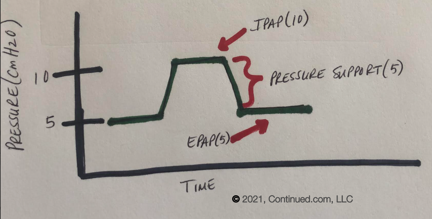
There is evidence suggesting the need for more aggressive settings. Research indicates that patients receiving high-intensity pressure support fare better than those receiving low-intensity pressure support. When prescribing settings for home use, it may be advantageous to opt for slightly higher pressure differentials between inhalation and exhalation, defining pressure support. This calls for a reconsideration of our approach, perhaps leaning towards a more proactive stance than previously adopted.
Hypercapnic COPD patients constitute a distinct phenotype driven by specific physiological factors, such as air trapping, which imposes a significant mechanical disadvantage. Many of these individuals experience nocturnal hyperventilation and are prone to sleep apnea, placing them at heightened risk for adverse health outcomes. They represent a priority target for intervention, as highlighted in the latest GOLD guidelines, which offer a comprehensive discussion of COPD phenotypes worth exploring.
Home non-invasive ventilation holds promise in addressing the underlying physiological issues contributing to hypercapnia. While older literature yielded mixed results regarding its efficacy in various populations, recent clinical trials have demonstrated significant benefits, including improvements in quality of life, reduced hospitalizations, and mortality rates. This emerging body of literature supports a more assertive approach towards its utilization. Additionally, advancements in non-invasive ventilation technology, particularly in optimizing inspiratory pressure, may account for the observed success in home settings.
Effective implementation necessitates a thorough understanding of patient selection criteria and familiarity with available device modes and titration strategies. While literature offers insights into these aspects, practical constraints often limit our options in device selection and mode availability, both in hospital and home settings. Providers typically operate within the confines of specific devices offered by their institution or supplier, thereby influencing treatment decisions. Nevertheless, advocating for access to appropriate devices and modes remains crucial in optimizing patient outcomes.
We are excited about the emergence of telemonitoring, as it addresses a key challenge in the management of patients on non-invasive ventilation transitioning from hospital to home care. In the hospital setting, we have the luxury of being present, continuously titrating the device, and monitoring patient response. However, once patients return home, this level of oversight diminishes significantly. While in-home visits are feasible, they cannot provide round-the-clock surveillance, making telemonitoring an invaluable tool.
Telemonitoring enables us to remotely adjust device settings, enhance patient adherence, troubleshoot issues, and even anticipate exacerbations. Its evolving capabilities hold promise in optimizing non-invasive ventilation outcomes in home settings. Studies have demonstrated the efficacy of non-invasive ventilation in prolonging exercise endurance in COPD patients. However, implementing this during routine activities outside clinical settings poses logistical challenges, as most devices lack portability and usability during normal activities.
Nevertheless, there are exceptions to this limitation, which we will delve into further. Overall, telemonitoring represents a significant advancement in the management of COPD patients on non-invasive ventilation, offering enhanced support and optimization beyond the confines of traditional care settings.
Thoracic Restrictive Disorders
Another group of conditions we frequently consider is what we refer to as thoracic restrictive disorders. This encompasses a broad spectrum of conditions characterized by hypercapnic respiratory failure, where ventilatory defects impede adequate air movement in and out of the chest, resulting in elevated CO2 levels exceeding 45 millimeters of mercury.
A prominent category within this group comprises neuromuscular disorders, including conditions such as ALS, spinal cord injuries, muscular dystrophy, diaphragm paralysis, and spina bifida. Additionally, certain congenital central hypoventilation syndromes may hinder sufficient air exchange. Chest wall deformities, notably kyphoscoliosis, also fall under this classification. Furthermore, ventilatory drive defects, where patients lack the innate urge to breathe adequately, are pertinent considerations.
It is crucial to note that interstitial lung diseases are not typically classified under thoracic restrictive disorders despite causing restrictive pulmonary dysfunction. Consequently, thorough assessment is imperative to rule out obstructive sleep apnea (OSA), which, although less common in these conditions compared to COPD, may still occur. Monitoring CO2 and oxygen levels during wakefulness and sleep is essential post-therapy initiation, with sleep testing reserved for cases suggestive of OSA. Non-invasive ventilation remains a viable therapeutic option for these patients.
Obesity Hypoventilation Syndrome (OHS)
Another disorder to consider is obesity hypoventilation syndrome (OHS), characterized by the combination of obesity (with a BMI greater than 30), daytime hypercapnia exceeding 45 millimeters of mercury for CO2, and a diagnosis of obstructive sleep apnea (OSA). It is crucial to exclude other potential causes of alveolar hypoventilation before confirming this diagnosis.
Given the co-existence of OSA in these patients, continuous positive airway pressure (CPAP) is typically the initial treatment choice. However, CPAP may prove ineffective or poorly tolerated in correcting the ventilation disorder, especially when patients are hypoventilating alongside their OSA. In such cases, non-invasive ventilation becomes necessary to address their respiratory needs effectively.
Careful titration is essential for these patients, particularly regarding pressure selection, to reduce the apnea-hypopnea index to normal levels (less than five). Additionally, treatment must aim to alleviate both hypoventilation and hypoxia. Patients with OHS require comprehensive monitoring and follow-up, including sleep lab titration and clinic visits to assess blood gases and device adherence. Typically, follow-up appointments are scheduled four to six weeks post-initiation of non-invasive ventilation, allowing patients sufficient time to adjust to and utilize the device properly.
Interstitial Lung Disease (ILD)
Interstitial lung disease (ILD) encompasses a diverse array of progressive and debilitating lung conditions characterized by similarities in lung pathology and radiographic findings. However, with hundreds of distinct ILDs identified, the patient population exhibits considerable heterogeneity.
Management of ILD often involves oxygen therapy to maintain adequate oxygen saturation levels, yet achieving this can be challenging due to various factors contributing to limited exercise tolerance in these patients. In instances where oxygen therapy alone proves insufficient, considering adjunctive non-invasive ventilation may be beneficial. Non-invasive ventilation can help improve the ventilation-perfusion (VQ) ratio, reduce respiratory effort, and enhance exercise tolerance, particularly during episodes of exercise-induced desaturation.
While evidence supporting the use of non-invasive ventilation in ILD remains limited compared to other respiratory conditions, preliminary studies have shown promising results. Some studies have demonstrated improved oxygen saturation levels during exercise with the use of non-invasive ventilation, while others have observed enhancements in physical functioning and quality of life following non-invasive ventilation therapy.
Though the evidence base for non-invasive ventilation in ILD is still evolving, it remains a viable option to consider, especially for patients experiencing significant functional impairment. While more research is needed to establish its efficacy definitively in this patient population, non-invasive ventilation stands as a potential intervention to enhance the management and quality of life for individuals with ILD. Now that we have identified the patient groups that may benefit from non-invasive ventilation. Let's discuss the practical aspects of its implementation.
Methods for Non-Invasive Ventilation
- Traditional positive pressure ventilation with various interfaces (NIV)
- Proportional Open Ventilation (POV®)
- Negative pressure ventilation (NPV) or Biphasic Cuirass Ventilation (BCV)
- Tidal Assist Ventilation (TAV®)
Our discussion briefly touches on traditional positive pressure ventilation, which is a commonly utilized method known as non-invasive ventilation. This approach involves delivering positive pressure to the airway through various interfaces, a technique frequently employed in hospitalized patients and increasingly utilized in home care settings.
Next, explore proportional open ventilation, a distinct technique offering notable advantages. Following that, we will review negative pressure ventilation, an older yet still relevant method historically associated with the iron lung, originally developed during the polio epidemics. Negative pressure ventilation, often referred to as Biphasic Cuirass Ventilation (BCV) by the sole provider of this technology, presents unique benefits worth considering. Finally, we will touch briefly on Tidal Assist Ventilation, a newer approach that may gain prominence in the future.
Non-Invasive Ventilation
Traditional NIV
Traditional non-invasive ventilation (NIV), is a widely employed method utilizing interfaces to deliver either volume or pressure ventilation to patients across all age groups, from neonates to adults. These interfaces are meticulously designed to provide a snug fit while allowing for minor leaks to prevent potential pressure ulcers. With hundreds of interface options available, the selection process is critical, considering factors such as patient comfort and tolerance.
One noteworthy interface worth mentioning is mouthpiece ventilation, which is often overlooked in acute care settings but offers substantial benefits to many patients. While some devices feature preset values, others, like AVAPs, offer auto-adjustment capabilities, ensuring consistent tidal volume delivery with each breath.
A primary advantage of NIV is its avoidance of invasive procedures like endotracheal intubation, which come with a host of potential complications. Patients previously tracheostomized due to prolonged invasive mechanical ventilation can potentially transition to NIV, a more tolerable option for many.
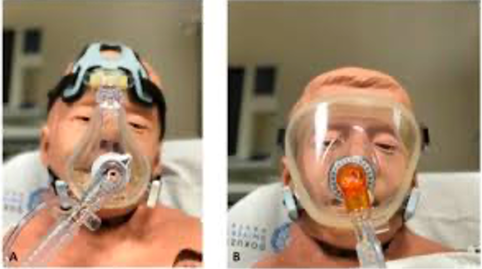
However, challenges persist, primarily centered around patient intolerance or discomfort with the interface. Addressing these issues often involves exploring various interface options and rotating between them to alleviate pressure points and irritation. Ensuring access to a diverse range of interfaces is essential, particularly in home care settings, where patient comfort and adherence are paramount.
While traditional NIV offers a plethora of interface options—from full face masks to nasal prongs and minimalist designs—it is crucial to recognize that access to every interface may be limited depending on the clinical setting. Nonetheless, prioritizing interface comfort and patient preference can significantly enhance the effectiveness and acceptance of NIV therapy, mirroring the challenges faced with CPAP therapy. Thus, a comprehensive approach that incorporates patient-centered care and interface variety is essential to optimize outcomes in NIV implementation.
Mouthpiece Ventilation (MPV)
Mouthpiece ventilation (MPV), often overlooked in acute care settings, serves as a vital daytime non-invasive respiratory support method, particularly for patients with neuromuscular disorders. With MPV, patients utilize a mouthpiece or straw to facilitate ventilation. During MPV, patients intentionally initiate a series of disconnections, whether partial or complete, from the ventilator, followed by reconnections. Essentially, with each inhalation, patients have the autonomy to determine the volume of air they wish to intake. By adjusting the seal with their lips on the mouthpiece, patients can modulate their breaths.
A tight, complete seal ensures nearly full tidal volume (approximately 100%), as dictated by the settings of the volume-cycled ventilator. Conversely, a more relaxed or incomplete seal results in either partial tidal volume or, in some instances, no tidal volume intake at all, ranging from 0 to 99%. This flexibility allows patients to tailor their ventilation to their comfort and respiratory needs, promoting a more personalized and effective approach to non-invasive respiratory support.
Devices
A Breas ventilator is equipped with a mouthpiece that simplifies mouthpiece ventilation. It is typically mounted on an adjustable arm for easy positioning directly in front of the patient's mouth. Theoretically, leaks during mouthpiece ventilation will not cause discomfort because they are determined and managed by the patient. However, this holds true specifically for volume-cycled modes of ventilation. In volume-cycled modes, patients have control over the amount of air they inhale, allowing them to adjust the seal with their lips on the mouthpiece to accommodate intentional leaks without discomfort.
Conversely, in pressure-cycled modes of ventilation, leaks can indeed result in discomfort. This discomfort stems from the ventilator's automatic compensation for leaks by delivering additional flow, which can be excessive and difficult for the patient to manage. As a result, patients may experience challenges in speaking and swallowing during MPV, highlighting the importance of utilizing volume-cycled modes to optimize patient comfort and adherence. Mouthpiece ventilation provides a range of devices, and while the following list is not exhaustive, it highlights the importance of identifying the most appropriate option for each patient.
- Vyaire LTV
- ResMed Astral
- Breas Vivo65
- Breas Vivo45
- PhillipsTrilogy EV300
- Ventec VOCSN
- Movair Luisa
- Phillips Trilogy 100
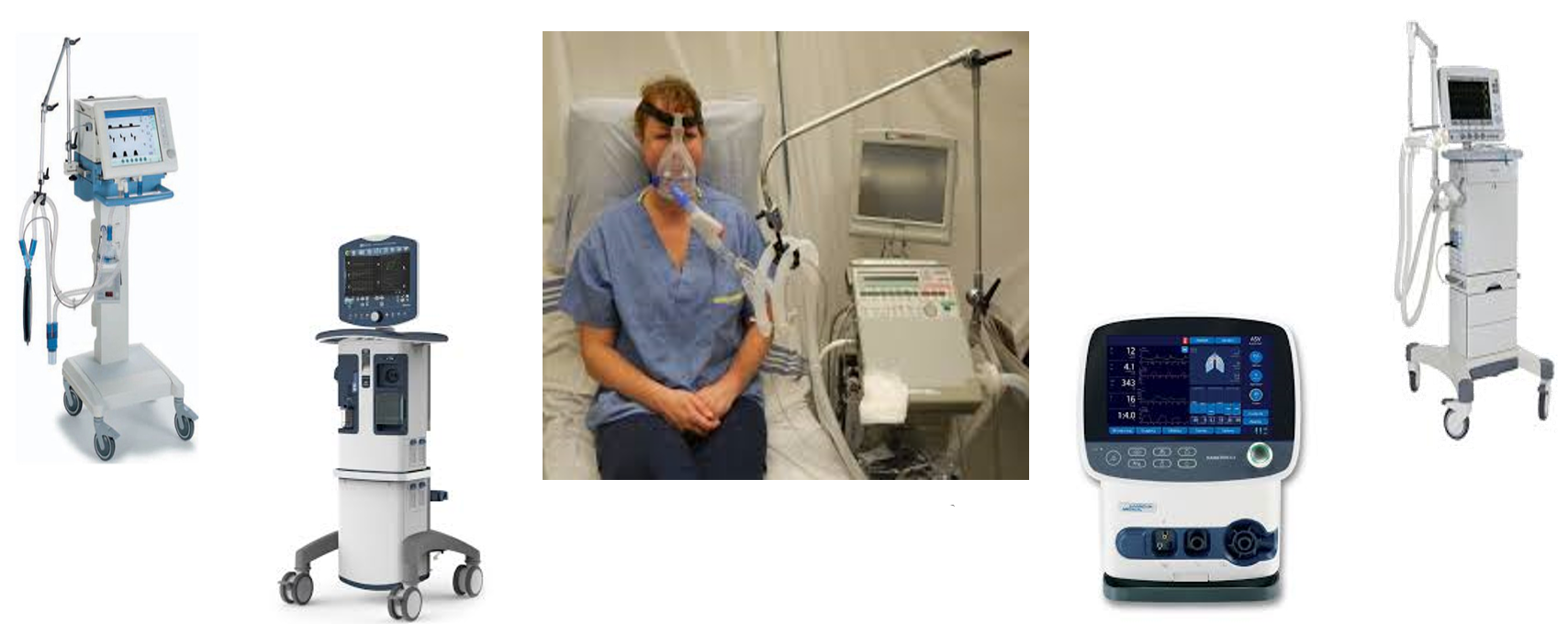
Common options include the Vyaire LTV, ResMed Astral, and various models from Breas, with notable features such as integrated end-tidal CO2 monitoring in Breas devices, enhancing patient care. The Phillips Trilogy and Ventec VOCSN are also prominent choices. The VOCSN, notable for its multifunctionality, integrates ventilation, oxygen concentrator, suction, and nebulizer functions into a single device, streamlining mobility for active patients.
For instance, without the Ventec VOCSN, patients relying on traditional devices like the Phillips Trilogy would need additional equipment for oxygen supply, suctioning, and nebulization, complicating mobility. The Movair Lusia is a newer addition generating considerable excitement, alongside established options like the Phillips Trilogy 100, commonly employed for home use.
While these examples offer insight, collaborating with durable medical suppliers remains crucial to explore available options tailored to individual needs in standard non-invasive ventilation. In addition to the devices mentioned, it is worth noting that advancements in non-invasive ventilation technology continue to evolve, offering enhanced features and capabilities. For instance, some newer models may incorporate innovative algorithms for more precise patient monitoring and therapy delivery. Features such as remote monitoring capabilities, wireless connectivity, and user-friendly interfaces are becoming increasingly common, facilitating seamless integration into patients' daily lives.
Furthermore, considerations such as portability, battery life, and ease of maintenance play pivotal roles in device selection, particularly for patients who lead active lifestyles or require frequent travel. As technology advances, the aim is to provide patients with greater autonomy, comfort, and flexibility in managing their respiratory conditions.
It is important for healthcare providers to stay abreast of the latest developments in non-invasive ventilation technology and collaborate closely with patients and durable medical equipment suppliers to ensure optimal device selection and patient outcomes. By leveraging the benefits of cutting-edge technology, healthcare professionals can enhance the quality of care and improve the overall experience for patients relying on non-invasive ventilation.
Novel Methods of Non-invasive Ventilation
Proportional Open Ventilation (POV)
Let's delve into one of the novel methods of non-invasive ventilation, known as proportional open ventilation (POV). Originally developed by Brief Technologies under the name Non-Invasive Open Ventilation (NIOV), this innovative approach has evolved and is now trademarked by Hillrom as proportional open ventilation or POV. While NILV may still be referenced in the literature as the original terminology, it is essential to recognize the transition to POV.
Proportional open ventilation synchronously delivers positive inspiratory pressure and supplemental oxygen, typically via a nasal pillow interface. It operates by augmenting tidal volume during spontaneous breathing, making it particularly suitable for individuals with limited tolerance for physical activity due to breathlessness. The device offers a myriad of programmable settings tailored to meet the unique needs of each patient. Notably, patients experiencing significant dyspnea during exertion may find proportional open ventilation beneficial.
Baxter Hillrom Life 2000 Ventilator
The interface of this ventilation method incorporates openings that leverage the Venturi principle to entrain room air, which combines with the preset volume from the device and the patient's own effort. The total delivered tidal volume results from the interplay of these factors. Currently available from Baxter Hillrom under the name Life 2000 Ventilator, this technology represents a promising advancement in non-invasive ventilation, offering enhanced support for patients with respiratory challenges.
The Baxter Hillrom Life 2000 Ventilator device has three patient-selectable prescription settings, denoted by pictograms on the right-hand side of the interface. The top pictogram illustrates someone walking, representing the highest level of support suitable for mobility. The middle pictogram depicts someone sitting, which is ideal for home activities and daily routines. The bottom pictogram is designated for restful states.
These three distinct settings offer patients considerable flexibility in tailoring their support levels to match their varying activities and ventilatory needs. Whether they require continuous support or wish to adjust settings periodically, the interface simplifies the process with the push of a button, adapting seamlessly to their breathing patterns.
This adaptability proves invaluable in both clinical and home settings. In the hospital, clinicians can readily titrate ventilation settings to accommodate patients' evolving needs. Conversely, in-home care settings, periodic adjustments by healthcare providers ensure optimal support, as patients may not have the expertise to modify settings independently.
Approved for both invasive and non-invasive use, this device holds promise not only for post-acute care but also for promoting mobility in acute care settings. Streamlining equipment and reducing bulk facilitates patient mobility with ease. Powered by a rechargeable lithium-ion battery, it weighs only about a pound, making it remarkably lightweight and portable—a stark contrast to traditional ventilator setups. Its compact size, akin to a large home telephone, further enhances its practicality and accessibility for patients.
- Clinician adjustable settings:
- PEEP: 0 to 10 cmH2O
- Breath Rate: 1 to 40 BPM
- Breath Timeout Period: 20 or 60 seconds
- Breath Timeout Action: 12 BPM or 3 LPM
- Delivered Volume: 50 mL to 750 mL
- Gas source: Oxygen or Air
- I-Time: 0.15 to 3.00 seconds
- Trigger Sensitivity
The clinician has the flexibility to fine-tune settings within each activity level—rest, sitting, and walking. These adjustable parameters include PEEP (positive end-expiratory pressure), breath rate, and breath timeout period—the duration the machine waits before detecting a cessation in breathing. In the event of breath absence, the device can be programmed to maintain a continuous flow of oxygen or revert to a predefined breath rate, typically 12 breaths per minute.
Furthermore, the clinician can customize settings such as delivered volume, gas source (air compressor or oxygen tank), and inspiratory time—a critical consideration, especially for patients with COPD. Ensuring the device accurately detects patient breaths is paramount, and sensitivity adjustments allow for precise responsiveness.
A comprehensive array of clinician-adjustable alarms is available, covering low respiratory limits, low and high-pressure thresholds, and the breath timeout period. These alarms serve as essential safeguards, alerting clinicians to deviations from desired parameters, including excessively high or low breath rates. Adjustments can then be made promptly to optimize patient comfort and safety—a standard feature across most ventilators.
- Clinician adjustable alarms:
- Low Respiratory Limit: 0 to 119 BPM
- High Respiratory Limit: 5 to 120 BPM
- Low PIP Limit: 0 to 15 cmH2O
- High PIP Limit: 5 to 40 cmH2O
- Breath Timeout Period: 20 or 60 seconds
- High Breath Rate: 5 to 120 BPM
- Low Breath Rate: 0 to 119 BPM
Then there are also some fixed alarms:
- Low/High Source Pressure
- Low Battery
- Low/High Delivery Pressure
- High PEEP Pressure
- System fault
The Life 2000 ventilator incorporates an extensive alarm system designed to alert clinicians to various potential issues. These alarms encompass discrepancies in source pressure, battery status, and delivery pressure, as well as high PEEP (positive end-expiratory pressure) levels, among others. This comprehensive alarm functionality distinguishes the Life 2000 as a full-fledged ventilator, ensuring thorough monitoring and prompt identification of critical situations. The interface commonly paired with the Life 2000 ventilator optimizes its functionality and usability, facilitating seamless integration into patient care protocols.
Breathe Pillows Entrainment Interface
The interface used with the Life 2000 ventilator, known as Breathe Pillows, embodies the principles of POV NIOV (proportional open ventilation, non-invasive open ventilation). It comprises cannulas equipped with tubes for delivering both oxygen and positive pressure alongside small air entrainment ports designed to remain open to allow the machine to draw in air as needed to meet the patient's ventilation requirements. Additionally, the cannulas feature sensing ports to detect the patient's breathing patterns. Despite its advanced functionality, the Breathe Pillows interface resembles a standard nasal cannula, albeit with slightly larger ports entering the nose, available in various sizes ranging from extra small to extra large. Moreover, users have the flexibility to connect this ventilator to alternative interfaces if desired.
Breathe Universal Circuit Connector
The Life 2000 ventilator features a universal circuit connector that offers compatibility with various types of interfaces. Whether the patient prefers using a mask typically utilized for CPAP or BiPAP therapy at night or requires a full face mask or tracheostomy tube during the day, this ventilator can be seamlessly connected to accommodate their needs. This versatility extends to patients with tracheostomies who may benefit from additional respiratory support during periods of activity. The ease of switching between different interfaces makes it convenient for healthcare providers to transition patients from the hospital or acute care setting to the home environment. This universal circuit connector enhances flexibility and adaptability in providing non-invasive ventilation tailored to each patient's requirements.
Versatile Configurations
The Life2000 Ventilation System offers three configurations to maximize versatility. The first is the stationary setup, where the patient remains at home, seated in their chair, connected to a compressor. Additionally, up to 50 feet of tubing can be attached to the compressor, allowing the patient to move around the home while still receiving respiratory support.
For patients requiring support during daily activities such as using the bathroom or preparing meals, the system can be worn while being active within the home environment. It can also be worn outside the home by connecting it to an alternate pressure source, typically an oxygen cylinder. The device is compact and attaches to the patient's waist, with a belt clip for easy carrying. A carrying case is also provided for added convenience.
Research indicates that the Life2000 system can enhance activities of daily living (ADL) performance at home by significantly increasing endurance time, improving oxygenation, and reducing dyspnea, fatigue, and discomfort. This practical solution proves beneficial for oxygen-dependent COPD patients seeking to improve their activity and exercise tolerance.
NIV has demonstrated effectiveness in improving exercise endurance in COPD patients. However, traditional NIV devices are often not designed for portability, making routine use outside of clinical settings challenging. With lightweight devices like POV, providing non-invasive ventilation during exercise becomes much more feasible and convenient for patients, thereby potentially enhancing their exercise tolerance and quality of life.
Novel Methods of Non-Invasive Ventilation
Negative Pressure Ventilation (NPV)
The next type of ventilation we will discuss is NPV or non-invasive positive pressure ventilation. Mechanical ventilation originally began with negative pressure ventilation in the early 1930s, particularly during the polio epidemics of the mid-20th century when many patients developed respiratory failure. Devices such as iron lungs were used, but they required patients to be enclosed, making care challenging. Despite this, negative pressure ventilation saw considerable success. Nowadays, more comfortable devices have been developed, leading to increased interest in using non-invasive negative pressure ventilation due to its improved convenience and comfort compared to earlier iterations.
Hayek Biphasic Cuirass ventilator (BCV)
The Hayek Biphasic Cuirass ventilator (BCV) is a non-invasive ventilator that has gained popularity for its efficacy and versatility. Unlike traditional positive pressure ventilation devices, the Hayek ventilator does not require a mask, eliminating issues related to mask discomfort or interface complications. It utilizes negative pressure ventilation, which helps with lung recruitment, volume expansion, and gas exchange improvement.
One notable feature of the Hayek ventilator is its ability to deliver both negative and positive pressure to the chest wall. This biphasic approach aids in promoting inspiration and expiration, making it particularly beneficial for patients struggling with elevated CO2 levels. With multifunction capabilities, including controlled ventilation, the Hayek ventilator offers clinicians flexibility in tailoring ventilation support to individual patient needs.
The Hayek Biphasic Cuirass ventilator offers several advanced features to enhance patient comfort and respiratory support. One notable feature is its ability to synchronize ventilation with the patient's spontaneous breathing, improving comfort and synchronicity. Additionally, the ventilator includes a built-in high-frequency chest wall oscillation function, which can aid in mobilizing secretions for patients with secretion clearance issues. It also incorporates a cough assist feature, providing additional support for patients with excessive secretions.
The device consists of a shell that is placed over the patient's chest, with a foam backing for comfort. It comes in various sizes to ensure proper fit and comfort for different patients. The ventilator itself is connected to the shell via a simple hose, through which settings such as negative and positive pressure, as well as respiratory rate, can be adjusted. Despite its advanced capabilities, the device is relatively straightforward to use, making it suitable for a wide range of patients, including pediatrics and even infants. Its ability to mimic natural breathing patterns with negative pressure on inspiration makes it particularly advantageous for patients with various respiratory conditions, including those with congenital heart defects who may require extensive surgeries.
The Hayek BCV offers significant advantages in promoting cardiac output and blood return to the chest, thereby supporting better cardiac function. Its multifunctional capabilities, combined with a single interface, simplify respiratory support for patients, eliminating the need to switch between different devices for chest wall oscillation or ventilation. This integrated approach reduces patient discomfort and the need for sedation, particularly in the acute care setting where sedation may be required due to endotracheal tube discomfort.
The synchronization of the ventilator with the patient's spontaneous breathing reduces the risk of hospital-acquired infections associated with intubated patients. Overall, the use of the Cuirass ventilator has been associated with shorter hospital stays, making it a valuable option to consider for patients who could benefit from non-invasive ventilation in both acute and post-acute care settings.
The use of negative pressure ventilation, such as with the Hayek Biphasic Cuirass ventilator, offers several advantages over positive pressure ventilation. Negative pressure ventilation provides more homogeneous lung inflation, ensuring that all parts of the lung receive adequate ventilation, unlike positive pressure ventilation, which may inflate only the already inflated areas. This approach is safer and more effective, reducing the need for lung recruitment maneuvers and potentially harmful interventions.
Negative pressure ventilation mimics the natural intrathoracic pressure wave of spontaneous respiration, promoting cardiac function by facilitating the return of blood to the chest cavity. In contrast, positive pressure ventilation, particularly with PEEP, may decrease cardiac output by impeding blood return to the chest. The biphasic thoracic ventilation provided by the Cuirass ventilator supports cardiac function similarly to spontaneous breathing, offering a significant advantage in patient care. Additionally, the cost-effectiveness and versatility of the device make it suitable for use in both hospital and home settings.
Nocturnal negative pressure ventilation is a highly effective method for preventing sleep-induced reductions in alveolar ventilation, particularly in patients experiencing elevated CO2 levels during sleep. This approach serves as a practical long-term management strategy for individuals with non-obstructive chronic respiratory failure. Unlike positive pressure techniques, negative pressure ventilation supports both inspiratory and expiratory work of breathing in a natural manner, offering patients the ability to eat, speak, and drink without interference.
Moreover, negative pressure ventilation minimizes the risks and complications associated with other ventilation methods, making it a preferred choice for many patients. It has demonstrated effectiveness across various patient groups, including those with neuromuscular disorders, COPD, cystic fibrosis, and even acute respiratory distress syndrome (ARDS). The versatility and favorable patient outcomes associated with negative pressure ventilation highlights its significance in respiratory care.
Inogen TAV
The Inogen Tidal Assist Ventilator (TAV) offers a compact and non-obtrusive solution for patients requiring respiratory support. The device is equipped with three settings: Tidal Assist, Pulse, and Constant. In the Tidal Assist mode, patients can select from five settings ranging from one to five, with each setting delivering varying volumes of air assistance. This mode is particularly beneficial for patients who require additional respiratory support during activities or periods of exertion.
The Pulse mode functions similarly to traditional pulse dose oxygen concentrators, delivering oxygen in short bursts based on the patient's inhalation. The Constant mode provides a continuous flow of oxygen as needed. Patients can adjust the settings based on their oxygen requirements and activity levels.
The device comes with a regulator and specific adapter to ensure compatibility and proper functioning. It offers flexibility and convenience for patients needing respiratory assistance in various situations, whether at home or on the go. The TAV is specifically designed for patients with COPD, providing assisted non-invasive ventilation in a portable and lightweight unit. Similar to the Life 2000 ventilator, it offers simplicity and ease of use. The device features settings ranging from one to five, where each setting delivers a different volume boost to the patient's breathing, ranging from 50 to 250 milliliters.
The technology is integrated into the nasal interface, utilizing two tiny discs surrounded by pillows to provide higher oxygen flow pressure, up to five times the volume per breath compared to oxygen alone. The device operates on a single AA battery, making it convenient for patients to carry with them, weighing only 3.8 ounces with the battery installed.
Although there is limited published literature and reimbursement challenges, studies have shown promising results. A study conducted at a pulmonary rehab center in California demonstrated that patients using oxygen therapy with the TAV were able to increase their exercise tolerance compared to those using oxygen therapy alone. This indicates the potential for the TAV to improve patient outcomes and increase exercise capacity in COPD and other respiratory conditions.
NIV Comparisons
Indeed, traditional non-invasive positive pressure ventilation (NIPPV) with a mask or other interface has several advantages:- Well-established: NIPPV has been extensively used and studied, making it a familiar and reliable option for healthcare providers.
- Hospital to home transition: Hospitals are proficient at setting up NIPPV for patients, facilitating a smooth transition to home care.
- Variety of interfaces and devices: There are numerous interface options available, allowing for customization to meet individual patient needs and preferences.
- Useful across multiple disorders: NIPPV has shown efficacy in various respiratory disorders, including COPD and thoracic restrictive disorders, based on extensive clinical experience and research.
On the other hand, proportional open ventilation (POV) offers unique advantages, particularly for activities of daily living (ADLs):
- Portability: POV devices are designed to be portable, allowing patients to engage in ADLs and other activities while receiving ventilation support.
- Adaptability: POV devices have multiple settings that can accommodate changes in activity level, providing flexibility for patients as they move about.
- Comfortable interface: The nasal pillows interface used with POV devices is comfortable and minimally obtrusive, enhancing patient comfort and compliance.
Overall, while traditional NIPPV is well-established and effective, POV offers additional advantages in terms of portability, adaptability, and comfort, particularly for patients engaging in daily activities. The choice between these options should be based on individual patient needs and preferences, as well as clinical considerations. Certainly, while non-invasive ventilation (NIV) offers many benefits, it also comes with some disadvantages:
- Cumbersome with activity: Standard NIV devices can be challenging to use during physical activity. Patients may require assistance, and the equipment can be cumbersome, hindering mobility.
- Difficulty in adjusting to the interface: Some patients find it challenging to adjust to the interface, such as masks or nasal cannulas used in traditional NIV. Discomfort or dislike of the interface may lead to non-compliance with treatment.
- Risk of pressure sores: Proper application of NIV devices is crucial to prevent leaks, which can lead to pressure sores or skin irritation. Achieving a tight seal without causing discomfort can be challenging.
- Minimal leak requirement: NIV devices typically require a minimal leak to maintain effective ventilation. Balancing the need for a seal with the risk of pressure sores can be challenging for healthcare providers.
In contrast, POV devices, such as the Breathe Pillows, offer a comfortable interface and may mitigate some of these disadvantages. The nasal pillow interface used with POV devices is designed for comfort and may be more tolerable for patients. Additionally, the ability to adjust settings and accommodate changing conditions provides flexibility and may improve patient compliance. Overall, while NIV has its challenges, advancements in technology, such as POV devices, aim to address some of these issues and improve patient comfort and compliance with non-invasive ventilation therapy.
We have encountered some pressure sore issues with NPV, where the pressure may build up on the chest due to the cuirass shell's contact with the skin. However, we have implemented effective strategies to mitigate this problem, so it is typically not a major concern. Negative pressure ventilation, or NPV, is not as widely embraced as other methods. Many healthcare professionals have moved away from NPV, as it may not be as comfortable or familiar as positive pressure ventilation. This highlights a disadvantage that warrants further consideration and exploration. NPV also necessitates good upper airway function, as patients must manage secretions and cough effectively. If a patient struggles with these aspects, NPV may not be suitable for them. Additionally, NPV lacks mobility, meaning it cannot be easily used while performing daily activities or moving around. In summary, I hope this overview has provided valuable insights into the various available non-invasive ventilation methods, along with important considerations for patient care.
Questions and Answers
How are hospitals adapting to the use of newer non-invasive ventilation devices in the post-acute care setting?
Hospitals face limitations in accessing a wide range of positive pressure devices due to budgetary constraints. While some hospitals may have a variety of options, many are restricted to one or two devices. This limitation necessitates careful consideration of available options and familiarity with patient needs. Additionally, hospitals may have access to programs like the hospital-to-home program, which assists in setting up patients for home use of specific devices.
What advice is offered for selecting the appropriate non-invasive ventilation device?
It is crucial to explore all available options and consider patient-specific needs when selecting a device. Understanding the patient population and their requirements is essential. However, certain devices, such as the Life 2000 POV, may require involvement from external companies like Hillrom for setup and support.
Are there specific considerations for non-invasive negative pressure ventilation?
Yes, non-invasive negative pressure ventilation requires specialized equipment and expertise. Companies like Hayek provide assistance in selecting and implementing these devices, offering training and ongoing support. This option presents a unique alternative for patients with specific respiratory needs.
How important is it to recognize that there is no one-size-fits-all approach in selecting non-invasive ventilation devices?
Recognizing the diversity of patient needs is paramount. Each patient may require a tailored approach, and what works for one individual may not be suitable for another. Therefore, understanding the available options and patient-specific requirements is crucial for effective care delivery.
References
Select references are listed here. A complete reference list is provided in the course handout.
Annunziata, A., Calabrese, C., Simioli, F., Coppola, A., Flora, M., Marotta, A., Di Spirito, V., Didonna, F., Cicalese, M., & Fiorentino, G. (2022). Negative-pressure ventilation in neuromuscular diseases in the acute setting. Journal of Clinical Medicine, 11(9), 2589.
Brochard, L. J. (2023). Mechanical ventilation: Negative to positive and back again. Critical Care Clinics, 39(3), 437–449.
Cooksey, J. A., & Sergew, A. (2020). Noninvasive ventilation in amyotrophic lateral sclerosis. Sleep Medicine Clinics, 15(4), 527–538.
Cuerpo, S., Palomo, M., Hernández-González, F., Francesqui, J., Albacar, N., Hernández, C., Blanco, I., Embid, C., & Sellares, J. (2020). Improving home oxygen therapy in patients with interstitial lung diseases: Application of a noninvasive ventilation device. Therapeutic Advances in Respiratory Disease, 14, 1753466620963027.
Dong, S., Lu, W., & Wang, Y. (2023). Positive- and negative-pressure ventilation characterized by local and global pulmonary mechanics. American Journal of Respiratory and Critical Care Medicine, 207(6), 800.
Dretzke, J., Wang, J., Yao, M., Guan, N., Ling, M., Zhang, E., Mukherjee, D., Hall, J., Jowett, S., Mukherjee, R., Moore, D. J., & Turner, A. M. (2022). Home non-invasive ventilation in COPD: A global systematic review. Chronic Obstructive Pulmonary Diseases, 9(2), 237–251.
Frazier, W., van Eijndhoven, E., Murphy, R., & Jena, A. B. (2021). Noninvasive ventilation at home reduces mortality in COPD with CRF. American Journal of Managed Care, 27(9), e308–e315.
Gong Y, Sankari A. Noninvasive Ventilation. [Updated 2022 Dec 11]. In: StatPearls [Internet]. Treasure Island (FL): StatPearls Publishing; 2024 Jan-. Available from: https://www.ncbi.nlm.nih.gov/books/NBK578188/
Citation
Aloan, Claire (2024). Non-invasive Ventilation in the Post-Acute Care Setting. Continued.com - Respiratory Therapy, Article 218. Available at www.continued.com/respiratory-therapyAug 28, 2023
1 CE HourHigh Flow Nasal Oxygen: What’s What!
Jessica Fino, RRT, EdD
Editor’s note: This text-based course is an edited transcript of the webinar, High Flow Nasal Oxygen: What's What!, presented by Jessica Fino, EdD, RRT.
Learning Outcomes
After this course, participants will be able to:
- Describe the different approaches to providing high flow oxygen.
- Identify the indications for use across high-flow devices.
- Recognize the role of respiratory therapists in device selection and progression of non-invasive support.
Introduction
I wanted to talk to you a little bit today about high flow nasal oxygen. There are a lot of terms that are tossed around. From my perspective, when working with students, I know there can be some confusion as some terms mean the same thing. For example, we have similar terms: high-flow nasal cannula, high-flow nasal oxygen, high-flow nasal therapy, heated high-flow nasal oxygen, heated high-flow nasal cannula. I have experience using these in acute care, the ICU, and the NICU.
What is High Flow Oxygen?
- High flow oxygen systems include those that can supply a flow that meets or exceed the patient’s peak inspiratory flow.
- These systems use air-entrainment or blending systems to create precise Fi02.
For oxygen devices, there are high-flow, low-flow, and reservoir systems. Many times, the terms high-flow and high liter flow all get mixed. When by definition, they are quite different. A high-flow oxygen therapy system includes those that supply a flow that meets or exceeds the patient's inspiratory flow demand. Many of these terms are used synonymously when they are very different. You might have learned the magic box or some other formula to calculate the total flow of an oxygen device. When we are talking about total flow, that is how we get a true high-flow device.
Although they can get to higher liter flows, traditional oxygen delivery devices are often diluted by room air, so the patient is breathing oxygen and room air. The FiO2 then becomes variable. These devices cannot meet the definition of true high-flow because they do not bring any additional flow from another source to increase the total flow of the device. Systems that do meet this are high-flow air entrainment and blending systems. An entrainment system, for example, would be a venturi mask. A blending system would be just like it sounds-- a machine that has a blender built inside of it so it can create a precise FiO2.
Who Needs High Flow Oxygen?
- Typically high flow oxygen is reserved for severe oxygenation issues.
- Patients with high inspiratory flow demands
- Patients who need help in C02 removal
- Can be used in all patient populations
What kind of patients need this type of therapy? Some patients potentially have severe oxygenation issues but also need to breathe spontaneously. However, they need to be able to protect their airway. They can do mild to moderate work of breathing but not severe work of breathing. Usually, by the time they have reached that level of muscle fatigue, it is too late in the game to try high-flow nasal oxygen.
Those with high inspiratory flow demand are another group that needs high flow oxygen. This is one of those terms that is not tossed around in everyday language when talking about taking care of patients. Terms that may be a little more familiar to you would be large tidal volumes, short inspiratory times, and increased minute ventilation. They have increased work breathing or an increased drive to breathe. Those are the type of patients you will see with increased or high inspiratory flow demands.
An aspect of high-flow nasal oxygen therapy is that it is also capable of CO2 removal. Sometimes, this device gets intertwined with non-invasive therapy, but the way CO2 is removed using this type of therapy is slightly different. We will discuss this a little bit later.
A considerable benefit to high-flow nasal oxygen and nasal cannular therapy is that it has been designed for all patient populations. Depending on the device or the model you are using, which I will also discuss in just a little bit, some therapies will work great for the neonatal population to the adult population and everything in between.
One question that I am frequently asked when teaching this to students or discussing high flow with someone who is not well versed in using it is, "Why cannot I just put a non-rebreather on this patient?" If we are looking at giving them higher FiO2, the non-rebreather mask goes up to 15 liters. However, in these cases, we need to provide them with more FiO2. It has been tossed around about starting with a non-rebreather mask first, but there are instances where you should and instances where you should not.
Alternative Types of Masks/Devices
Non-Rebreather Masks
- Non-rebreather masks are capable of higher liter flows.
- They are not high flow (total flow) devices.
- Fi02 is variable
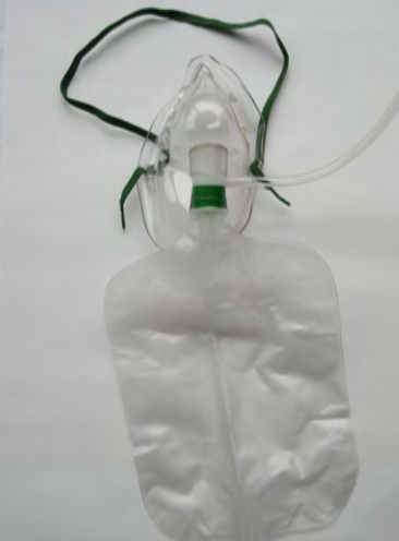
Here is a quick review of the non-rebreather mask. It runs up to 15 liters a minute. It does have a high liter flow, but it is not a high flow total flow device.
Figure 1. Non-rebreather mask.
When we look at total flow, we have to have some kind of entrainment of air/oxygen mixture. This is where we are increasing the total output that the device is capable of delivering. The non-rebreather does not do that. The non-rebreather can provide approximately up to 70% FiO2, but again it is variable. The reasons why it is variable depends on the way the patient is breathing. It also depends on how well the mask fits the patient's face. You could see two patients put on the same settings of a non-rebreather, but if you were capable of measuring the FiO2 being delivered, it could be pretty different even if they were both on a 15-liter non-rebreather mask.
There are quite a few variabilities in the non-rebreather. This is not saying it is not a suitable device or should not be used, but it is in a different category. It is sometimes intertwined with high flow oxygen when people talk about delivering higher FiO2s and higher liter flows.
Venturi Masks
- Venturi masks/ AEM masks are true high flow (total flow) devices.
- The air - entrainment increases the total output of flow.
- Deliverable FiO2 is lower but generally more precise.
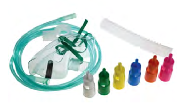
There is the venturi mask (Figure 2).
Figure 2. Venturi mask.
The venturi mask is sometimes referred to as the air entrainment mask. This is a true high-flow device. Remember, it can meet or exceed the patient's inspiratory flow because it mixes the air and oxygen to increase a total flow and a greater output. The deliverable FiO2 is precise, and the color-coded devices give a specific FiO2. A downside to these is that most devices, or at least the most familiar ones, max out at about 50%. As there are patients that need more than 50% FiO2, what do we do in these situations? We need to give them a total flow device to meet that inspiratory demand. They need more than 50% FiO2, thus creating the need for high-flow nasal cannular devices.
High (Liter) Flow Cannula
- High (liter) flow cannulas deliver oxygen at greater flow rates than a standard nasal cannula.
- Most max out at a flow of 15 lpm and require humidification.
- In most situations, this is not a high flow device and the Fi02 is variable.
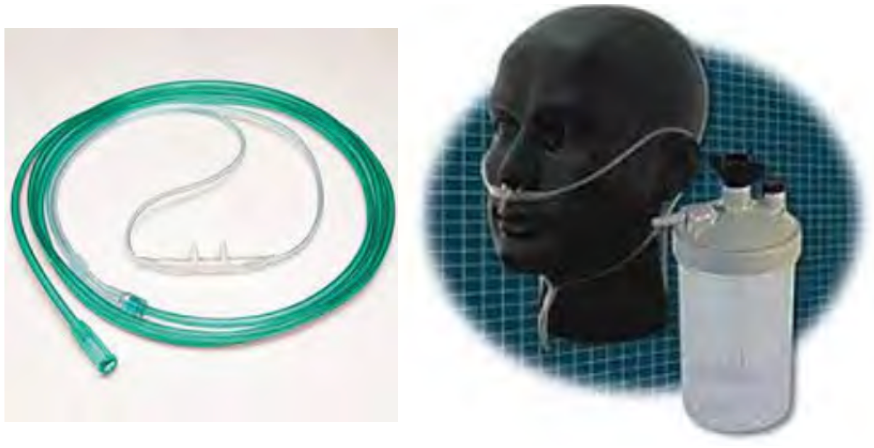
This is another one that I have seen out there that can be confusing. This particular device is referred to as a high flow nasal cannula, as seen in the two examples in Figure 3.
Figure 3. Examples high flow cannulas.
It is not only a high flow nasal cannula, but it is a high liter float nasal cannula. If you are familiar with using this particular cannula, it will go up to approximately 15 liters. I used this cannula several years ago when I first started practicing, and then it disappeared. It has resurfaced again in recent times. This cannula is designed like a traditional cannula, but it does not max out at that 6-8 liters that a conventional nasal cannula will. This one will go up to 15 liters per minute, but it does require humidification. You can see the refillable humidifier device in the right picture. A downside to this is there is a potential for cross-contamination when you use refillable devices. If this device works well for your patient, that is great.
A higher FiO2 will meet some client's oxygen needs. Again, this is still a variable FiO2 variable device, and it is not a total flow device. It is a high liter flow device because the patient will still be able to breathe room air around the prongs, around their nares, and through their open mouth. The amount of oxygen that they are taking in will be diluted, and the FiO2 will be variable.
When These Are Not Enough
- High inspiratory flow
- High tidal volume
- Fast rate of breathing
- Large minute ventilation
- Short inspiratory time
There are some instances when the devices I have just talked about are not enough. What is going to be the right approach? How do you know when you should start with a high-flow nasal cannula versus some of the other devices that I just mentioned? It is not that those devices are harmful. Except for the air entrainment or the venturi mask, all of the others are genuinely low-flow devices. There are instances where we would start a patient on a high flow nasal cannula instead of some low flow devices. They include someone who does have a high inspiratory flow, a high or a large tidal volume, or an individual with a fast breathing rate, so the respiratory rate is elevated. Another way of describing that is large minute ventilation with a short or a quick inspiratory time. These specific conditions make using a high flow nasal cannula more favorable to meet the patient's inspiratory needs.
High Flow Nasal Cannula (HFNC) Review
- High flow nasal cannulas offer flow rates up to 60 lpm with precise FiO2 concentrations up to 100%.
- Humidification is often incorporated into the device setup.
- Indications are continually evolving with numerous benefits in both adult and pediatric populations.
- New devices are being developed frequently to meet the demand for this type of device.
Here is a quick review of high flow nasal cannula therapy before breaking down some of the devices and looking in a little more detail. In general, high flow nasal therapy or high flow nasal cannulas offer flow rates up to 60 liters per minute while offering a precise FiO2. Most of the time, those concentrations will go up to a hundred percent. One of the tricks that I teach my students is the rule of four, where one liter is approximately 24% oxygen, two liters is about 28, 3 liters is 32, and so on. High flow nasal cannulas have a blender system already incorporated into the device. The air and oxygen are blended manually. I can offer a precise FiO2 that is no longer dependent on liter flow with this device. For example, I can deliver 50% oxygen at 40 liters. I can offer 100% oxygen at five liters in a NICU setting. Oxygen concentration and amount of liters are no longer dependent upon each other in a high flow nasal cannula, differentiating it from some of the other oxygen therapy systems.
Humidification is often incorporated in the high-flow nasal cannula setup. I will talk to you in a minute about why that is so important. The indications of HFNC use are continually evolving, with numerous benefits in pediatric and adult populations. There are new devices that are constantly and frequently being developed. High-flow nasal cannula therapy is gaining widespread use in the treatment of COVID, especially in the early-onset trying to avoid intubation. There are several benefits that I will break down and talk about in just a few minutes.
The Role of Humidity
- All humidity is NOT equal!
- Heated humidity is required for all HFNC.
- The flow rates generated by HFNC can cause the isothermic saturation boundary to shift distally.
- This can lead to further complications.
The reason why humidity is so essential in HFNC is that all moisture is not equal. Some of the traditional devices, like the ones I just showed you, are not heated. If we are using a high-flow nasal cannular therapy, it has to have heated humidity.
Isothermic Saturation Boundary
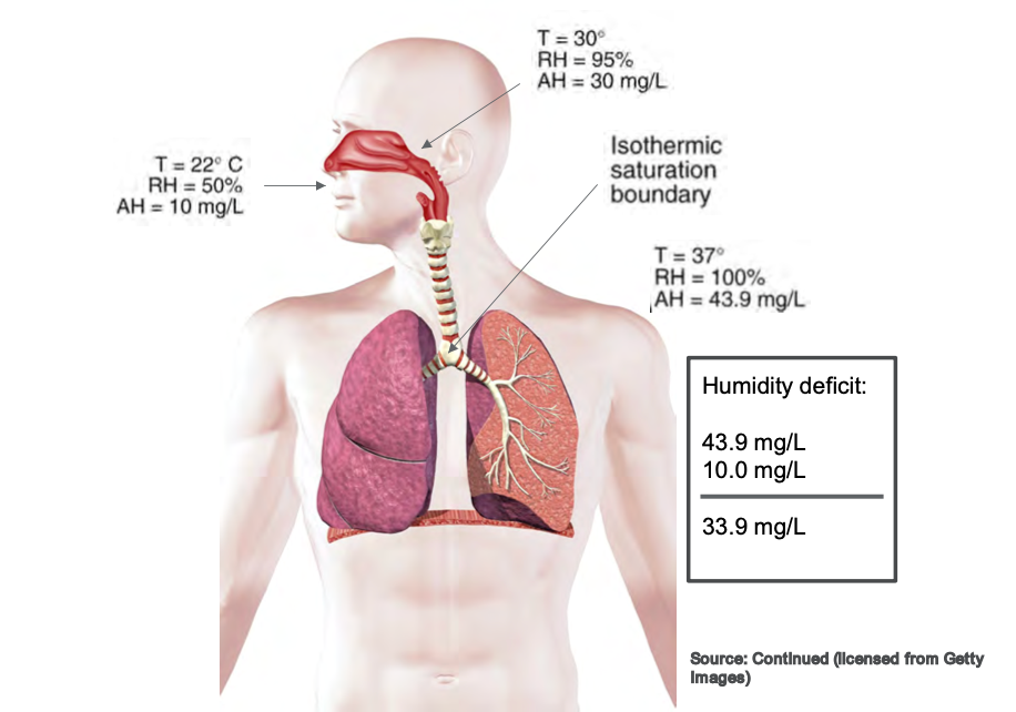
The flow rates generated by a high flow nasal cannula can cause the isothermic saturation boundary to shift. When that occurs, we can cause all sorts of complications in the airway, and the isothermic saturation boundaries shifts. This can cause mucosal trauma, dried secretions, and impaired mucociliary escalator function. It results in a myriad of complications that we do not want in the upper airway because we want our patients to cough, clear their airways, and maintain normal function of the upper airway. Whenever we use high flow therapy, those flows are too significant for the regular operation of the isothermic saturation boundary to overcome. This is why we have to add heat so that we can help balance that out. Figure 4 shows a picture of the isothermal saturation boundary.
Figure 4. Illustration of the isothermic saturation boundary. Click to enlarge the image.
The primary function of the nose is to warm and humidify inspired gas. If the temperature was approximately 22 degrees Celsius, the relative humidity would be about 50%. If we increase the temperature by the time it reaches the oropharynx at the back of the throat, we are looking at a higher temperature of about 30 degrees Celsius and relative humidity of 95%. Movement from the nose to the oropharynx causes a significant increase in temperature and relative humidity. By the time it reaches the isothermic saturation boundary around the carina before it enters the lung, we want it to be full relative humidity at a hundred percent saturated and our average body temperature of 37 degrees Celsius. That is a whole lot of words to say that the nose warms the inspired air typically. When we put a nasal cannula in someone's nose, we are potentially disrupting that normal function.
When we use heated high flow therapy, we are generating flows that far exceed the patient's normal inspiratory flow, and the nose cannot warm those gases fast enough. It is imperative that we use heated humidity for high flow therapy because we do not want to negatively impact the humidification and the normal function of the upper airway itself.
HFNC: How It Works
- HFNC improves oxygenation by:
- Washing CO2 from end expiration
- Reducing dead space
- Providing positive end-expiratory pressure
- Optimal PEEP levels are directly linked to a regular breathing pattern and a closed mouth
(Dres and Demoule, 2017)
It improves oxygenation in these three ways. First, it washes out the CO2 from the end expiration. Every time a person exhales, they have end-expiratory gas that remains in the upper airway. Whenever they exhale, there is a little bit of CO2 that is left in that end expiration. Whenever we use higher flows, we wash out CO2 to have a more oxygen-rich environment when the patient goes to inspire, and there are fewer traces of those end-expiratory gases. Ultimately, this increases the oxygen that is being delivered to the patient.
We can also reduce dead space. Dead space is anywhere that oxygen exchange cannot occur. Oxygen exchange cannot happen in any upper airways like the nose, sinuses, or trachea. Oxygen exchange cannot occur until deep in the lungs, where it crosses over in the alveoli and membranes there. If we can reduce dead space by generating flows that get the oxygen down to where it can be absorbed, not only are we going to improve oxygenation, but chances are we will also improve the work of breathing. The patient is not having to work as hard to overcome their normal anatomical dead space to get that oxygen down into their lungs where it needs to be.
Over the last few years, there have been varying studies about whether high flow therapy provides PEEP or positive end-expiratory pressure. Dres and Demoule (2017) reported that optimal PEEP levels were directly linked to a regular breathing pattern and a closed mouth. This means that if the patient has a normal rate and pattern, they can get up to approximately seven centimeters of pressure. However, we know that that is not always the case. Sometimes our patients are talking, eating, or taking medications, and their mouth is open. We cannot get them to keep their mouth closed at all times. So yes, PEEP is possible.
We know that PEEP increases the alveoli's surface area, creating lung expansion and improving the potential for oxygenation. However, it is a catch-22 when we look at it as a non-invasive support device. I will talk about that in a little bit more detail later. Another interesting aspect I found about this study is that they also reported not only positive end-expiratory pressure but also for every 10 liters of the flow used on a high flow therapy device, there was an increase in central airway pressures. This was a .69 increase if the patient's mouth was closed and a .35 increase for the same user if the patient's mouth was open. These are not huge increases, but if you have a patient with high main airway pressures, you need to be mindful of this.
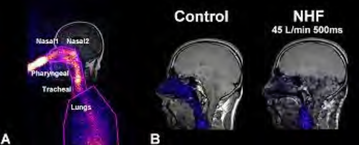
Looking at the CO2 and nasal washout, this is an excellent illustration in Figure 5 that Moller et al. (2017) saw in their study.
Figure 5. CO2 and nasal washout using tracer gases. Click here to enlarge the image.
They had the patient inhale a tracer gas to see a visual of the nasal washout. Looking at the first picture, the gas is inhaled so that it can be seen. In the middle, you can see that the end-exhalation gases remain in the nasal and upper airways in the control subject. However, with the individual on high flow therapy (45 liters per minute), you can see that those gases are almost non-existent in the nasal airway. There is a drastic improvement in oxygenation because we have washed out CO2 and primed that nasal cavity for higher oxygen concentrations. This is why it is particularly effective in individuals who have hypoxemic acute respiratory failure.
Types of HFNC
Let's now look at the different types of high-flow nasal cannulas. I am just going to talk about a few. I do not claim to be inclusive of all devices that are on the market. I spoke with a few therapists that I know, and these are the main ones that everyone seems to be currently using.
Optiflow
- Provides flowrates from 2-60 lpm
- With Airvo 2, this device can be used during transport
- Several interfaces available to increase patient comfort and compliance
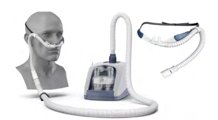
The Optiflow is one of them. This version is made by Fisher & Paykel, and this device uses flow rates of 2-60 liters per minute. It has several patient interfaces to fit the needs of various patient populations. A benefit to using this device is that it has compatibility with the Airvo 2. The Airvo 2 is a humidification integration device that has a built-in humidification and flow generator. It generates its flow, making it easier to set up and transport per the feedback I got from many therapists. Figure 6 shows an example of this.
Figure 6. Example of an HFNC device with a humidification integration device.
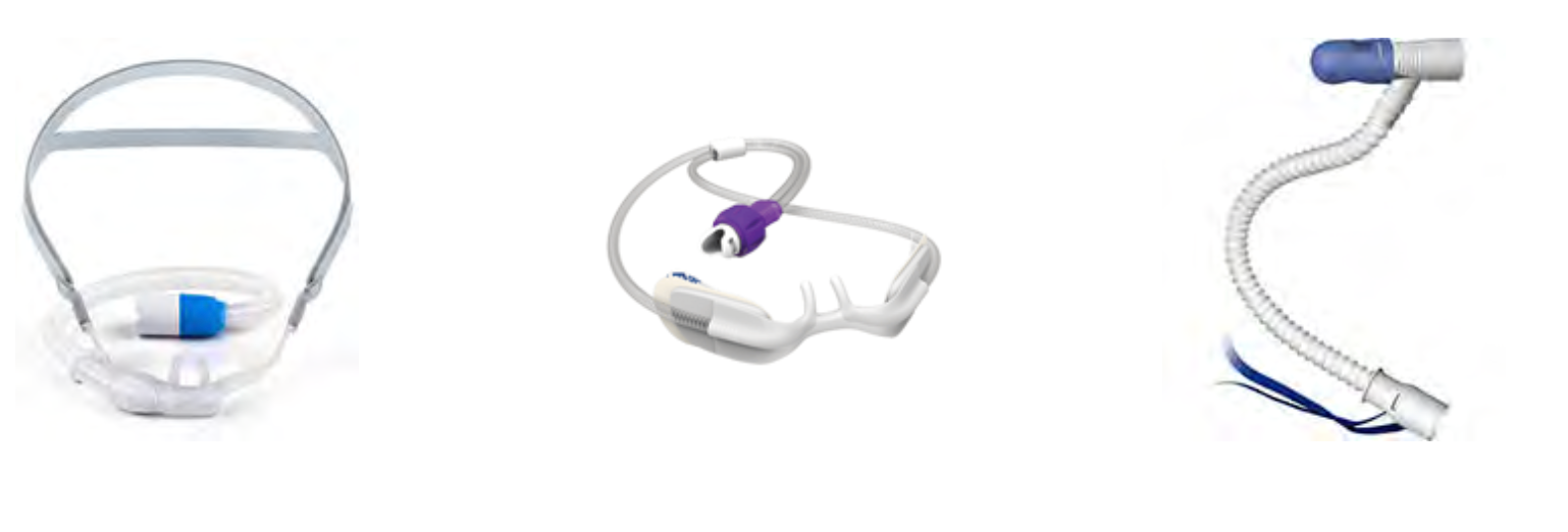
It also improves the accessibility of this device. In one older hospital where I worked, some floors did not have multiple oxygen outlets, or some of them only had oxygen outlets and no air outlets. I know this may still be the case in some areas. Ideally, all hospitals should have multiple oxygen and air wall outlets for multiple device access. The Optiflow has its flow generator, which helps with accessibility. Figure 7 shows several interfaces available to increase patient comfort and compliance.
Figure 7. Examples of Optiflow patient interfaces.
These can range in sizes from infants to adults. Adult models come in small, medium, and large, and there are infant and pediatric sizes and trach attachments. If you get on the Fisher & Paykel website and look up their Optiflow, there was also a mask interface. I could not find much information or any patient testimonials, but there is an aerosol-type mask that will also work with the Optiflow. I asked around, but I do not know anyone who has used it.
Vapotherm
- Flow rates of 1-40 lpm
- Can be integrated into EMR and nurse call systems.
- HeliOx compatible for 80/20 mixture
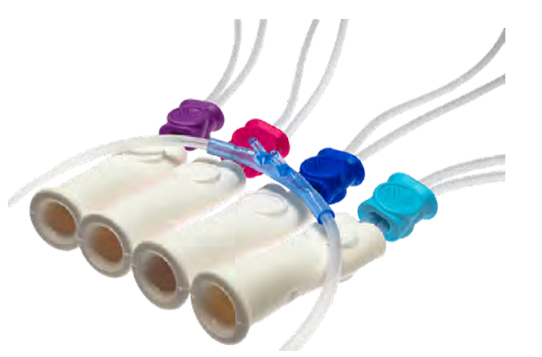
- Multiple cannula sizes are available (premature, infant, pediatric, adult, adult large).
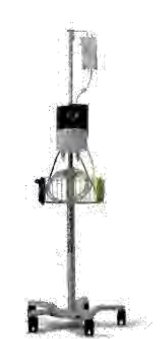
Vapotherm is the HFNC that I am well-versed in as I used it quite frequently. This particular device has flow rates of 1-40 liters per minute, so a little bit lower than the Optiflow (Figure 8).
Figure 8. Vapotherm on a rolling stand.
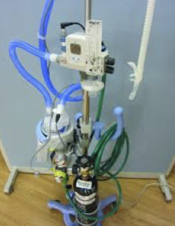
The newer models can be integrated into the EMR, and the nurse call light systems. This is something new that I found out when preparing for this lecture. If they were to run out of humidification in the past, you would have to be in pretty close proximity to hear it alarming. There was also a concern that these devices might overheat. It is a gravity-feed liquid system, but it just has to be watched and replaced from time to time to ensure that the humidifier continues to function the way it is supposed to. Now, this system can be integrated into the existing call light, which is a huge benefit to this device. It is also compatible with HeliOx with an 80/20 mixture. I searched, and I did not find many patient studies about the use of a high flow HeliOx therapy, but it could be because it is brand new. This is an area that I will also continue to look at to see how it is integrated into practice.
The Vapotherm also offers quite a few interfaces, even more so than the Optiflow (Figure 9).
Figure 9. Various size interfaces of the Vapotherm.
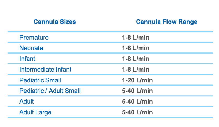
There are cannulas for preemies, infants, children, adults, and even adults large. Eight different interfaces are available on the market through the Vapotherm site. There are various settings, as shown in Figure 10.
Figure 10. Cannula sizes and flow range chart for various ages.
The premature style ranges from 1-8 liters per minute; neonate, infant, and intermediate infant all have that same liter flow range. The spacing and the size of the prongs help you to determine how best to fit a client. The pediatric setting is a flow of 1-20 liters. Once we get up to the adult small, can crossover from pediatric, the flow range is 5-40 liters per minute.
Why are there so many different sizes? Everyone's nose is different, like the spacing of their nares and how far apart they are. I know this was my experience. It is essential, especially in the NICU with the preemies, that it fits correctly. Even in the adult setting, there was a lot of variability in nose structures. As we know, compliance is dependent on patient comfort.
HFNC Convertible Ventilators
- Some ventilator models have HFNC capabilities integrated into the vent.
- ▪ Hamilton C-6, G-5, C3, C1, and T1
- ▪ Offers more complex features in addition to
- HFNC
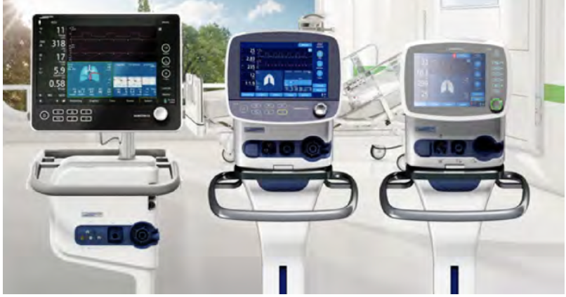
A few ventilators on the market today are convertible to high-flow nasal cannular devices (Figure 11).
Figure 11. Ventilators that convert to HFNC.
In particular, the Hamilton models (C-6, G-5, C3, C1, and T1) can be converted. I have not used any of these. I know at Midwest State, where I teach, we have the Hamilton G-5. This is a ventilator with widespread use in many of my clinical affiliates. It offers a lot of additional monitoring. High-flow nasal cannulas provide a built-in SpO2 monitor. The Hamilton is not only a ventilator that can convert to a high-flow, but it also has SpO2 capabilities.
The flow rates of these devices are up to 60 liters per minute. The G-5, in particular, offers integration with the Aerogen nebulizer. You can also incorporate the nebulizer capabilities with this device to be delivered through the high flow nasal cannula. The interfaces that are available with this device include adult, pediatric, and neonatal varieties and trach attachments.
These are all of the high flow nasal cannula studies that I could find.
HFNC Vs. NIV
- HFNC does generate PEEP however it varies across patients.
- When compared to traditional oxygen masks and NIV, the outcomes were similar.
- However, when initiated early in patients with hypoxemic ARF intubation rates were significantly lower.
(Dres and Demoule, 2017)
How do you know when to use high-flow nasal cannula therapy versus non-invasive? Or, when should you have started non-invasive instead of high flow? We do offer the PEEP and expansion, so there are some of the same similarities here. How do you know which one is best? Unfortunately, there is not a hard, fast rule. In my opinion, they offer some similar features, but it is like comparing apples and oranges.
The high flow nasal therapy does offer PEEP. However, it varies across patients and interfaces. As mentioned, depending on if your patient has their mouth open or closed, their PEEP will vary all day long. Whereas, with NIV, you can set the PEEP and inspiratory/expiratory pressure depending on the modes you are using. This is pretty much what you will get with the high flow if they are opening their mouth, talking, awake, sleeping, etc.Compared to traditional oxygen masks and non-invasive ventilation, HFNC had very similar outcomes in a study by Dres and Demoule (2017). I want to discuss that a little bit. They evaluated patients who were admitted primarily for pneumonia. Once the patients were admitted, they were assigned to traditional oxygen masks, NIV, or HFNC. Initially, the study found that the outcomes were similar in almost half of the subjects. Across all three categories, subjects failed and had to be intubated. Most of the time, high flow nasal cannula therapy failure is marked by the need for intubation, not the progression to non-invasive. This is where I think a lot of therapists ask the question, "If high-flow nasal cannula therapy fails, do I go to non-invasive, or do I go to intubation?" All of the studies that I found marked high-flow nasal cannula therapy failure as the movement to intubation, not the progression to non-invasive. For individuals who presented with severe hypoxemic acute respiratory failure and used high flow nasal cannula therapy, their failure rate was significantly lower. So, when you look at the patients who not only had pneumonia but also had a hypoxemic acute respiratory failure with frequent or early-onset initiation of high-flow nasal cannula therapy, they did have a favorable outcome. Their intubation rate was lower compared to individuals who were put on non-invasive or traditional oxygen masks.
Recommendations for Clinical Practice
- Assessment of Need: Determined by measurement of inadequate oxygen tensions and/or saturation, and the presence of clinical indicators.
- AARC Clinical Practice Guidelines for Oxygen Therapy
- Adults: Acute hypoxemic respiratory failure,
- Postoperative support, pre- intubation,
- post-extubation respiratory failure, DNR/DNI
- Pediatric: Asthma, bronchiolitis, pneumonia
- Neonatal: RDS and stabilization of preterm infant, post-extubation
Recommendations for clinical practice were somewhat challenging to find. I think many therapists would agree that there are no cut-and-dry recommendations for high flow nasal cannula therapy. There are significant variations in how it is used, when it should be initiated, how to determine its failing, when it should be started, when it is appropriate, early initiation versus late initiation, and how to determine if it is failing? I began to look at some studies to see any trends in clinical practice or types of appropriate patients.
The AARC has clinical practice guidelines for oxygen therapy. For one, it has to be determined by a measurement of inadequate oxygen tensions and/or saturation, so either blood gases or monitoring in the presence of a clinical indicator. It also looks at the work of increased breathing, hypoxemia, dusky appearance, and all of those things where a client needs oxygen.
High flow nasal cannula therapy was indicated in acute hypoxemic respiratory failure, in postoperative support, and in particular, postoperative support of cardiothoracic surgeries, which I will discuss in the next slide.
In pre-intubation, setting up a whole high flow system is not the norm while preparing to intubate someone. Still, it was found that high flow nasal therapy was successful or adequate in providing oxygenation to an individual who was already on it but clinically needed to be intubated. So, there are instances in which it will meet the oxygenation needs while preparing for intubation
Post-extubation respiratory failure is another indication. This is for an individual who has been successfully or relatively successfully weaned from a ventilator but still needs a little bit of support. This is a great example where high-flow could offer some assistance for PEEP to ease and assist with work of breathing while also offering appropriate FiO2s. This is where we are waiting for them to regain their strength and control and the ability to breathe on their own.
Additionally, there are instances in which it is appropriate for our "do not resuscitate" and "do not intubate" clients. If you have a patient who is adamant that they do not want to be intubated, but they are presenting with some oxygenation needs, many times you will find that families and patients are more accepting of the high-flow nasal cannula as it looks similar to a traditional nasal cannula. Although we know that it is not and has additional functions, it is frequently more accepted.
In pediatric patients, individuals who are suffering from an asthma exacerbation may benefit from HFNC treatment. This supports them with increased flow and oxygenation through the higher FiO2 capabilities. This is similarly true for bronchiolitis and pneumonia. It is a necessary treatment in the neonatal setting, particularly with respiratory distress syndrome and the stabilization of preterm infants. High flow treatment is different from C-PAP as the neonate can begin to eat and take oral food when using it. If they were on C-PAP and positive pressure devices, they would have to wait and have different forms of nutrition. High-flow nasal cannular therapy, if the patient tolerates it, is a favorable option. And for post-extubation, it offers the babies additional support versus just putting them on a traditional low-flow nasal cannula.
Early Intervention
- Early intervention shows promising results in HFNC therapy
- When compared to conventional low-flow devices, HFNC was associated with reduced escalation of therapy/ length of stay.
(Wang, Huang, Ni, & Liang, 2020)
There is an argument that early intervention is vital. It states that the earlier you get clients on high-flow nasal cannula therapy, the more favorable the outcomes are going to be. In this particular study, they compared conventional low-flow devices to a high-flow nasal cannula. They found that HFNC was associated with reduced escalation of therapy, and ultimately, a decreased length of stay. They also evaluated the initiation of high flow nasal cannular therapy following extubation of these cardiothoracic surgical patients. Most of them I believe had a CABG. High-flow nasal cannular therapy met their oxygenation needs and got them off the ventilator. It got them back to using their own muscle strength, but it also eased their work of breathing. After the thoracic area is opened in these types of procedures, they are usually very painful. If we can offer that little bit of PEEP to help keep their oxygen levels up and provide lung expansion, this will promote breathing and reduce the risk of developing postoperative pneumonia. So, it was very favorable in that setting. It not only reduced the need to escalate to either non-invasive or reintubation, but it also helped with decreasing their overall length of stay in the hospital and ICU.
Too Good To Be True?
- Absolute clinical practice guidelines are still very limited.
- HFNC failure was associated with the delayed application of NIV support or intubation.
- Another concern is that HFNC is repeatedly being initiated directly from low-flow devices with no documentation of other O2 device attempts.
(Kim et al., 2020)
On the other hand, there are arguments out there that claim that high-flow nasal cannula therapy is too good to be true. Their belief is that there are no clear guidelines for when we should take it off the HFNC or when we should determine that they have failed and need either non-invasive support or intubation. There is the argument that early initiation is how patients are more successful. The other argument is that signs of failure are not being recognized quickly enough in patients on high-flow nasal cannula. We are delaying therapy that could be more beneficial increasing the potential for complications and increased lengths of stay. There are both sides of the story here.
This study compared the failure rates of high flow nasal cannula and found that the cases were widely varied. I think all therapists would agree there is no set path for certain signs and symptoms. Again, there are not those hard, fast rules for HFNC use, but there are criteria for low-flow. So, they looked at the particular group of patients and found that there was such varied use that it was hard to even compare them. However, one finding from this study is that high-flow is often initiated with no documentation of any other O2 attempts. There is also no use of reservoir or low-flow devices used. Instead, clients are immediately put on high-flow. Again, there was not a lot of information on that. Another conclusion that they found was that failure was associated with who prescribed the high-flow. For example, there was a higher failure rate among residents versus pulmonologists. Thus, if residents were the ones prescribing the high-flow therapy, there was a higher failure rate. I do not know why this is other than to say I would hope that pulmonologists would be better at recommending equipment as that is their specialty. It also could be dependent upon the patients and environment.
Spo2/FIO2: Breathing Frequency
- ROX Index- indicator for HFNC failure
- ROX= Spo2/FIO2
RR
- It has been used to predict failure at 2, 6, and 12 hrs.
- Success is determined when the ROX index stays above 4.88.
(Roca et al., 2019)
There is a model for determining high flow nasal cannula failure called the ROX Index. I found a few studies that used this as a clinical indicator for outcomes. For more information, I would encourage you to review the F&P Optiflow website. They have a section that is called Flow Matters. This study is linked on the site, and this calculator is linked at the bottom of that page. This calculation tells us the patient's SpO2 as compared to their FiO2 in relation to what their breathing frequency is or respiratory rate. We plug these numbers into the above formula, and we get what is called a ROX Index. For example, a patient has a SpO2 of 95, their FiO2 is 21% on room air, and have a normal respiratory rate of 12. Using this formula, their ROX Index is probably going to be somewhere in the 30s. That would be a normal index. This particular calculation is evaluated upon initiation at 2 hours, at six hours, and then at 12 hours of use. If we see a steady decline in those numbers, that indicates that the high flow therapy is essentially failing. If at any point we get below a 4.88, we should start looking at intubation. Actually, the scale that I found was anything greater than 4.88, there is little risk for intubation; a score between 3.85 to 4.87 is an increased risk for intubation; and at 2.85 to 3.84, we should monitor them in the ICU setting if at all possible as they have a highly increased risk of intubation. If their ROX score is less than 2.85, we should consider intubation at that time and not wait. And, if you get less than a 2.85 at two hours, you would consider it a failure and not continue to measure for 6 hours.
Role of RT
- Progression and Weaning
- The success of HFNC is undeniable although the devices have limitations
- Specific to patient condition and underlying cause
- Monitoring is Key
What is our role? What is the role of the respiratory therapist when working with high-flow? There are lots of areas that we need to be involved in, and I understand that in current times we are pulled in every direction. I feel like that is all of healthcare in general, but there are some areas that we should specifically focus on in order to determine and best optimize the use of high-flow nasal cannula therapy.
Should you wean FiO2 first, or should you wean flow first? I really do like that ROX indicator score. It has not been validated widespread, but there I have seen studies on its use as early as 2019, a couple in 2020, and one in 2021. I think it has promising outcomes when using it to assess the progression of high-flow nasal cannular therapy and knowing when it has been successful or when it has failed. In my experience, I always weaned flow first and then FiO2 taking a stair-step approach. For instance, if they could tolerate less flow and did not have an increase in work of breathing, then I would drop the FiO2. I would look for the same signs and symptoms and continue to stair-step this down until we could wean them off of it.
It is very important that the respiratory therapist is involved in the weaning of high flow because no one understands oxygen therapy quite as well. Sure, there are great allied health professionals, nurses, and doctors that understand what an oxygen device does, but no one is going to understand all the treatments options like low-flow, high flow, reservoir, cannula, trach, humidity, etc.
The success of high-flow nasal cannula therapy has shown promise in the treatment of COVID. If we can avoid intubation in those patients while meeting their oxygen needs, that is the best for this patient population.
It has limitations. If the patient begins to have an increased work of breathing or muscle fatigue, high flow nasal cannula therapy is not going to be enough. We need to recognize the signs and symptoms so that we are able to intervene appropriately. Their oxygen saturation may still be 95 or even 90, but if we see that they are retracting and using accessory muscle, we know that it is time to make a change.
There may be underlying causes that we have to consider. The most successful application of high flow nasal cannula therapy is for acute respiratory failure associated with hypoxemia. We also have to consider what other comorbidities and conditions they have. As I said in the beginning, we need to know if they spontaneously breathing and are able to protect their airway. We also need to know if they have been huffing and puffing for a long time, and they are ready to give out because they are so tired. We need to continuously monitor these patients, which I know is easier said than done because we can be pulled in so many directions.
Again, we are so well versed in oxygen therapy that we know the limitations, indications, and different parts and pieces of this equipment. We need to hold our ground and make our presence known when it comes to the use of these types of devices.
In conclusion, I wanted to let you know is that the ROX Index is available in an app and can be downloaded through Google Play or Apple Stores. I also want to encourage you to review the existing literature on high flow nasal cannulas. I found tons of literature just in the year 2020 especially for the treatment of COVID. I could find the arguments for both sides of whether it was successful or not. I particularly one study on the Fisher & Paykel for Optiflow website that is included here.
I want to thank you for your time today. I hope that you learned something new and interesting about the use of high flow nasal cannula therapy.
References
Available in the handout.
Citation
Fino, J. (2021). High flow nasal oxygen: What's what!. Continued.com - Respiratory Therapy, Article 106. Available at www.continued.com/respiratory-therapy
Sep 16, 2021
1 CE HourAcute Non-Invasive Ventilation
Rena Laliberte, RRT, CPFT, BS
Editor’s note: This text-based course is a transcript of the webinar, Acute Non-Invasive Ventilation, presented by Rena Laliberte, BS, RRT, Clinical Education/Emergency Medicine Specialist.
Learning Outcomes
After this course, participants will be able to:
- Discuss the primary reasons NPPV fails despite its strong evidence-based research
- Recognize patient selection criteria and response to therapy
- Identify modes and parameter selection when using BiPAP or CPAP
- List potential complications and titration strategies for weaning
- Discuss the differences between HHFNC, and other available equipment
Introduction
This has been a passion of mine for a very long time. I am looking forward to teaching you something that is very near and dear to my heart.
Non-Invasive Positive Pressure Ventilation (NPPV)
- Non-Invasive Positive Pressure Ventilation (the absence of an artificial airway, utilizing a mask or other interface) has been used since the 1980s
- Since then, it has been the subject of numerous randomized clinical trials and its use in Acute Hypercapnic Respiratory and Hypoxemia Failure has been well documented.
- For the purposes of this lecture, we will focus on the acute aspects of the use of NIV.
- Despite the most current evidence with supporting clinical practice updates and guidelines, NIV remains a technically difficult therapy to truly learn well enough to utilize to its fullest potential for success in our patients.
- Knowledgeable Respiratory Therapists and other Licensed Healthcare providers can be instrumental in proper patient selection, initiation, and management of Acute NIV.
Non-invasive positive pressure ventilation has been around for a long time, since the 1980s. Since then, there have been numerous randomized clinical trials and research projects, primarily regarding hypercapnic and hypoxemic respiratory failure. All of these studies and trials have been extremely well-documented.
For the purposes of this lecture, we are going to focus on the acute aspects of non-invasive ventilation, not the chronic aspects like obstructive sleep apnea. Despite the most current evidence and support for its use, this type of ventilation remains very technically difficult for people. People have a difficult time learning it. Due to this, it is not used to its full potential and not used very successfully.
Knowledgeable respiratory therapists and other licensed healthcare providers can be very instrumental in proper patient selection, initiation, and management of acute non-invasive ventilation.
Why Does NPPV Fail?
Why does non-invasive positive pressure ventilation fail? I have given this talk, or a version of it, numerous times over the past 10 to 15 years. I personally already know the answer to this question, but for the purposes of this talk today, I wanted to do a quick Google Scholar search and share the results. I just typed in, "lack of knowledge in non-invasive ventilation."
- Articles (Google Scholar search for “lack of knowledge in Non-Invasive Ventilation”)
- About 85,600 results (0.08 sec)
What does the evidence show?
I received almost 86,000 results in 8/10s of a second. What does the evidence show? Here are some studies I found.
Utilization of Noninvasive Ventilation in Acute Care Hospitals: A Regional Survey Chest (Maheshwari et al., 2006)
- Results
- We obtained responses from 71 of the 82 hospitals (88%)
- The overall utilization rate for NPPV was 20% of ventilator starts, but we found enormous variation in the estimated utilization rates among different hospitals, from none to > 50%
- The top two reasons given for lower utilization rates were a lack of physician knowledge and inadequate equipment
- In the 19 hospitals that provided detailed information, COPD and congestive heart failure constituted 82% of the diagnoses of patients receiving NPPV, but NPPV was still used in only 33% of patients with these diagnoses receiving any form of mechanical ventilation
The particular article that I pulled up was a regional survey that was done by Dr. Maheshwari entitled, "Utilization of Non-Invasive Ventilation in Acute Care Hospitals." He sent out a survey to 82 hospitals and got responses back from 71, or 88% of hospitals responded.
They were looking at the overall utilization rate for non-invasive positive pressure ventilation. In all ventilator starts, only 20% of those were non-invasive positive pressure ventilation. And even amongst those, he found an enormous variation in the utilization rates between different hospitals, ranging everywhere from not at all up to 50%.
The top two reasons given in this survey for the low utilization rates were lack of physician knowledge of non-invasive ventilation and inadequate equipment. Nineteen of those hospitals provided very detailed information about the use of non-invasive positive pressure ventilation, and COPD and congestive heart failure constituted 82% of the diagnoses of patients receiving it. However, non-invasive positive pressure ventilation was still only used in 33% of those patients needing any form of mechanical ventilation.
What that means and what the doctor mentioned in this paper is that if the hospital did not understand non-invasive positive pressure ventilation, these patients were placed right away on invasive mechanical ventilation. Instead of even trying non-invasive positive pressure ventilation, these doctors were intubating these patients.
Evidence-based Utilization of Noninvasive Ventilation and Patient Outcomes (Mehta et al., 2017)
The results of this study are summarized as follows:
- Among 22,706 hospitalizations with NIV as the initial ventilatory strategy, 6,820 (30.0%) had SECs.
- Patients with SECs had a lower risk of NIV failure than patients with weak evidence conditions (8.1 vs. 18.2%, P < 0.0001).
- Regardless of underlying diagnosis, patients admitted to hospitals with greater use of NIV for SECs had a lower risk of NIV failure (Quartile 4 vs. Quartile 1 adjusted odds ratio = 0.62; 95% CI = 0.49–0.80).
- Even patients without an SEC benefited from admission to hospitals that used NIV more often for patients with SECs (Quartile 4 vs. Quartile 1 adjusted odds ratio for NIV failure = 0.68; 95% CI = 0.52–0.88)
I really like this paper, "Evidence-based Utilization of Noninvasive Ventilation and Patient Outcomes," and have used it quite a bit. This was written by Dr. Mehta in 2017. This is a huge study where they looked at almost 23,000 hospitalizations retrospectively. Of those hospitalizations, 6,800 people that were put on non-invasive ventilation as the initial strategy had strong evidence conditions (SEC). These patients with SEC also had a very low risk of failure as compared to patients that were perhaps chosen, for whatever reason, were placed on non-invasive ventilation to increase the work of breathing. However, regardless of any underlying condition, patients who were admitted to hospitals that used non-invasive ventilation a good majority of the time did better and had a very low risk of failure. This was even true in the clients without a strong evidence condition and did not meet the initial criteria. I am going to talk about that in one second.
The conclusions are as follows:
- Most patients who received NIV did not have conditions with strong supporting evidence for its use with wide institutional variation in patient selection for NIV.
- Surprisingly, we found that all patients, even those without an SEC, benefited from admission to hospitals with greater evidence-based utilization of NIV, suggesting a “hospital effect” that is synergistic with patient selection
Evidence-based utilization of non-invasive ventilation in these patients showed that most patients who received non-invasive ventilation did not have conditions with strong supporting evidence for its use with wide institutional variation in patient selection. Surprisingly, they found again that in those hospitals without a strong evidence condition, people benefited from admissions to hospitals with greater evidence-based utilization of the non-invasive method, which suggested a hospital effect that is synergistic with that patient selection. In particular, hospitals, where they had a very strong knowledge base of non-invasive ventilation, tried it. The clients may have presented with an extreme work of breathing, an unknown code status, or no intubation information in the chart. Regardless of the reason why they chose non-invasive ventilation, the health professionals gave it a try based on their existing knowledge. And, if these patients were not going to fare well, they knew when to call it quits. They knew when to intubate. I still believe that these institutions had a strong knowledge base going in, and that is why these patients did so well.
The patient outcomes are as follows:
- Overall, 15.2% of patients failed initial treatment with NIV and subsequently required IMV
- Patients with SECs had significantly lower risks for NIV failure (8.1 vs. 18.2%; adjusted OR [aOR] = 0.21; 95% CI = 0.14–0.32)
- A patient’s risk of NIV failure was lower when admitted to hospitals with higher NIV-SEC rates (Quartile 4 vs. Quartile 1 aOR = 0.65; 95% CI = 0.50–0.83)
- In a subgroup analysis, patients without an SEC also had a lower risk of NIV failure when admitted to hospitals with higher NIV-SEC rates (Quartile 4 vs. Quartile 1 aOR = 0.68; 95% CI = 0.52–0.88)
Previously, I mentioned that 30% of the time non-invasive ventilation was used. Here it is saying that 15.2% of the patients failed initial treatments with non-invasive ventilation and subsequently required invasive mechanical ventilation or IMV. That sounds a little depressing, but that is about average. If this was used properly from the start, I think that you would see that this number would not be so bad and would actually drop off. The failure rate is lower in hospitals that utilize non-invasive ventilation and use it as it should be used with strong evidence conditions.
They also performed subgroup analysis where they looked at clients without a strong evidence condition. When they were admitted to hospitals that utilized non-invasive ventilation to its full potential, the patients ultimately did much better.
- Mortality among patients who suffered NIV failure was higher than those treated initially with IMV (39.4 vs. 31.0%; aOR = 1.49; 95% CI = 1.38–1.62)
- Among patients who failed initial NIV treatment, those with SECs had lower hospital mortality compared with patients with conditions with weak evidence for NIV use (11.1 vs. 28.7%; aOR = 0.44; 95% CI = 0.29–0.65)
- Patients who received NIV at hospitals with higher NIV-SEC rates tended to have lower hospital mortality rates than patients receiving NIV at low NIV-SEC rate hospitals (Quartile 4 vs. Quartile 1 aOR = 0.83; 95% CI = 0.68–1.00)
Mortality among patients who had a non-invasive ventilation failure was higher than patients that were initially treated with invasive ventilation (IMV). And among patients who failed initial non-invasive ventilation, those with a strong evidence condition had a lower risk of mortality as compared with patients that had weak evidence conditions for non-invasive ventilation use. So, if you are choosing the right patients and those patients are doing well on non-invasive ventilation because you are using the right criteria for patient selection, those patients' mortalities are going to be a lot less than if you are choosing to put a patient on with very weak evidence. Thus, depending on your patient population, you may have some failures despite your best efforts. But, if you are choosing the right patients at the right time, you are going to see those mortality rates drop off.
In this bottom statement, patients who receive non-invasive ventilation at hospitals with higher non-invasive ventilation/strong evidence condition rates tended to have lower hospital mortalities than patients at another facility that may not be using it properly. I know a lot of these numbers are a little complicated and if you are not used to reading research or understanding what quartiles are, it can be confusing. The bottom line is that those hospitals, that understand how to utilize non-invasive ventilation to its full potential compared to those hospitals that may be smaller or where the providers are not digging in and using non-invasive ventilation to its fullest potential, are going to do better. It just makes sense. Unfortunately, they may not fare well at other facilities that do not either have enough exposure, enough patients, or enough education regarding non-invasive ventilation.
Patient Selection
Key points on patient selection are as follows:
- Best Evidence (Strong Evidence Condition)
- Exacerbation of COPD (Impending Respiratory Failure, hypercapnia, respiratory acidosis)
- Congestive Heart Failure
- Post extubation support
- Obesity and obesity hyperventilation syndrome
- Chest wall abnormalities and Neuromuscular diseases, Palliative Care, DNI patients
- Low Level of Evidence
- Hypoxemic Failure (**Covid 19)
- Sepsis
- Pneumonia
- Asthma
There is a ton of information on patient selection. I have managed to condense some of this information for you. The best evidence or the best strong evidence condition that I have been talking about is the exacerbation of COPD. These are the clients with impending respiratory failure, hypercapnia, and present in respiratory acidosis. Another strong evidence condition is congestive heart failure. Once we extubate a patient off of a ventilator, this is somebody that either has a known lung condition or somebody that has been on mechanical ventilation and just needs a little bit of extra support post-extubation. This is very well documented. Obesity and obesity hypoventilation syndrome are others. Finally, the strong evidence conditions include anybody that has a chest wall abnormality. These are patients that present with scoliosis or kyphoscoliosis, have neuromuscular diseases such as ALS, Guillain-Barre, on palliative care, or patients that have a "do not intubate" order on their file. For all of these patients, there is a ton of evidence for using non-invasive ventilation.
As we are at the one-year anniversary of COVID-19, I will mention with a couple of asterisks that hypoxemic failure in COVID-19 is an area where the evidence is growing towards this use. Physicians are still in the middle of doing their research and evidence regarding these patients. Stay tuned on that one. I think as time goes on, you are going to see more evidence towards the use of non-invasive ventilation in COVID-19. For patients that have sepsis, pneumonia, and severe asthma, there is still not good evidence regarding its use, mainly because you are not going to be fixing something in a matter of a few hours, and sometimes not even a few days.
Absolute contraindications are:
- Shock
- Hemodynamic instability/cardiac ischemia/arrhythmias
- Multi-organ failure
- ARDS
- Severe Hypoxemia
- Recent Upper Airway or GI surgery
- Impaired Swallow
- Unable to protect the airway
- Unable to tolerate therapy
Contraindications for non-invasive ventilation would be patients that are suffering from shock, patients that have hemodynamic instability, multi-organ failure, and acute respiratory distress syndrome (ARDS). Other issues that are contraindicated are severe refractory hypoxemia, any recent upper airway or GI surgery, and an impaired swallow. Patients who are unable to protect their airway or tolerate the ventilation. I am going to spend some more time talking about those types of patients as well.
Important Considerations Before Choosing Mask Non-Invasive Ventilation
- Patient's level of consciousness
- Must be able to remove mask unassisted
- Must be able to control secretions
- The use of restraints should be an absolute contraindication
- There is no true “apnea” ventilation – Only a backup rate (explained later in presentation)
- You should not “mask” ventilate patients – For this situation, intubation and IMV should be considered
- The condition should be treatable and reversible within hours or a couple of days
- Vomiting, overwhelming pulmonary edema, or coughing up blood are all considered contraindications and put patients at risk for aspiration
- The patient must be able to tolerate therapy (claustrophobia and anxiety may be an issue)
There are some other important considerations before choosing mask non-invasive ventilation. One is the patient's level of consciousness. Sometimes we get asked to place patients on non-invasive ventilation, and I like to call them "opey-dopey." They are sleepy and lethargic, and sometimes they are very confused. If a patient has an altered level of consciousness and not really awake, they may not be able to understand what to do or be able to protect their airway.
They must be able to remove their mask unassisted if they get into any trouble. I instruct my patients how to put on a mask and remove it if I need to step away and they need to cough, feel nauseated, or need to throw up. They have to be able to do that, and if they are not awake or alert enough, this might not be the thing for them. They need to be able to also control their secretions.
We should never use restraints, and it should be an absolute contraindication. Policies can be different between facilities. I can only speak for myself and my institution. However, if a patient is confused or does not understand necessitating the need for restraints, this is not safe.
Most of the equipment that we use for non-invasive ventilation does not have true apnea ventilation. It only has a backup rate that I am going to be talking about later. Apnea ventilation is a big one. If your patient stops breathing, there is no true apnea backup so that has to be a consideration.
We should not be mask ventilating our patients. I should not be putting a mask on a patient, treating them acutely, setting a rate, and expecting a rate to be doing my job. We only usually use backup rates for those patients that are using non-invasive ventilation for neuromuscular things like ALS. If we are trying to correct a blood gas or something like that, setting a backup rate and mask ventilating these patients is not the best idea.
Any condition that we are treating should be reversible, if not within a few hours, at least within a day or two. For many of these patients, once we start treating them, we can give them little trials off. As their condition improves over a few days, they may be going on and off and on and off which is fine.
Vomiting, overwhelming pulmonary edema, or coughing up blood are considered contraindications and put our patients at great risk for aspiration. We should not be using it on those patients.
And again, the patient has to be able to tolerate it. They may have feelings of claustrophobia and extreme anxiety which can become an issue. We have to really work harder with those patients.
BiPAP Versus CPAP
- CPAP
- Continuous positive airway pressure consisting of some level of PEEP and a FI02
- The choice for Hypoxemic or “Fluid Events” as in Congestive Heart Failure/ Similar presentation in patients who have missed hemodialysis
- BiPAP
- Bi (two) positive airway pressures, EPAP (PEEP) end positive airway pressure and IPAP inspiratory positive airway pressure
- The Delta pressure between IPAP and EPAP is the level of pressure support that augments tidal volume/minute ventilation
- The choice for hypercapnia, when Tidal Volume is LOW, RR High and needs to be augmented as in COPD exacerbations
When we are using non-invasive ventilation, there are two modes that are used primarily, BiPAP and CPAP.
CPAP is continuous positive airway pressure. We are setting some level of PEEP, or the positive end expiratory pressure. This is the term that we primarily use in a mechanical ventilator for invasive ventilation. This is continuous positive airway pressure over time with some level of positive end expiratory pressure and a FiO2, a fraction of inspired oxygen. CPAP would be the choice for hypoxemic respiratory failure or fluid events as you would see in congestive heart failure, flash pulmonary edema, or someone who has missed hemodialysis. These patients can be extremely fluid-overloaded, and this is a great adjunct to treat shortness of breath in these patients that are suffering from that.
BiPAP is two positive airway pressures. You have expiratory positive airway pressure (EPAP) and inspiratory positive airway pressure (IPAP). I am going to show you this represented in a graph later on. The delta pressure between the inspiratory and expiratory pressures is the level of pressure support that is augmenting your tidal volume or minute ventilation. This would be the choice for a hypercapnic patient.
When you have a COPD patient that is presenting with acute failure, they have a very low tidal volume and a very high respiratory rate. Taking those two things, tidal volume times respiratory rate gives us minute ventilation. Patients present with high minute ventilation, and they cannot continue to breathe like that for an extended period of time.
What do we do? We need to augment that. We need to give them a higher volume, lower their rate, and lower their minute ventilation to treat the hypercapnia. Let the patient rest, and let them breathe more normally. This is what we do for COPD exacerbations, and I am going to talk about that when I focus on BiPAP by itself.
CPAP (Continuous Positive Airway Pressure)
Airway Pressure on CPAP
On this slide, I am showing you graphically what it looks like when a patient is on CPAP (Figure 1).
Figure 1. Airway pressure support on CPAP (Adapted from Pillai, 2009).
With this, a patient is breathing in and out with contiguous pressure. It is like sitting in front of a fan or driving down the freeway with your head out the window. You have the constant pressure of the air coming at you. The higher it is, the stronger you are going to feel that flow and pressure. Imagine putting a mask on your client, strapping it around their head, and then increasing and decreasing this pressure depending on what they need. When you see them inspire, you are going to see a little dip in that pressure-time scalar. The only time you really see that big hump is during expiration is if the patient is actively exhaling or pushing against that pressure. It is not usually so pronounced as the graph. Ultimately, this is just a graphical presentation or explanation of what is happening. Whether the patient is inhaling or exhaling, that pressure is always going to be there. It never lets up.
Decision to Choose CPAP
- The decision to choose CPAP ventilation is based on evidence and patient presentation
- Specific pathology, patient assessment, and clinical criteria can all be used in decision making
- Most patients requiring CPAP are those patients who present with acute hypoxemic failure with minimal or poor response to continuous oxygen therapy (COT), and long with the following:
- Orthopnea
- Pulmonary Congestion on X-ray or CT
- Elevated BNP
- Congested (crackles) lung auscultation
- PaO2/FI02<300mmHg
- Pa02 <60mmHg ( Normal or Alkalotic PH)
The decision to choose CPAP is based on not only the evidence but your patient. You have to look at your patient and see how they are presenting when they come into your facility. They can be in the emergency room, the ICU, or even on a general floor with an acute change in their ventilatory status. Most patients, as I said, present with acute hypoxemic failure or may have minimal or a very poor response to continuous oxygen therapy. They may be desaturating and you first try them on a nasal cannula. If they do not get any better, you may go to a non-rebreather with a little bit of improvement but their saturations are still not where you would like them to be. You may see that the patient still has trouble breathing and look uncomfortable. If you feel like they are not progressing and are having this poor response to conventional oxygen therapy, you have to start thinking about something else. You have to rely on the presentation of the patient because it takes time to get these other clinical signs and reports such as the results of a chest X-ray, CAT scan, or lab work. And, if you see an elevated BNP, you would question whether they have congestive heart failure. You could auscultate breath sounds to hear if they have fluid onboard. Are you hearing crackles or coarse rhonchi? The P/F ratio in these patients is usually less than 300, and if I take a blood gas, venous or arterial, they are going to have a low PO2. The interesting thing about these patients is the pH of those gases is either going to be within your normal limits, or they may be on the alkalotic side because these patients are not retaining CO2. They can be very tachypneic, as you would see with a PCO2 that is lower and more on the alkalotic side. If we determine that our patient meets our strong evidence-based criteria and choose CPAP, we are going to want to use mask ventilation on these patients and the mode is CPAP.
CPAP Parameters
- Mode is Chosen, Where Do We Start?
- Always best to begin at tolerable pressures and titrated to need
- Begin at 100% and 5-8cmH20
- As FI02 is weaned down – if saturation begins to drop, increase the CPAP level in 2cm H20 increments
- Continue until FI02 is <60%
- Avoid hyperoxia
- Monitor patients closely for responsiveness to all therapy
- RT assesses for tolerance and synchrony
- All patients should be assessed every hour for the first 3 hours. If the patient is not responding to therapy and continues to deteriorate, do not delay in moving to IMV.
It is always best to begin at a tolerable pressure, and then titrate as needed. This is one of the things that I am going to tell you right now and where knowledge is a problem. I may see a patient that is struggling. Think about what your patients look like. Are they hypoxemic or diaphoretic? If they are afraid, they can be very hypoxemic. They may be telling you, "I can't breathe. I am not going to make it." You have to gain their trust and put a mask on this patient. It is going to blowing air all over the place, and they are not going to like it. While I know ultimately I am going to need a higher pressure, I am not going to start there.
We are going to start off at low tolerable pressure and at 100%. You do not want to put the patient on too much oxygen, but you also do not want them to have hyperoxia. You want to start out at 100% at low pressure. As you wean down the oxygen, you are going to slowly increase the CPAP level. You want to get to the level that your provider sets like a saturation greater than 92%. You want to continue to wean until your patient not only looks better but is getting some decent numbers. You want to see that their heart rate is going down, their blood pressure looks good, their saturation is good, and that they look comfortable. You have to get that buy-in and explain to them what is going to happen. In order to do that, you have to give them time, get them comfortable, and most importantly, monitor them closely for responsiveness to all therapy.
Then, we have to look for tolerance and synchrony. All patients should be assessed at least every hour. Check them frequently. You cannot set them up and leave. You have to stay on top of these patients, and it can be a lot of work. If you see that there is no real change and the patient continues to deteriorate, you need to be right there to make adjustments. Sometimes, despite our best effort, these patients continue to deteriorate. If that is the case, we need to alert the provider of the need to move to more invasive mechanical ventilation.
CPAP Treatment
- Work of breathing in these patients is directly correlated to the length of time and degree of hypoxemia
- Correction of hypoxemia usually will correct work of breathing
- BiPAP has been used, however, if VENTILATION is excessive in these patients, they are at risk for self-induced lung injury (SILI)
- On minimal settings, tidal volumes can easily exceed > 800ml and have been noted as well over 1 Liter, which places these patients in the high risk of failure group
The work of breathing in these patients is usually directly correlated to how long they have been hypoxic. Sometimes, patients know that if they are not feeling well and go to the hospital, they are going to be admitted. Due to this, they will stay at home and come in very late. We have to be cognizant of that.
We also get requests, because of the work of breathing, to put these patients on BiPAP, not CPAP. This may not be the best choice. The providers want to address the work of breathing in these patients due to this hypoxemia and give these patients pressure support. Even on the most minimal settings on BiPAP, tidal volumes in these patients can exceed over 800 ml to over a liter. Not only does this put these patients at risk for a self-induced lung injury but also in a high risk of failure group. We have to be very careful with that. You never want to see a patient on a tidal volume this high.
- Patients can be combative with very high anxiety, Ativan or other medications can be considered; however, they can cause adverse effects including hypoventilation and nausea.
- Heated High Flow Nasal Cannula can also be considered for ‘comfort” (no mask) and the ability to use high FI02 and flow rates up to 60L/M (device dependent)
- Patient also maintains the ability to cough and clear secretions in the event pulmonary edema are overwhelming
Patients can also be very combative and high anxiety. You may want to talk to the provider to see if they have an existing order for Ativan or something to relax them as long as it is not making those patients hypoventilate or nauseated. Once in a while, I resort to medications, but most of the time, I can talk them down. "It's you and me together. I'm going to help you. You have to trust me." You have to talk to them on layman's terms. You cannot use medical jargon to communicate with these patients. You have to keep it as simple as possible.
If they still cannot tolerate it, you can try a heated high-flow nasal cannula. I will discuss this a little later. You have to know all the tools in your toolbox and what other things you can utilize to help these particular patients out. If you do use a heated high-flow nasal cannula, one of the great things, especially with clients that have pulmonary edema, their mouth is clear so they are able to cough and clear secretions. Many times, the pressure, that we are giving with CPAP and non-invasive ventilation, helps push that fluid out of the parenchyma, and the patients stop producing so many secretions. It depends on the level of pressure that you are using. A heated high-flow may be a good choice for those patients.
CPAP Weaning
The decision to wean a patient from CPAP is determined by the following:
- Improvement in hemodynamics
- Positive response to diuretics (or completion of dialysis)
- Improvement in breath sounds
- Resolution of productive cough
- Successful weaning of FI02, CPAP level or both
- Ability to tolerate a trial off mask ventilation without a significant increase in work of breathing or recurrence of hypoxia
When we wean a patient, we have to monitor them. Do they look better? Are they doing better? Is there considerable improvement from when they first came in? Has their hemodynamics improved? If it is congestive heart failure or pulmonary edema and they have been receiving Lasix, have they had a positive response to that? Did they receive their dialysis? How are their breath sounds? What are you seeing as far as their productivity with their cough? Has that pulmonary edema gone down quite a bit? Were we able to successfully wean our CPAP and FiO2 down? Are we down below 50%, 40%, 30%? Can we now manage them off of non-invasive ventilation with standard oxygen therapy? Ultimately, it is important to give them a trial off and see how they do. Sometimes, they do really well. We can then just put them on a cannula. The other thing is that once a patient starts asking me for things to eat and drink, I know I have done my job. However, they may be off for a little while and you may see issues coming back. At that point, go ahead and put them back on as they are not quite ready. Patient assessment is instrumental.
BiPAP (Two Levels of Positive Airway Pressure)
Definition
- Like CPAP patient selection is based on the strongest evidence
- Acute Exacerbation of COPD
- Acute Respiratory Failure with PH <7.35, PaC02 >45mmHg
Now, let's talk about BiPAP. We are going back to our pressure-time scalar so you can see that there are two levels of support. You have the EPAP or the expiratory positive airway pressure, and the IPAP, inspiratory positive airway pressure.
Figure 2. Airway pressure support with BiPAP. (Adapted from Britt, 2017).
For BiPAP, providers order eight over four, 10 over five, 12 over six, or whatever. For example (as shown in Figure 2) if the max pressure is 10 and the minimum is five, the difference between the two is the level of pressure support. In this example, I would have pressure support of 5 for this particular patient. Every time the patient takes an inspiratory effort, you go up to your IPAP level of 10, then when the patient spontaneously exhales, they do not go any lower than five. The pressure support is there every time. Remember, this is a spontaneous inspiratory effort. We are not mask ventilating nor are we setting a rate. Every time the patient takes a spontaneous breath, up to 10, they are getting the five of pressure support. They are never falling below that five on their EPAP. As with CPAP patients, the selection is based on strong evidence. This shows that the best pathology to use for BiPAP is an acute exacerbation of COPD. These are patients that are presenting with acute respiratory failure and usually have a pH of less than 7.3 and a PCO2 greater than 45.
- Patients with PH <7.25 should still be considered to avoid ETI (endotracheal intubation) these patients are at greater risk for failure if not monitored closely
- NIV (BiPAP) is NOT recommended for acute exacerbation of COPD who present with hypercapnia without acidosis
- Target should be a lower oxygen saturation (88-92%) as Hyperoxia in this patient population can be the catalyst for acidosis and elevation of PC02 above the patient's “normal” range
Even patients with a very low pH, 7.25 should still be considered to avoid endotracheal intubation at they are at greater risk for failure if you do not monitor them closely. For example, let's look at a COPD patient that has frequent exacerbations and has been on non-invasive ventilation. Every time we intubate them, they run the risk of not being extubatable. We need to give them a chance.
Non-invasive ventilation BiPAP is not recommended for an acute exacerbation with hypercapnia without an acidosis. You can have a patient that comes in with a COPD exacerbation, but when you draw a blood gas on that patient, they may have a high CO2, but they are corrected. If the pH is within a normal range, you can look at their bicarb. This would be a fully compensated patient. We do not want to now uncompensate them and put them on a machine that is going to change where they live normally. We can address their work of breathing and add bronchodilators, but they may not necessarily need non-invasive ventilation. If their condition changes, their CO2 starts going up, and now the pH is becoming acidotic, then we can consider non-invasive ventilation. These patients often have a hypoxic drive and can tolerate a lower saturation. Thus, we do not want to give these patients more oxygen than what they need because this can be a catalyst for acidosis and elevate their PCO2 if we take them out of their normal range.
BiPAP Treatment
- Work of breathing in these patients is directly related to the degree of obstruction, air trapping, and the amount of time of accessory muscle usage from the onset of exacerbation
- Decreasing work of breathing
- Increasing Tidal Volumes and decreasing respiratory rate while correcting hypercapnia and “normalizing” pH
- Patient comfort and synchrony
- The use of anti-anxiety medications should not be discouraged provided they do not suppress respiration
- Patients must be closely monitored for signs of improvement or deterioration
- Aggressive alternative therapeutics must be also considered if signs of infection are present
- For example Solumedrol, bronchodilators, antibiotics
The work of breathing in these patients is directly related to the degree of obstruction and air trapping. They have a lot of accessory muscle usage, right from the onset of that exacerbation. Our goal is that we want to decrease this work of breathing. We want to increase their tidal volume, decrease their spontaneous rate, and correct that hypercapnia while normalizing their pH. Remember, we do not want to make it normal per our normal; we want to make it normal for that particular patient. Anti-anxiety medications should not be discouraged in a patient that already uses anti-anxiety medications, but we do not want the medication to suppress respiration. Patients should be closely monitored. Keep your eyes on these patients. We also want to aggressively treat them with any therapeutics, like Solu-Medrol, bronchodilators, and antibiotics. You have to hit them and hit them with these treatments because we want to get them turned around. We do not want to leave them on non-invasive for a long period of time.
BiPAP Parameters
Mode is Chosen, Where Do We Start?
- Like CPAP it is always best to start out with lower pressures and low delta and quickly titrate as the patient acclimates to therapy
- Typical BEGINNING pressures 8/4, 10/5, 12/6 (minimum Delta P is 4cmH20)
- Avoid hyperoxia, especially in exacerbations that lack hypoxemia
- EPAP levels are titrated to assist with airway patency elimination of trapped air, not necessarily oxygenation
Where do we start? It is no different from CPAP. You have to start out with lower pressures and titrate up. You have to look at the equipment that you use and your manufacturer. The minimum delta pressure is four centimeters of water. Between that IPAP and that EPAP, that minimum delta should be four. Start out low, and we are going to titrate up. You should be titrating immediately. If you started at eight over four and the patient does not look any better and there is no big change in their minute ventilation, you can start titrating up right away. What you want to see is a respiratory rate that comes down, a tidal volume that goes up, and a resolution of that minute ventilation. You want to see everything look better and you want to see them start to relax. That is the key. Again, we are going to avoid hyperoxia, especially in patients in an exacerbation that are not hypoxemic. We can titrate EPAP levels to assist not only with airway patency but also the elimination of that trapped air. You want to open up those airways and get some of that trapped gas out.
- IPAP levels are titrated to adequate (lung protective) tidal volumes, decreasing respiratory rate and work of breathing while maintaining adequate minute ventilation for correction of PC02
- Blanket orders for these patients are not appropriate and titration must be performed on every patient based on need/response to therapy
- If a patient does not appear significantly improved after initiation and titration of NIV, assess the need for pharmacologic therapy, further adjustments in settings, address synchrony, or anxiety.
- If the patient condition does not improve or continues to decline, do not delay in initiation of IMV
IPAP levels are titrated. You have to keep the lung-protective strategies in mind. We want to increase tidal volume, decrease that rate, and again, maintain adequate minimum ventilation for clearance of that CO2. Blanket orders in these patients are not ever appropriate. This is where the knowledge level of the provider is important as they have to give you an order. We cannot do things without orders unless you have a protocol that states you can. Ultimately, we are going back to our providers and telling them where our patients are and what we have set them on. They may tell you to put a client on BiPAP and then just pull some numbers out of the sky because they have to write something. They may say, "Start the patient on 12 over five, and let's get a gas in an hour and see how they do." Well, if I start a patient on 12 over five, and the patient does not look any better than they did before I put that mask on them, what do you think is going to happen in an hour? They are going to still be breathing exactly the same as they did before. Their minute ventilation will be out of control with a horrible tidal volume. I am going to get a blood gas measurement in an hour, and they are going to say that non-invasive ventilation failed. They are then going to want to intubate the patient. They had to write an order and come up with something. However, if a clinician puts a patient on non-invasive ventilation, and they do not look any better after an hour, that is not enough. They have to titrate those patients up by increasing IPAP to get a better tidal volume and do all those things I told you to do before. Pay attention to the patient. How do they look? When they start looking better, smiling, and giving you the thumbs up, then you know you have done your job. You cannot (and should not) with good conscience put a patient on a blanket order and walk away.
If they do not appear significantly improved after initiation, you have to do something more. Give them their treatments and titrate those pressures. If there are synchrony issues or anxiety, you have to address all of those things before you step away. If the patient's condition does not improve, we have to be the ones to go to the provider and say we are not going to win this one. We cannot delay the initiation of invasive mechanical ventilation if that is what that patient truly needs.
- Patients are closely monitored
- Mask fit, comfort, and patient synchrony are crucial for success
- Respiratory rate and inspiratory time are “settings” but only backup rates – They are not “synchronized” to spontaneous breaths, not guaranteed and are only active if the patient's spontaneous breaths taken by the patient fall below set parameters.
- For example:
- The set rate is 8, the patient’s spontaneous rate is 10 – ZERO breaths are “delivered by the ventilator
- The set rate is 8 patient’s spontaneous rate is now 4, there are 4 additional “breaths” delivered by the ventilator
- For example:
Mask fit, comfort, and synchrony are all crucial for success. If any of these fail, your patient is not going to do well. On some equipment, there are respiratory rates and inspiratory time settings, but these are only for backup rates. They are not synchronized like on a ventilator to any spontaneous breaths and are not guaranteed. They are only active if your patient stops breathing. For example, if I set a rate of eight and the patient's spontaneous rate is 10, no breaths are delivered by the ventilator. They are all patient spontaneous breaths. However, if I set a rate to eight and the patient has episodes of apnea with a spontaneous rate of four, four of those breaths are going to be delivered by the ventilator and four are going to be the patient's spontaneous breaths. Is this a good thing? No, and you should be doing something about that. You have to understand every piece of equipment to use it properly.
- If the patient is not improving within the first 1-3 hours or becomes less responsive, initiate NIV
- Titrations may include changes in EPAP for oxygenation issues or attempts to open airways
- Tidal volume titrations are the resultant of IPAP increases (Delta P)
- In the event of EPAP increases, IPAP needs to be increased in the same increments if Delta P is adequate (Decreasing the Delta P would result in a decrease in Tidal Volume)
If your patients are not improving within the first one to three hours and they become less responsive, then we have to consider invasive mechanical ventilation. Titrations that you will make may include changes in EPAP for oxygenation or attempts to keep those airways patent. Tidal volume titrations are a result of that delta pressure. If you are increasing or decreasing the other parameters, but want to maintain that delta pressure, you have to remember that you have to increase either your IPAP or EPAP levels in the same increments. For example, if you like where your delta pressure is and increase EPAP by two, you may need to go up on the IPAP by two to maintain that delta pressure between the two of them.
Closed Loop Ventilation
- AVAPS = average volume assured pressure support (Phillips V60 ventilator)
- A positive pressure mode with a volume target
- Pressure limits are set (high and low limits) with an average tidal volume “target”
- Primary use ALS, neuromuscular patients, patients with normal/near normal lung compliance, also used in COPD exacerbations.
- Cautionary usage in patients with low lung compliance. Patients with decrease compliance where upper pressure limitations have been reached, LOSE tidal volume that can no longer be guaranteed
- Other examples of Closed Loop Ventilation include:
- Proportional Assist, NAVA, MCC, Intellivent ASV, Smart Care
- There is no supporting evidence for the use of closed loop ventilation in acute NIV
Some facilities use an automated mode. One of them is on the Phillips V60 ventilator called average volume-assured pressure or AVAPS. It is a pressure mode with a volume target. You are setting high and low-pressure limits, and you are telling the machine the volumes you would like to get (e.g. 415 ml). The machine will titrate the pressures up and down to guarantee you a certain tidal volume. Its primary use is for ALS and neuromuscular patients. I would use it with caution in these patients because a COPD patient in exacerbation has very low lung compliance. Depending on where you are setting your upper pressure limit, if the patient's lung compliance changes or gets worse and you hit that upper pressure limit, you are no longer guaranteed that volume. And, if you have stepped away or have not seen that patient in a while, you may have lost that tidal volume (unless you have set an alarm on that piece of equipment). You may lose the tidal volume, and your patient might not be doing as well as you thought they were. I would caution usage in automated modes.
There are other things on ventilators besides closed loop. There are proportional assist, NAVA, MCC, Intellivent ASV, and Smart Care. This is all for another lecture. We are going to stick primarily to CPAP and BiPAP. I just wanted to mention it for informational purposes. There really is no supporting evidence out there right now for the use of these automated modes in acute non-invasive ventilation.
BiPAP Weaning
Key points:
- Improvement in ventilation
- Improvement in breath sounds (increase in aeration, may or may not hear more pronounced wheezing, but will hear improved airflow)
- Improvement in blood gases
- Improvement in use of accessory muscles
- If tidal volumes have increased, IPAP and EPAP pressures need to be titrated down or the patient is given a trial off
- If the patient still requires BiPAP (needs more time) titrate pressures down and continue therapy, monitoring frequently
How do we wean? The same way we wean CPAP. Does your patient look better? Is there an improvement in ventilation, breath sounds, or blood gases? How is their accessory muscle usage? If all looks good, give them a trial off. You can either wean them down on their pressures or just pop the patient off and look at them. Ask them how they feel. How are you feeling? Are you feeling better? Do you want some of those Lorna Doones in that snack pack? You may know it as soon as you take them off for a trial, or you may need to check on them in a half-hour to an hour to see how they are doing. Frequently, they may need to go on and off, but that is okay. You are still making progress.
Caution! Do not allow patients to over ventilate or overcorrect blood gases!
- COPD patients present with compensated respiratory acidosis (elevated PC02 and HC03 with normal PH)
- When overcorrected COPD patients may have apnea episodes or drop their spontaneous rate (less triggering)
- Assess patients to make sure they remain rousable, if so- wean settings or trial off. If not, prepare for intubation and IMV
Do not allow these patients to over ventilate. We can over-correct their blood gases and when we do. A COPD patient absolutely loves to not have to work to breathe. As they improve, remember, they are on non-invasive ventilation and we are supporting them. If I have a tidal volume that was averaging around 450 and I see they are moving toward 6,700 ml of volume, I know they are doing a lot better. However, I do not want them to continue breathing like that because I am going to over-correct their CO2 and may head toward the alkalotic side. When a COPD patient does that, they start going into apnea. They have little apnea episodes, and they now are sleeping like babies. They love it. If you go back to your patient and they are snoring away, but you are seeing high volumes, you have to make sure those patients remain rousable. Every time you assess your patient, give them a little shake. If they wake up right away, they are still rousable, then I know that I have over-corrected and it is time to wean or give them a trial off. If I am rousing the patient and they are not waking up, they may have gotten worse. Now, I have to think about preparing to intubate that patient. Over-ventilation is not good either, and they are pretty acute at that point. They get disappointed then when I tell them that they have to go back to breathing on their own.
Synchrony Adjuncts
- Rise time/slope – can be adjusted to patient comfort
- How fast or slow pressure is achieved during the patient's spontaneous breath
- E Cycle, % Inspiratory Flow Termination, Cycle Off
- Ventilators are preset to terminate inspiratory flow at 75% (25% remaining) of PIF (peak inspiratory flow) can be adjusted based on each patient
Many ventilators for non-invasive protocols have synchrony adjuncts with rise times/slope that can be adjusted for patient comfort. Typically, a COPD patient likes to feel the air going in very rapidly. You can adjust that rise time as to how fast or how slow that ventilator is going to reach that pressure during a patient's spontaneous breath. The other thing is the E cycle or the percent of inspiratory flow termination or cycling off can be adjusted based on each patient.
- Easily identified in Pressure/Time Scalars as a “peak” and pressure overshoot at the beginning of exhalation.
- V60 ventilator has a C flex option for CPAP
- A deceleration of flow and slight “dip” in CPAP pressure of 1, 2, or 3cmH20 during exhalation
- Ramp option
- Should not be used in acute NIV, reserved for slow, timed, “ramp” to pressure while a chronic NIV patient falls asleep (in some equipment ramp times are up to 45 minutes)
This is a free article where you can see a pressure-time scalar (Gentile, 2011). I am not looking at the inspiratory portion of the breath, but rather I am looking at the expiratory. You can see a little pressure overshoot in the graphic in Figure 3.
Figure 3. Asynchrony at the transition to expiration (Adapted from Gentile, 2011).
When that pressure overshoots at the end of the breath, that means the patient is actively exhaling or pushing against that flow that is contiguous. You can change that as you do not want your patients actively exhaling.
The V60 ventilator has something called a C-Flex option for CPAP, so that decelerates the flow and gives a little dip in that CPAP pressure of either one, two, or three centimeters of pressure, only during exhalation. It decreases a little bit and dips into that CPAP. You can choose one, two, or three, and it allows that patient to more passively exhale to get a little break from that constant flow.
On some equipment, you are going to see an option called ramp. This is used for chronic patients to ramp up pressure for 45 minutes while the patient is falling asleep. It starts out very low and over 45 minutes, it gradually goes up to their ultimate therapy pressure that was prescribed by the sleep doctor. You never want to use this ramp option in an acute patient. It that option is there, ignore it.
There is a graph in the above-referenced article where you would see that pressure overshoot on the E cycle. This is where the ventilator now is transitioning flow into exhalation. In the chart, you can see at 10% that the breath is essentially over. Then, you have exhalation at 25% and at 50%. You can change that based on where your patient is and matching that to what the patient is doing. If you see that pressure overshoot or active exhalation, sometimes I put my hand on my patient's abdomen to see if I can feel their diaphragm contracting during exhalation or to feel if they are bearing down (pushing against that flow trying to exhale). If you have this option and activate it, you can examine your graph to see if you need to make any further adjustments to get them to relax. Ultimately, you do not want to feel active exhalation in these patients. It is a sweet feature on your equipment if you have it and are able to utilize it.
Figure 4 shows a graph of what C-Flex looks like on a piece of equipment. It shows you the flow and pressure dip that you are able to adjust.
Figure 4. C-Flex option on a monitor.
Equipment
Acute vs. Chronic Non-Invasive Ventilators
There are a ton of different equipment out there (Figure 5).
Figure 5. Types of non-invasive ventilators.
Some are meant for more chronic use. The devices over on the right-hand side are for chronic obstructive sleep apnea patients; whereas, there are a few acute ventilators that are typically only used in hospital ICUs. Not all are appropriate for use in acute non-invasive ventilation, but now many ICU ventilators now are absolutely terrific. They have great leak compensation and non-invasive modes.
Invasive Ventilators
Figure 6. Examples of invasive ventilators.
ICU ventilators for mask ventilation work tremendously well. All of the ones shown on the screen are pieces of equipment that I have at my facility. They all have that percent of inspiratory termination where I am able to do make adjustments during the expiratory cycle.
When we are choosing masks for non-invasive ventilation, they are meant to leak so stop over-tightening them. If you are using a mask and the leaking does not seem to be appropriate, it may not be sized appropriately or the right type. You are running a risk of failure in these particular patients if not fitted appropriately. Each manufacturer and every different mask has different sizing charts so make sure you know that you are fitting your patients properly.
Mask fitting should be an essential part of departmental orientation and instruction. When you are teaching new therapists or providers about non-invasive ventilation, you need to show them how to appropriately size and fit a mask. We should not be using nasal masks or nasal pillows to treat an acute patient as there is too much air escaping through the patient's mouth, even if you are using chin straps. With leaks, the tidal volume and minute ventilation variables are too great. Unfortunately, for acute non-invasive ventilation, you want to stick to a full face mask. If your patient is going to be on non-invasive for a longer length of time, you have to monitor their skin integrity. And if your patient cannot tolerate a full face mask, you can try to use a heated high-flow nasal cannula.
Mask Choices
- When treating patients for acute hypoxic or hypercapnic failure you should only use full face or total face masks.
- The goal is to have a minimal leak and to maintain both pressures and volume
- Each manufacturer has different sizing charts and proper mask “fit” should be part of departmental orientation and instructed for each type of mask available
- Nasal masks or nasal pillows allow to much air to escape through the patient’s mouth, even when chin straps are used. This leakage makes tidal volume/minute ventilation too variable
- If the patient cannot tolerate a full-face mask, your next best option is to attempt to use a heated high flow nasal cannula
- If the patient is going to be on NIV for a length of time, skin integrity should be carefully monitored
You should either be using a full face mask or a total face mask for non-invasive ventilation. We want to make sure that we are controlling ventilation in these patients and getting as much tidal volume as we can. We also want to be able to measure it and do not want to lose anything from someone's mouth or out the side of the mask. You have to use something that covers the patient's nose and mouth as in Figure 7.
Figure 7. Example of mask options.
At our facility, these are our choices and that is Henry, a mannequin.
Complications
- Masks are meant to leak should not be over-tightened to create a “better seal”
- Protective skin barriers are available to avoid/treat injured tissue
- If the fit does not seem appropriate, fit a different size or type of mask
- DOCUMENT, treat, and possibly rotate masks and or devices.
- HHFNC is an option
- If a patient has a skin breakdown the likelihood of NIV failure increases due to discomfort
The masks are meant to have some degree of leakage. They cannot and should not be over-tightened to create a better seal. This article goes over some issues with skin breakdown from masks. (Maruccia, M., Ruggieri, M., & Onesti, M. G. (2013). Facial skin breakdown in patients with non-invasive ventilation devices: report of two cases and indications for treatment and prevention. International Wound Journal,12(4), 451–455. https://doi.org/10.1111/iwj.12135
There are several skin barrier protectants that are available on the market, and they should be utilized to treat and avoid tissue injury. If a mask does not seem like it is fitting, choose something different. The other thing that you can do is you can rotate the types of masks or devices frequently. You do not have to put a full or a total face mask on somebody and stick to that. Perhaps you can put them on mask ventilation for a little while, and then change them over to a heated high-flow for a little while. If a patient has a skin breakdown, the likelihood of failure of the non-invasive ventilation is greatly increased because they are not going to be comfortable.
Use of Heated High Flow Nasal Cannulas
- Specific FI02
- Heated humidification
- Flows up to 60 L/M
- Improved mucociliary movement
- PC02 clearance (dead space ventilation)
- Generation of PEEP (small amounts not sustainable, but useful)
- Considerable evidence comparing HHFNC to NIV – more RCT is needed
I wanted to go over the heated high-flow nasal cannula for a little bit. There are several different types of heated high-flow nasal cannulas on the market. This just happens to be the one that we use at our facility in Figure 9.
Figure 9. Example of a heated high-flow nasal cannula.
With this, you are able to set a specific oxygen percentage. There is also heated humidification (we should be humidifying our patients anyway). You want to make sure to keep the lung parenchyma moist when you are putting a patient either on mask ventilation or heated high-flow. There is increased mucociliary movement. This device has flows up to 60 liters a minute. This is not too dissimilar from what you would see on a more invasive piece of equipment like a non-invasive device or even a ventilator with a very high flow rate.
The evidence for these particular devices shows that there is some CO2 clearance as well with flushing out of dead space ventilation. This is a talk for another time. I could give a whole hour lecture on just heated high-flow. It does have some minor CO2 clearance, and it does generate small amounts of PEEP, positive end expiratory pressure. The evidence shows anywhere between two, three, or up to five, but it is a small amount and not sustainable. There is a lot of evidence if you want to look it up comparing the use of a heated high-flow nasal cannula to non-invasive ventilation. However, more randomized clinical trials are needed.
Patient Story: Importance of Monitoring and Education
Using an example, I wanted to show the importance of monitoring and educating when it comes to non-invasive ventilation. I got a call that we were going to be intubating this particular patient. Here were some of the indications for that in Figure 10.
Figure 10. Example of a monitor screen.
You can see the respiratory rate is at 40. The tidal volume is over a liter and the minute ventilation is at 40.4 L/min. I think you can get a picture of what this patient looks like. As I was coming on shift, they called me and said that they needed me to come as they were getting ready to intubate. I walked into the room and saw these parameters on the monitor. This client was tachypneic with a very high work of breathing. Figure 11 shows the numerical data.
Figure 11. Example of ventilator graphics.
This is the ventilator graphic on my machine where you can see that the tidal volume very high and the flow rate is all over the place. In the flow-time scalar, you can see the flow rates were almost 190 liters a minute. This patient was generating not only significant amounts of inspiratory flow but expiratory flow. This particular patient was on BiPAP. If you remember the graphic that I showed you earlier, the EPAP should remain constant. As you can see, this is all over the place. The patient is pushing to exhale and pulling in to inhale as they were very air hungry. I want you to zero in on the dots at the top. What you are going to see is the IPAP and the EPAP. Earlier in the lecture, I told you that the minimum should be a delta pressure (between IPAP and EPAP- four should be the minimum.) In this particular patient, his delta pressure was set at two. The importance of that delta is for CO2 clearance and making sure that there is enough contiguous flow going through that circuit.
This is a prime example of when if we do not have that knowledge base and that education, we do not set the right parameters. This order was given by a persnickety provider and only set at two. Ultimately, he was over-ventilating. I took him off, and he was so happy to see me. The first thing he asked me was my name and said, "Woo," he said, "I was having more trouble breathing through it now than I did earlier." I am sure he did. This was a wonderful patient outcome, but it could have gone a different direction. These are the things that you need to pay attention to and why this education is so important. We have to learn it, share it, and use it.
Conclusion
- There is considerable research on the success of NIV in the discussed patient populations
- As technology improves and more research is conducted, I am sure more uses will be discovered
- Initially, during the initial Covid pandemic surge in spring 2020, NIV was avoided due to the potential of fugitive aerosols and the assumption that ETI with IMV was the best immediate treatment
- Since then, several papers have been published which debunks that original theory and now we use NIV along with HHFNC to hopefully avoid IMV, which has been linked to poor outcomes
- A strong knowledge base of the use of NIV is the number one key to successful outcomes in our patients
- At the very base of that knowledge is Education (which we can never have enough of)
In conclusion, there is so much research when it comes to using non-invasive ventilation, and especially in those patient populations that I reviewed like COPD and congestive heart failure. We need to come armed with evidence-based medicine in order to advocate for patients the best that we can. As technology improves and more research is done, I am sure there are going to be even more uses for non-invasive ventilation. During the COVID pandemic last spring, we were not using non-invasive ventilation we were afraid of fugitive aerosols and COVID being blown all over a room. We were rushing to intubate everybody and maybe that was not the best thing. Several papers have now been published which debunks that original theory about not using non-invasive ventilation.
A strong knowledge base is really key with the use of non-invasive ventilation and for a successful outcome. It is our job as clinicians to avoid intubation when we can and to use everything we can to make sure our patients are safe. Even if we have to intubate them, we can rest assured knowing that we did everything we could to avoid that situation.
Thank you for taking this course. Do some legwork on your own, be educated, and please share the information with your colleagues.
References
Arulkumaran, N., Brealey, D., Howell, D., & Singer, M. (2020). Use of non-invasive ventilation for patients with COVID-19: a cause for concern? The Lancet. Respiratory Medicine, 8(6), e45. https://doi.org/10.1016/S2213-2600(20)30181-8
Britt, D. (2017). BiPAP essentials for prehospital providers [Graph]. Available from https://www.emsworld.com/218042/ce-article-bipap-essentials-prehospital-providers
Cortegiani, A., Longhini, F., Carlucci, A., Scala, R., Groff, P., Bruni, A., Garofalo, E., Taliani, M. R., Maccari, U., Vetrugno, L., Lupia, E., Misseri, G., Comellini, V., Giarratano, A., Nava, S., Navalesi, P., & Gregoretti, C. (2019). High-flow nasal therapy versus noninvasive ventilation in COPD patients with mild-to-moderate hypercapnic acute respiratory failure: study protocol for a noninferiority randomized clinical trial. Trials, 20(1), 450. https://doi.org/10.1186/s13063-019-3514-1
Gentile M. A. (2011). Cycling of the mechanical ventilator breath. Respiratory Care, 56(1), 52–60. https://doi.org/10.4187/respcare.01088
Maheshwari, V., Paioli, D., Rothaar, R., & Hill, N. S. (2006). Utilization of noninvasive ventilation in acute care hospitals: a regional survey. Chest, 129(5), 1226–1233. https://doi.org/10.1378/chest.129.5.1226
Mehta, A. B., Douglas, I. S., & Walkey, A. J. (2017). Evidence-based utilization of noninvasive ventilation and patient outcomes. Annals of the American Thoracic Society, 14(11), 1667–1673. https://doi.org/10.1513/AnnalsATS.201703-208OC
Masip, J., Peacock, W. F., Price, S., Cullen, L., Martin-Sanchez, F. J., Seferovic, P., Maisel, A. S., Miro, O., Filippatos, G., Vrints, C., Christ, M., Cowie, M., Platz, E., McMurray, J., DiSomma, S., Zeymer, U., Bueno, H., Gale, C. P., Lettino, M., Tavares, M. (2018). Acute Heart Failure Study Group of the Acute Cardiovascular Care Association and the Committee on Acute Heart Failure of the Heart Failure Association of the European Society of Cardiology. Indications and practical approach to non-invasive ventilation in acute heart failure. European Heart Journal, 39(1), 17–25. https://doi.org/10.1093/eurheartj/ehx580
Pillai, S. A. (2009). Mechanical ventilation made easy (1/e ed.). Macmillan Publishers. https://doi.org/10.5005/jp/books/10511_9
Scala, R., & Pisani, L. (2018). Noninvasive ventilation in acute respiratory failure: which recipe for success?. European Respiratory Review, 27(149), 180029. https://doi.org/10.1183/16000617.0029-2018
Citation
Laliberte, R. (2021). Acute non-invasive ventilation. Continued - Respiratory Therapy, Article 39. Available at www.continued.com/respiratory-therapy
Mar 15, 2021
1.5 CE Hours