Course Articles
MAP vs. NAP: The Impact of Mechanical Ventilation on Hemodynamics
Evan Richards, Advanced Practice Clinical Consultant, BSc, RT
Editor’s note: This text-based course is an edited transcript of the webinar, MAP vs. NAP: The Impact of Mechanical Ventilation on Hemodynamics, presented by Evan Richards, Advanced Practice Clinical Consultant, BSc, RT.
It is recommended that you download the course handout to supplement this text format.
Learning Outcomes
After this course, participants will be able to:
- Describe areas of potential physiological interaction with positive pressure ventilation
- Identify the safest of the five techniques for increasing MAP
- Describe the impact of non-invasive ventilation on hemodynamics
- List ventilator strategies that may reduce the negative impact of positive pressure
The Impact of Ventilation on Hemodynamics
I always look forward to these sessions. I have done this for 35 years, and not once have I taken for granted the fact that people take time out of their day to learn. I really respect the fact that you are here, and I am going to try to pay you back with something that might be immediately relevant to your clinical practice. The effect of mean airway pressure (MAP) on pulmonary blood flow and systemic blood flow can be observed in various ways. In this discussion, I will address the consequences of MAP, focusing on both its impact and the strategies to mitigate its negative effects. Additionally, I will briefly explore what I consider the most effective approaches to recruiting and stabilizing lungs. Finally, I will share an anecdote about how a trip to the produce section with my wife provided unexpected insights into pulmonary mechanics, and that is how I will conclude today's presentation.
Areas of Potential Physiological Interaction with MEAN Airway Pressure
- Define MAP and NAP
- Methods of increasing MAP
- MAP and pulmonary blood flow
- Address the consequences of MAP
- Best way to recruit and stabilize lungs
- Learning pulmonary mechanics from the produce section of the grocery store
The 60/90 Rule
Before I end, I want to remind you of the 60/90 rule, which I first learned about on NPR. Hearing it was a sobering moment, as I realized I am often the biggest culprit. According to the rule, 60 minutes after a lecture, people forget 90% of what was said—just like that, it is gone. This isn't about placing blame; it is simply human nature. I empathize because I, too, struggle to retain everything shared during a presentation. After each section, I will summarize the key take-home message. That way, you'll have the 10% of what I said that truly matters. This presentation covers a lot of information, and my goal is to ensure you leave with the most important insights.
Updated Definitions
First, let's begin with MAP versus NAP. I will introduce you to these updated definitions, which I coined myself—they may be entirely new to you. MAP emphasizes the word "mean," referring to any potentially counterproductive pressure applied to the airways. I call it "mean" airway pressure because, quite frankly, it is not very nice. In contrast, NAP stands for Nice Airway Pressure. NAP is the lowest amount of pressure possible, tailored to the patient’s specific pathophysiology.
This distinction may seem like a playful alliteration—with all those P's—but it is also a serious concept. Our goal is to apply just the right amount of airway pressure to avoid harm while still effectively ventilating the patient. As we proceed, we will aim to identify the optimal Nice Airway Pressure (NAP) and steer clear of the counterproductive Mean Airway Pressure (MAP).
What Is the Most Dangerous Component of Mechanical Ventilation?
As I prepared for this talk, I asked myself: What is the most dangerous component of mechanical ventilation? Initially, I was convinced the answer had to be volume. After all, volume trauma is a well-documented risk, and its dangers are emphasized frequently. I also recognized the peril of inspiratory time (or "I-time") for patients with air leaks, where prolonged pressures can exacerbate the damage.
Despite my assumptions, my research revealed a surprising consensus: the most dangerous component of mechanical ventilation is not volume, pressure, or time—it is the act of intubation itself. This realization reframed my perspective on ventilator-induced lung injury (VILI). If we aim to prevent such injury, the best strategy is often to avoid intubation altogether. A patient cannot experience ventilator-induced harm without a ventilator in the first place.
Of course, non-invasive ventilation has its own complexities, but the damage associated with mechanical ventilation begins with the endotracheal tube. The moment that the tube is placed, the dynamics between the patient and the ventilator fundamentally change. Intubation inherently increases mean airway pressure and, with it, the risk of elevated intrathoracic pressure. These pressures, regardless of how carefully they are managed, alter the physiology of the lungs.
The most challenging aspect of mechanical ventilation, however, is not just mitigating pressure or volume—it’s achieving effective gas distribution within the alveoli. Positive pressure ventilation can unevenly inflate the lungs, leading to regions of overdistension while others remain under-ventilated. This uneven distribution not only complicates oxygenation but also increases the risk of lung injury over time.
In summary, mechanical ventilation is a powerful tool, but it comes with inherent risks. While volume, pressure, and timing are all critical considerations, the act of intubation itself sets the stage for potential harm. As clinicians, we must remain vigilant in balancing these factors, seeking opportunities to minimize invasive interventions whenever possible.
This is why so many patients sustain harm during mechanical ventilation. In an attempt to ventilate the poorly compliant or collapsed alveoli—the “bad guys”—we often overventilate the healthy ones. This uneven volume distribution increases the risk of stretch injury to the functional alveoli, compounding the damage.
Stretch injury occurs when the alveoli are forced to expand beyond their physiological limits. This can lead to stress fractures in the delicate capillary-alveolar interface, disrupting the integrity of the lung’s microarchitecture. For example, stress fractures in the capillaries allow red blood cells to leak into the air spaces, triggering an inflammatory cascade. This process can progress to conditions like interstitial emphysema, where air infiltrates tissue spaces, exacerbating inflammation and further impairing lung function.
Overinflation can also compress the pulmonary capillary beds, reducing blood flow and impairing gas exchange. Conversely, underinflation may cause capillaries to become extravascular, disrupting the balance needed for effective ventilation and perfusion. Either scenario—overinflation or underinflation—disrupts pulmonary capillary blood flow, further complicating patient management.
In my experience working in an animal lab with Dr. Kurt Albertine at the University of Utah, I observed how sensitive the lungs are to ventilation-perfusion (V/Q) mismatches. The body has mechanisms to detect and respond to suboptimal V/Q matching, but when mechanical ventilation exacerbates these mismatches, the result is often detrimental. It is a delicate balance, and achieving proper ventilation without causing harm remains one of the greatest challenges in respiratory care.
When alveoli are not in contact with open, compliant neighbors, the body naturally redirects blood flow to areas with greater potential for gas exchange, optimizing ventilation-perfusion (V/Q) matching. However, both overinflation and underinflation can disrupt this delicate balance, impairing pulmonary capillary blood flow and creating further complications.
This phenomenon has been vividly demonstrated in research labs, such as Gary Neiman’s lab in Syracuse, New York. Using advanced imaging techniques, they have shown how a conventional ventilator breath can disrupt pulmonary capillary blood flow. These disruptions, visible as interruptions in the "sparkling" patterns of blood flow around the alveoli, illustrate how even a single ventilator breath can interfere with microcirculation. This effect, though unavoidable to some extent, should be minimized to reduce harm.
One critical component of lung function affected by ventilation is surfactant, a substance essential for reducing alveolar surface tension. When ventilation damages the alveolar walls, or when protein-rich edema fluid accumulates, surfactant becomes inactivated. This breakdown in surfactant function leads to further alveolar instability and exacerbates lung injury. In neonates and infants, who often produce surfactant in limited quantities, this disruption can be particularly catastrophic.
Beyond the lungs, mechanical ventilation can have systemic effects. Excessive mean airway pressure compresses the heart, reducing venous return and impairing cardiac output. The relationship is simple: the heart can only pump as much blood as it receives. Elevated pressures can also impact other organs, including the liver, kidneys, and brain, underscoring the far-reaching consequences of improper ventilator management.
The take-home message is clear: ventilation is an incredibly powerful tool, but it must be used judiciously. While it can save lives, mechanical ventilation has the potential to cause significant harm if not carefully managed. By understanding and addressing these risks, we can improve outcomes and minimize injury to our patients.
Least Invasive Method of Ventilating
When placing a patient on a mechanical ventilator, it is important to remember that this is not how we were naturally designed to breathe. This realization led me to reflect: if mechanical ventilation is not natural, how are we meant to breathe? The answer lies in negative pressure ventilation.
Unlike mechanical ventilation, which uses positive pressure to force air into the lungs, our bodies are designed to breathe by creating negative pressure. When we inhale, the diaphragm contracts and drops, while other respiratory muscles activate. This action expands the chest cavity, creating a pressure gradient that draws air into the lungs to equalize thoracic pressure. It is an elegant, efficient system.
Historically, negative-pressure ventilators like the iron lung mimicked this natural process. One well-known example is the iron lung, which was developed in the mid-20th century at Sick Children’s Hospital in Toronto, Canada. These devices encased the patient’s body and created a vacuum that allowed the chest to expand and air to flow into the lungs. They were remarkably effective and far less invasive than modern positive-pressure ventilation.
So why don’t we still use iron lungs if they align more closely with our natural physiology? The simple answer is practicality. Iron lungs are cumbersome, require specialized environments, and lack the versatility of modern ventilators. While they were lifesaving during their time—especially during the polio epidemics—they have been largely replaced by more portable and adaptable technologies.
Still, the principle behind the iron lung serves as a reminder of how our bodies are designed to function. Understanding the natural mechanics of breathing can inform how we approach mechanical ventilation, helping us to minimize harm and support patients in the safest way possible.
A Brief Tribute to the Iron Lung
Managing patients in an iron lung was notoriously challenging, but I want to take a moment to acknowledge the remarkable legacy of Paul Alexander, a man who lived most of his life in an iron lung. Paul passed away on March 11, 2024, at the age of 78. His life was nothing short of extraordinary.
Despite the limitations of living in an iron lung, Paul taught himself to breathe outside the device for up to three hours at a time. His determination and resilience were unmatched. He earned two master’s degrees, authored three books, and became an inspiration to many. Paul often expressed his appreciation for the iron lung, noting that it breathed the way humans are naturally meant to—with negative pressure.
For those unfamiliar with the iron lung, it is a fascinating device. The patient's body is encased inside the cylindrical chamber, with only their head protruding. The design includes portholes for caregivers to access and manipulate the patient as needed, as well as a window to visually monitor the patient. At its core, the iron lung uses a motor-driven piston to create alternating pressure within the chamber. This shift between negative and positive pressure allows the diaphragm to move, mimicking the natural mechanics of breathing.
The Iron Lung and Negative Pressure
Paul’s story is a powerful reminder of the iron lung’s significance in medical history. While it is no longer practical for modern use, the device served as a lifeline for many, enabling a natural form of breathing that aligns with our physiology. It also stands as a testament to human ingenuity and the resilience of the human spirit.
The iron lung even included a mirror so patients could see themselves, a small but meaningful touch for those who spent much of their lives inside the device. For those who relied on it, the iron lung was remarkable—it allowed them to breathe the way nature intended, with negative pressure. While it may seem unimaginable to live in such a device, patients often expressed gratitude for its ability to sustain their lives in a way that mimicked natural respiration.
I once encountered an unexpected illustration of this principle while working in a lab. An engineer had modified a standard laboratory flask by cutting off the bottom, inserting a tube through a cork, and attaching a Y-connector with two balloons. Curious, I commented, “You have reinvented the iron lung.”
The engineer looked puzzled and asked what I meant. I explained that his setup was essentially a negative-pressure breathing model. The rubber glove stretched across the open end of the flask represented a diaphragm. When the glove was pulled downward, the balloons inside inflated, just as the lungs do when the diaphragm contracts to create negative pressure.
When the rubber glove at the base of the flask is pulled downward, it simulates the diaphragm contracting, creating negative pressure inside the flask. This causes air to rush through the tube and inflate the attached balloons, mimicking how the lungs expand during natural breathing. When the glove is pushed back up, the pressure inside the flask increases, forcing air out of the tube, much like the process of exhalation.
Spontaneous vs. Mechanical Breathing
This mechanism mirrors how human beings breathe. Fortunately, our lungs maintain enough functional residual capacity (FRC) to prevent collapse at the end of exhalation, unlike the simulated system. If you have ever experienced having "the wind knocked out of you," you understand how crucial this reserve air is for maintaining breathing stability.
Negative pressure breathing offers significant physiological benefits. During inspiration, it assists venous return by reducing intrathoracic pressure, which facilitates blood flow back to the heart. It also reduces pressure on the pulmonary capillaries, promoting better circulation and gas exchange.
In contrast, positive pressure ventilation increases intrathoracic pressure during inspiration. This has the opposite effect: it restricts venous return, decreases right ventricular output, and makes it more challenging for blood to flow through the lungs. By effectively compressing the heart and pulmonary vessels, positive pressure ventilation can impair pulmonary blood flow and place additional strain on the cardiovascular system.
Despite these physiological drawbacks, positive pressure ventilation is widely used because it is far more practical and adaptable than negative pressure systems like the iron lung. Understanding these differences allows us to better manage ventilation and minimize potential harm to patients.
This simple experiment highlighted the elegance of natural respiration and served as a hands-on demonstration of the principles behind devices like the iron lung. it is fascinating how even the most basic tools can mirror complex physiological processes and remind us of the ingenuity of early medical devices.
Ventilation and Hemodynamics
- In normal breathing, the negative pressure phase of inspiration:
- Assists venous return
- Alleviates pressure on the pulmonary capillaries
- Encourages good blood flow
- In PPV, the intrathoracic pressure increases during inspiration, causing:
- Decreased venous return
- Decreased right ventricular output
- Restricted pulmonary blood flow
Let’s take a moment to discuss ventilation and hemodynamics. Hemodynamics, as we know, is the study of blood flow and its properties. Maintaining stable hemodynamics is critical because any disruption can compromise blood flow and, ultimately, organ function. Two key components of hemodynamics are venous return and cardiac output. Venous return refers to the amount of blood returning to the heart, while cardiac output is the volume of blood the heart pumps out. Both of these processes depend on two critical factors: preload and afterload.
Preload
- The amount of blood in the heart just before it contracts
- Depends on how much is coming back (Venous Return)
- Decreased pressure on the heart from MAP
- Reduced preload will tend to decrease cardiac output
- Increased preload is good
When I first started exploring this topic, I realized I needed a better understanding of preload. Like many, I found it a challenging concept at first, so I did some research. Preload is the volume of blood in the heart at the end of diastole, just before the heart contracts. It is directly influenced by venous return—how much blood is flowing back to the heart—and can be reduced by factors that apply pressure to the heart, such as excessive mean airway pressure from mechanical ventilation.
Interestingly, managing preload can significantly impact cardiac output. By optimizing preload—ensuring the heart is not overloaded or underloaded with blood before contraction—you can improve cardiac output. However, cardiac output may suffer when preload is decreased by external pressures, such as those caused by positive pressure ventilation.
When preload is reduced, cardiac output decreases because the heart simply doesnot have enough blood to pump effectively. Factors like excessive mean airway pressure from mechanical ventilation can compress the heart, reducing venous return and subsequently decreasing preload. This disruption impairs the heart’s ability to maintain adequate cardiac output.
Conversely, increased preload is beneficial. It ensures the heart receives more blood, allowing it to send more oxygenated blood to the body. The key takeaway is that reduced preload is generally detrimental, as it leads to reduced cardiac output, while increased preload supports efficient circulation.
For example, turning up the mean airway pressure too high can "squeeze" the heart, particularly the right side, making it harder for blood to return to the heart. Since cardiac output is directly dependent on venous return—essentially, the heart can only pump out as much blood as it receives—disrupting venous return disrupts the entire system.
Preload plays a vital role in maintaining cardiac output. Managing ventilatory pressures to avoid excessive compression of the heart and ensuring adequate venous return are key strategies for optimizing preload and supporting overall cardiovascular function.
Understanding Increased Afterload
- The pressure the heart must overcome to eject blood to the rest of the body
- Increases when blood vessels are compressed or restricted (e.g., excessive MAP)
- Reducing afterload will tend to improve cardiac output
- Increased afterload is bad
Afterload, unlike preload, refers to the resistance the heart must overcome to pump blood into circulation. It is the pressure against which the heart works to eject blood. Increased afterload occurs when blood vessels are compressed or restricted, which can happen with excessive mean airway pressure.
Reducing afterload generally improves cardiac output because it decreases the resistance the heart has to overcome. Here's a simple rule to keep in mind: increased preload is beneficial, but increased afterload is detrimental. Increased preload ensures more blood returns to the heart for pumping, while increased afterload creates more resistance to blood flow out of the heart, reducing efficiency.
Increased afterload can manifest in different ways. On the left side of the heart, it might result from high resistance in the systemic circulation, such as compression of the aorta or other vessels due to elevated mean airway pressure. While a cardiologist might point out that squeezing the heart can improve ejection fraction, the heart still struggles to push blood against narrowed or restricted vessels, which increases afterload.
On the right side of the heart, increased afterload often results from elevated pulmonary vascular resistance. This can happen when alveoli are overinflated, compressing pulmonary capillaries and creating resistance to blood flow. Conversely, underinflation of alveoli can also increase pulmonary vascular resistance, as the body may shut down blood flow to vessels that aren’t in contact with adequately inflated alveoli.
Balancing Pulmonary Vascular Resistance
Both overinflation and underinflation of alveoli can disrupt pulmonary vascular blood flow. Overinflation compresses pulmonary vessels, while underinflation fails to provide adequate contact between blood vessels and open alveoli. As a respiratory therapist, your job is to find the “sweet spot” in ventilation—a balance where alveoli are optimally inflated without overdistension or collapse. This balance ensures maximum surface area for gas exchange, with the greatest number of vessels in contact with open alveoli.
The Goldilocks Approach
The take-home message is that hemodynamics can be compromised by either too much or too little mean airway pressure. As respiratory therapists, we excel at finding this balance because we are attentive and meticulous. Think of it as the "Goldilocks principle": too much or too little of something is not ideal, but when it is just right, the lungs function optimally. By carefully optimizing mean airway pressure, we ensure the best outcomes for our patients, providing stable hemodynamics and efficient gas exchange.
How Is MAP Increased?
To effectively manage mean airway pressure (MAP) during mechanical ventilation, it is essential to understand the various strategies available and their physiological implications. Here are five key methods to increase MAP:
- Shortening the Rise Time: By accelerating the time it takes to reach peak inspiratory pressure, you can enhance MAP. This rapid attainment of pressure allows for more immediate recruitment of alveoli, improving oxygenation. However, caution is necessary, as overly aggressive rise times may lead to patient discomfort or barotrauma.
- Increasing Peak Inspiratory Pressure (PIP): Elevating PIP directly raises the pressure delivered to the lungs, thereby increasing MAP. While this can improve oxygenation, it is crucial to monitor for potential lung injury due to higher pressures, especially in patients with compromised lung compliance.
- Extending Inspiratory Time (I-time): Prolonging the duration of inspiration maintains the lungs at a higher pressure for a longer period, which increases MAP. This strategy can enhance gas exchange but may also reduce the time available for exhalation, potentially leading to air trapping or hyperinflation if not carefully managed.
- Increasing Respiratory Rate: Raising the number of breaths per minute reduces the time allocated for exhalation. This can elevate MAP by maintaining higher average pressures. However, an excessive respiratory rate may cause incomplete exhalation, resulting in air trapping and increased intrathoracic pressure, which could adversely affect hemodynamics.
- Raising Positive End-Expiratory Pressure (PEEP): Elevating PEEP prevents alveolar collapse at the end of expiration, thereby increasing MAP. This method is often considered the most effective and safest way to enhance oxygenation, as it promotes alveolar recruitment and improves functional residual capacity. Nonetheless, excessive PEEP can lead to overdistension and impede venous return, so it must be titrated carefully.
A pivotal study by Dr. Marcelo Amato and colleagues (2014) examined the impact of ventilator management strategies on patient outcomes. The study introduced the concept of relative risk of death associated with different ventilatory approaches:
- Raising PIP without Increasing PEEP: This approach was associated with a significant increase in the relative risk of death. Elevating PIP alone can lead to overdistension and ventilator-induced lung injury, especially when PEEP is not adjusted to maintain alveolar stability.
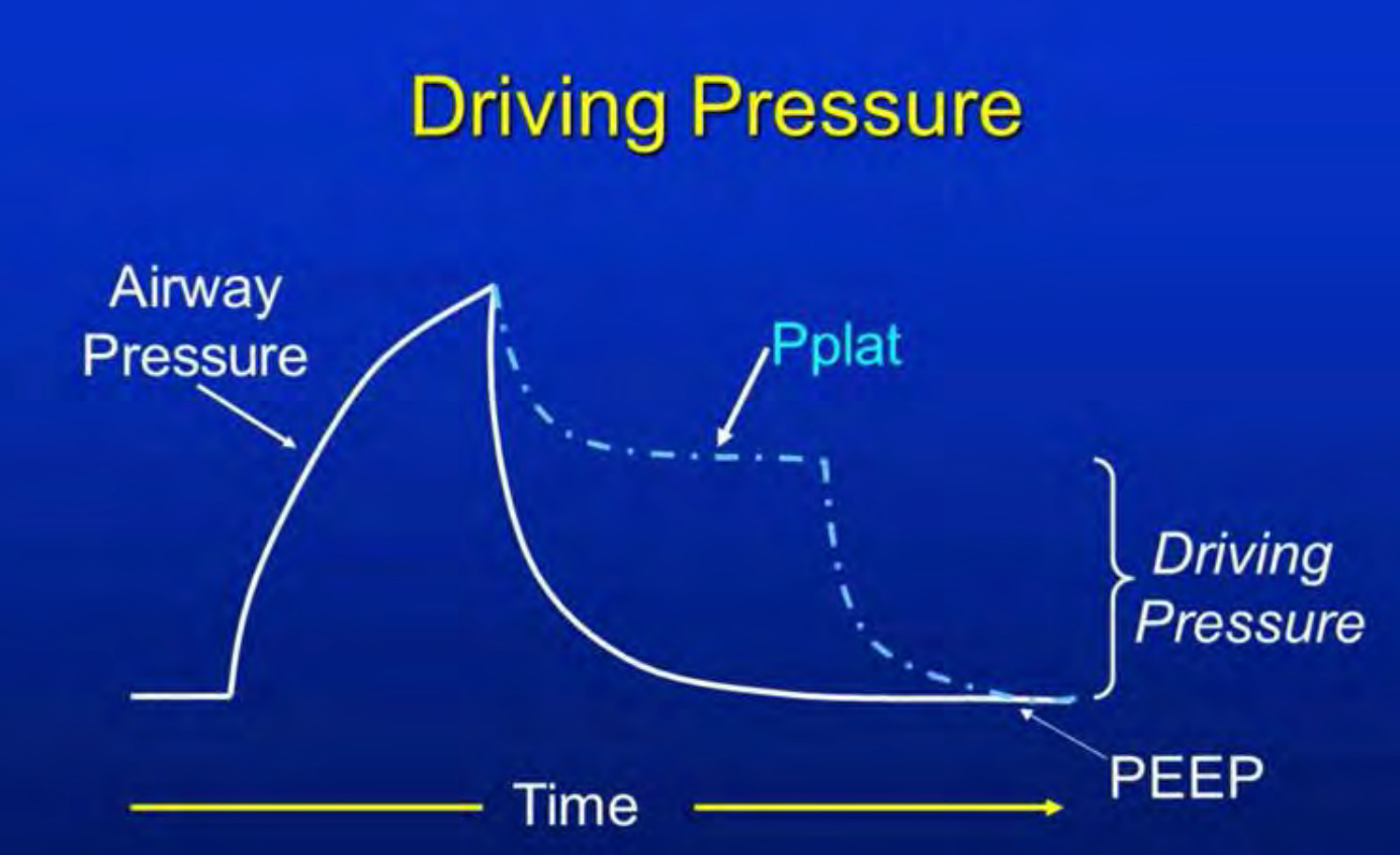
- Raising Both PIP and PEEP: Combining increases in PIP and PEEP showed a moderate improvement in outcomes compared to raising PIP alone. However, in patients with severe illness, this strategy still poses substantial risks. Maintaining an appropriate driving pressure (the difference between PIP and PEEP) was crucial in mitigating potential harm.
- Raising PEEP Alone: This method resulted in the most significant reduction in the relative risk of death. Elevating PEEP alone provided a stable and protective pressure, enhancing alveolar recruitment without the detrimental effects associated with high PIP.
The study underscores the importance of driving pressure as a critical determinant of patient outcomes. Driving pressure, calculated as the difference between PIP and PEEP, reflects the cyclic stress applied to the lungs during ventilation. Maintaining a lower driving pressure has been associated with improved survival in patients with acute respiratory distress syndrome (Amato et al, 2014).
In clinical practice, the goal is to optimize MAP to improve oxygenation while minimizing the risk of ventilator-induced lung injury. Among the strategies discussed, adjusting PEEP is often the most effective and safest method. However, individual patient factors must be considered, and ventilator settings should be tailored to achieve the best possible outcomes.
In summary, while several methods exist to increase MAP, careful consideration of their physiological impacts is essential. Elevating PEEP stands out as a particularly effective strategy, but it should be implemented judiciously within the context of a comprehensive ventilatory management plan.
Impact of MAP on Cerebral Circulation
Positive end-expiratory pressure (PEEP) is a critical component in mechanical ventilation, particularly for patients with acute respiratory distress syndrome (ARDS). By maintaining alveolar recruitment and improving oxygenation, PEEP plays a vital role in patient care. However, its application requires careful consideration due to potential systemic effects, especially concerning cerebral circulation.
Impact on Cerebral Circulation
Elevated PEEP levels can increase intrathoracic pressure, which may impede venous return from the brain, leading to elevated intracranial pressure (ICP). This rise in ICP can reduce cerebral perfusion pressure (CPP), potentially compromising cerebral blood flow and increasing the risk of hypoxic or ischemic injuries. Studies have shown that while moderate PEEP levels may have minimal impact, higher levels can significantly affect ICP and CPP.
Clinical Considerations
In patients with brain injuries or compromised cerebral autoregulation, the application of PEEP necessitates meticulous monitoring. The balance between optimizing oxygenation and maintaining stable cerebral hemodynamics is delicate. Adjustments to PEEP should be individualized, taking into account the patient's respiratory mechanics, intracranial dynamics, and overall clinical status. Continuous monitoring of ICP and CPP is advisable when applying higher levels of PEEP in such patients.
Recommendations for Practice
- Individualized PEEP Titration: Tailor PEEP levels to the patient's specific needs, considering both pulmonary and cerebral parameters.
- Monitoring: Employ invasive monitoring techniques to assess ICP and CPP in patients at risk of cerebral complications.
- Interdisciplinary Approach: Collaborate with neurology and critical care specialists to optimize ventilatory strategies for patients with concurrent respiratory and neurological concerns.
Understanding the nuances between hypoxic and ischemic brain injuries is crucial for accurate diagnosis and treatment.
Hypoxic Brain Injury
This occurs when the brain receives blood that is inadequately oxygenated. Despite sufficient blood flow, the reduced oxygen content leads to cellular dysfunction and damage. Common causes include respiratory failure, carbon monoxide poisoning, and high altitudes. Depending on the duration and severity of oxygen deprivation, symptoms can range from mild cognitive impairments to severe neurological deficits.
Ischemic Brain Injury
In contrast, ischemic injury arises when there is an obstruction or reduction in blood flow to the brain despite the blood being oxygen-rich. This deprivation prevents brain tissues from receiving necessary oxygen and nutrients, leading to cell death. Causes often include blood clots, strokes, or cardiac arrest. The resulting damage can lead to motor deficits, speech difficulties, and other neurological impairments. Both types of injuries can result in significant neurological impairments, and their management requires precise monitoring of both oxygenation and perfusion.
Near-Infrared Spectroscopy (NIRS)
NIRS is a non-invasive monitoring technique that offers real-time insights into tissue oxygenation and perfusion. By emitting near-infrared light into tissues and measuring the absorption spectra, NIRS can determine the balance between oxygenated and deoxygenated hemoglobin. This provides continuous data on both oxygen delivery and utilization in specific regions, making it invaluable in settings like cardiac surgery and critical care.
The NIRS device operates by placing sensors on the skin's surface. A light source emits near-infrared light, which penetrates tissues and is absorbed by hemoglobin. Two detectors measure the intensity of light after it has traversed the tissue:
- Shallow Detector: Captures light that has traveled a shorter path, providing information about superficial tissue layers.
- Deep Detector: Measures light that has penetrated deeper, offering data on underlying tissues and organs.
By analyzing the differential absorption at these depths, NIRS calculates regional oxygen saturation and assesses tissue perfusion. This dual capability distinguishes it from standard pulse oximetry, which primarily measures arterial oxygen saturation without providing direct information on tissue perfusion.
In clinical practice, NIRS has proven beneficial in various scenarios:
- Cardiac Surgery: Monitoring cerebral oxygenation to prevent neurological complications.
- Neonatal Care: Assessing brain oxygenation in preterm infants to detect and prevent hypoxic-ischemic injuries.
- Critical Care: Evaluating tissue perfusion in patients with circulatory shock or traumatic brain injuries.
By providing continuous, real-time data, NIRS aids clinicians in making informed decisions to optimize patient outcomes. In summary, distinguishing between hypoxic and ischemic brain injuries is essential for targeted therapeutic interventions. Utilizing advanced monitoring tools like NIRS enhances the ability to assess and manage these conditions effectively, ultimately improving patient care.
This device is remarkable because it monitors both oxygen delivery and perfusion simultaneously, providing critical real-time insights into a patient’s condition. Its utility lies not just in what it measures but in how it empowers clinicians to act proactively. Unlike many diagnostic tools—such as X-rays, CT scans, MRIs, or ultrasounds—this device doesn’t simply confirm whether harm has occurred. While these technologies are invaluable for diagnosing existing issues, they are typically reactive, informing clinicians only after an injury or complication has already taken place.
What sets this device apart is its ability to alert clinicians to potential harm before it happens. Rather than waiting for signs of an adverse event to appear, it provides early warnings, enabling timely interventions that can prevent injury altogether. This proactive functionality represents a significant shift in how we approach patient care, moving us closer to truly preventive medicine. By identifying and addressing risks in real-time, clinicians can reduce complications, improve outcomes, and potentially save lives.
The device monitors critical metrics related to organ saturation, such as how well oxygen is reaching the kidneys and brain—two organs particularly vulnerable to oxygen deprivation. These metrics are essential because they reflect how effectively the body is maintaining vital functions. A decline in saturation values signals potential organ dysfunction, allowing clinicians to act immediately, whether by adjusting oxygen delivery, optimizing perfusion, or addressing other underlying issues.
In one instance, a drop in both cerebral and renal saturation levels was identified within a few hours. The clinical team traced the issue to excessive mean airway pressure. They responded by suctioning the patient and switching from a device that utilized higher mean airway pressure to one that used less. This change led to a rapid improvement in oxygenation, preventing brain injury before it occurred. The ability to detect and address such problems early highlights the profound impact of this technology.
What’s remarkable is how this device facilitates real-time monitoring of both cerebral and renal saturations. By providing continuous feedback, it bridges the gap between reactive diagnostics and preventive care, offering precision and foresight that redefine patient safety standards. This capability ensures that clinicians can make informed, timely decisions in critical care settings, where even slight delays can have serious consequences.
The key takeaway here is that mean airway pressure can significantly impact cerebral circulation, making its careful management critical. Another takeaway is more general but equally important: attention to detail matters, whether you’re managing a clinical device or inflating a raft. Small adjustments can have big impacts, and proactive care can make all the difference in patient outcomes.
Consequences of MAP
I have personally experienced lightheadedness when blowing up something for my grandkids, which serves as a reminder to be cautious. Overexertion, even in seemingly simple tasks, can have consequences—similar to the clinical risks associated with excessive mean airway pressure. Understanding this concept is critical because the implications can be significant, as highlighted in a fascinating study.
This study, titled Cardiovascular Effects of HFOV with Optimal Lung Volume Strategy in Term Neonates with ARDS (Acherman, R.J., Siassi, B., deLemos, R., Lewis, A.B., & Ramanathan, R.), examined the cardiovascular effects of high-frequency oscillatory ventilation (HFOV) using optimal lung volume strategies. The collaboration included notable figures in the field: Dr. Reuben Acherman, a cardiologist; Dr. Ramanathan, the inventor of the RAM cannula; and the late Dr. deLemos, known as the "grandfather" of the SensorMedics oscillator. This team was based at LA Children’s Hospital, a hub for groundbreaking pediatric care.
Dr. Acherman, intrigued by the team's approach, visited Dr. deLemos to discuss their use of high mean airway pressures during oscillation. Dr. deLemos explained that using HFOV allowed for higher mean airway pressures with improved safety compared to conventional ventilation methods. The reduced risk of overdistension, as confirmed by X-ray imaging, supported their strategy.
However, Dr. Acherman, ever cautious, expressed concerns about the potential effects of mean airway pressure on the cardiovascular system. He proposed using his ultrasound machine to investigate further. The team welcomed his input, as they had not observed evidence of overdistension on imaging but were open to additional insights.
The primary challenge with oscillation and jet ventilation lies in their mechanics. Both devices essentially function as CPAP systems with added oscillatory movement, often referred to as “CPAP with a wiggle.” They operate at higher mean airway pressures while maintaining lower peak inspiratory pressures, which can obscure traditional signs of overdistension. For example, on a conventional ventilator, overinflation often causes visible changes like diaphragm flattening or rib expansion on X-rays. These indicators are typically absent in oscillatory ventilation, making it harder to detect underlying issues.
Dr. Acherman became concerned when he observed that patients on these devices, particularly at higher mean airway pressures, were showing signs of systemic cardiovascular stress. Despite interventions to maintain blood pressure and cardiac output, these patients continued to exhibit troubling symptoms. Using his ultrasound machine, Dr. Acherman investigated further and made a critical discovery: many of these patients had a significantly dilated inferior vena cava (IVC), elevated central venous pressure (CVP), and widespread edema.
His findings were striking. While the heart and lungs initially appeared normal on imaging, the inferior vena cava below the diaphragm was dilated, in some cases up to three times its normal size. This indicated that venous return to the heart was being obstructed. Both the superior and inferior vena cava were affected, contributing to increased intracranial pressure, which can have severe neurological consequences. Additionally, the hepatic veins, which drain blood from the liver, were also significantly dilated—approximately three times their normal size. This systemic venous congestion confirmed that elevated mean airway pressure was creating significant hemodynamic strain.
Dr. Acherman concluded that the elevated mean airway pressures were not just ventilating the lungs but also exerting pressure on the heart and pulmonary vasculature. This compression impaired venous return and increased resistance within the pulmonary and systemic circulations. These findings were critical because they highlighted a hidden risk of oscillatory ventilation that could not be readily detected with traditional methods like X-rays.
The study conducted at LA Children’s Hospital brought together several pioneers in neonatal and pediatric care, including Dr. Acherman, Dr. Ramanathan (inventor of the RAM cannula), and the late Dr. deLemos, known as the "grandfather" of the SensorMedics oscillator. Their work illustrated the complex interplay between ventilation strategies and cardiovascular physiology. While HFOV was deemed safer than conventional ventilation in certain cases, Dr. Ackerman’s findings underscored the importance of monitoring not just pulmonary parameters but also cardiovascular effects.
One of the most illustrative demonstrations of HFOV mechanics involved placing a pig or sheep lung inside a sealed bottle. When high mean airway pressures were applied, the lung inflated while maintaining low internal pressures, a feature often touted as an advantage of HFOV. However, Dr. Acherman noted a critical limitation of this model: it did not account for the constraints of an intact thoracic cavity. In a living patient, the rib cage, diaphragm, and surrounding structures limit lung expansion. This restriction is especially pronounced in adults but can also affect pediatric patients. Consequently, the pressures that seemed safe in an isolated lung model could, in practice, result in significant hemodynamic consequences when applied in vivo.
At LA Children’s Hospital, the clinical team observed that patients with high mean airway pressures did not exhibit traditional signs of overdistension, such as diaphragm flattening or rib spacing changes, commonly seen on X-rays. However, the elevated pressures were compressing the heart and pulmonary vasculature, leading to increased CVP, venous congestion, and impaired cardiac function. These effects were subtle yet profound, illustrating the need for advanced diagnostic tools like ultrasound to detect and address such complications.
This study demonstrated the importance of interdisciplinary collaboration in improving patient care. By combining the expertise of cardiologists, neonatologists, and pulmonary specialists, the team was able to identify and mitigate risks associated with HFOV. It also highlighted the need for careful consideration of mean airway pressures, even when using “safer” ventilation strategies. The findings emphasize that while advanced technologies can offer significant benefits, they also require a thorough understanding of their potential systemic impacts.
In clinical practice, this case serves as a reminder to monitor not just respiratory parameters but also the broader physiological effects of ventilation strategies. The insights gained from this study continue to inform how we approach high-frequency ventilation, emphasizing the importance of vigilance, comprehensive assessment, and early intervention to prevent complications.
Consequences of Too Much MAP
Managing mean airway pressure is critical, whether using a conventional ventilator or a high-frequency ventilator because elevated pressures can have serious consequences. The question then becomes: what can be done to address the problem? At Children’s Hospital Los Angeles, under the guidance of Dr. Philippe Friedlich, an experienced neonatologist, the team approached this challenge with innovation and collaboration. Dr. Friedlich and his team worked closely with biomedical technicians to design practical solutions that address real clinical needs, not just theoretical ones.
During a visit to speak with his group, I discussed the process of developing medical devices, emphasizing that they must solve real problems in the ICU, NICU, or PICU. Before my talk, Dr. Friedlich and I discussed a pivotal study conducted at his hospital (Marked Reduction in MAP and OI Using HFJV in Neonates with Severe Refractory Hypoxemic Respiratory Failure). This study focused on addressing mean airway pressure and overdistension in neonates, particularly those who had failed conventional ventilation and were deteriorating on oscillators after missing their ECMO window.
The Problem: Overdistension and Gas Trapping
In these cases, the neonates were experiencing gas trapping and hyperinflation, which contributed to elevated mean airway pressures. These pressures, in turn, exacerbated issues like barotrauma—or as Dr. Friedlich coined it, "mapotrauma." The team hypothesized that gas trapping was the primary cause of the elevated pressures, leading to poor oxygenation and ventilation. They realized that conventional ventilators with a 1:2 inspiratory-to-expiratory (I:E) ratio were not sufficient to alleviate this problem. This led them to revisit high-frequency jet ventilation (HFJV) to explore its potential in these critical cases.
The Solution: Extending Expiratory Time
Using HFJV, the team was able to adjust the I:E ratio to as much as 1:6 or 1:12, significantly increasing the time available for exhalation. This approach helped to minimize gas trapping and, subsequently, reduce mean airway pressure. The goal was to resolve hyperinflation, improve oxygenation, and ultimately stabilize these fragile patients.
The Results: Rapid Improvement
The first ten patients in the study had failed conventional ventilators and oscillators. These neonates were critically ill, with oxygen indexes (OI) averaging 30, indicating severe respiratory failure. Over 50 days, they had been stuck on oscillation without significant improvement. Once switched to HFJV, the results were striking:
- Reduction in Mean Airway Pressure: Within four to five hours of starting HFJV, the mean airway pressure dropped from 14 to below 10 cm H₂O
- Improved Oxygenation: As mean airway pressure decreased, the fraction of inspired oxygen (FiO₂) could be weaned from nearly 90% to below 50%
- Improved Oxygen Index: The OI, which reflects the severity of respiratory failure, fell from 30 to 10 within hours and remained low over the next several days
This rapid improvement allowed the team to extubate the patients and eventually send them home. Of the ten initial patients, nine survived and were discharged. Tragically, one patient suffering from sepsis did not survive. However, the surviving neonates—all with chronic lung disease at the time of intervention—were able to leave the NICU and go home on oxygen.
The Impact: Better Outcomes and Reduced Costs
While some neonates required supplemental oxygen at discharge, Dr. Friedlich emphasized that sending them home was a success. Each day a patient spends in the NICU costs $10,000 to $20,000. By reducing the time spent on mechanical ventilation and in the NICU, the team not only improved patient outcomes but also reduced financial burdens for families and healthcare systems. On average, after the intervention, patients were on mechanical ventilation for just seven more days before being weaned.
Lessons Learned
Dr. Friedlich and his team concluded that lowering mean airway pressure was the key to success. By resolving hyperinflation and gas trapping, they achieved better oxygenation and reduced the risk of complications. The study also highlighted the importance of avoiding hyperinflation in the first place. Prolonged expiratory times, achieved through HFJV, proved to be a critical strategy for minimizing barotrauma and reducing mean airway pressures.
MAP During Noninvasive (NIV)
Mean airway pressure has long been a focus of discussion in mechanical ventilation, but I had initially thought it wasn’t an issue during noninvasive ventilation (NIV). My reasoning was simple: during NIV, patients typically breathe in a way that mimics natural physiology. Humans are designed to breathe with negative pressure, not positive pressure. Unlike invasive ventilation, which requires mechanical assistance, NIV generally relies on the patient’s normal breathing mechanisms unless supplemental breaths are delivered. Because of this, I assumed mean airway pressure wouldn’t play a significant role in NIV. I was wrong.
Over time, I began to question what might be better for patients: delivering breaths through the nose using noninvasive positive pressure ventilation (NIPPV) or maintaining continuous positive airway pressure (CPAP) through the nose. Which approach was more effective? Was one safer than the other? To answer this question, I turned to a pivotal study by Dr. Haresh Kirpalani, a Canadian physician who researched this topic during his time in Washington, D.C. The study, published in The New England Journal of Medicine in 2013 (A Trial Comparing Noninvasive Ventilation Strategies in Preterm Infants), sought to compare these two strategies in preterm infants.
Key Findings of the Study
Dr. Kirpalani’s study explored whether NIPPV—delivering breaths through the nose—or CPAP—maintaining a continuous positive pressure—offered better outcomes for preterm infants. The findings were intriguing. There was no substantial difference between the two strategies in terms of preventing complications. There was a slight advantage for avoiding breaths through the nose, but the difference was minimal, just a few percentage points. Importantly, the rates of bronchopulmonary dysplasia (BPD) were nearly the same in both groups.
This was surprising to me. BPD, a chronic lung condition, is commonly associated with invasive mechanical ventilation and prolonged exposure to high oxygen concentrations. These preterm infants, however, had never been intubated. How could they develop BPD? This challenged my understanding of the condition and highlighted the complexity of its underlying mechanisms.
Rethinking Assumptions
Dr. Marty Kessler once made a thought-provoking observation about our attachment to terminology in respiratory care. He said, “I think we’re misled by these terms.” Terms like continuous positive airway pressure, high-flow nasal cannula, and noninvasive ventilation create an impression of minimal intervention. They suggest that these methods are inherently safer or less likely to cause harm compared to invasive approaches. Yet, as this study demonstrated, even noninvasive techniques can contribute to conditions like BPD.
The Implications
The findings of this study—and my reflections on it—serve as an important reminder of the nuanced nature of respiratory support. While NIV strategies like NIPPV and CPAP are invaluable tools, they are not without risks. Mean airway pressure, even during noninvasive support, can play a critical role in patient outcomes. it is not just about delivering the right therapy; it is about understanding the potential consequences of each approach and tailoring care to the individual needs of the patient.
Dr. Marty Kessler made an important observation during a conference we attended together. He pointed out that regardless of the terminology we use—whether it is CPAP, high-flow nasal cannula, or noninvasive ventilation—all these techniques share three critical components: pressure, flow, and ventilation. Even in noninvasive strategies, maintaining sufficient pressure and flow is essential to adequately ventilate the patient. Without these, complications can arise.
The Challenge of Maintaining Pressure
Dr. Kessler emphasized that a CPAP level must be sufficient to keep the lung open, even when the patient is not intubated. This principle ties into the concept of lung protection. If the lung collapses, particularly in a surfactant-deficient state, reopening it becomes more challenging and can cause injury. Dr. Jack Emerson, an early pioneer of the iron lung, referred to this phenomenon as “sticky lung syndrome.” He described how surfactant-deficient alveoli tend to stick together when they collapse. Reopening these collapsed alveoli can result in shear stress and lung injury, even with noninvasive techniques.
Dr. Emerson’s insight underscores an important reality: just as patients can experience ventilator-induced lung injury (VILI) with invasive strategies, they can also suffer lung injury from noninvasive methods if pressure and flow are inadequate. Persistent attempts to maintain noninvasive support in a struggling patient can sometimes do more harm than good.
When to Transition to Intubation
One of the key lessons from these discussions is knowing when to transition from noninvasive to invasive ventilation. Dr. Kessler explained that while noninvasive strategies are often preferable, there are cases where intubation and low-pressure, low-volume lung-protective ventilation may be the safer and more effective option. Administering a dose of surfactant in such scenarios can help stabilize the lung, making it easier to extubate the patient later.
A colleague in Canada shared a compelling example of this principle in action. They had a 24-week-old preterm infant who was started on nasal prongs immediately after birth. Initially, the infant responded well and defied the odds, maintaining stability on noninvasive support. However, over time, the team struggled to maintain adequate ventilation. Despite their best efforts—repositioning the prongs, switching systems, and adjusting the patient—they couldn’t achieve stability. After 24 days of noninvasive ventilation, they decided to intubate the infant.
What was striking was the infant’s first chest X-ray post-intubation, which revealed chronic lung disease (CLD). This was surprising, as the patient had never been intubated prior to that point. The cause? Insufficient or unstable mean airway pressure during noninvasive support. The lesson was clear: sufficient mean airway pressure is critical to maintaining functional residual capacity (FRC) and alveolar inflation. Without it, the lung remains unstable, even in a noninvasive strategy.
Noninvasive Ventilation: Not Always "Noninvasive"
Dr. Kessler’s reflections challenge the notion that noninvasive ventilation is entirely “noninvasive.” While it avoids the physical invasiveness of endotracheal intubation, it can still cause harm if mean airway pressure is insufficient to maintain stable lung function. In this way, “noninvasive” can be a misnomer. The absence of an endotracheal tube doesn’t negate the physiological invasiveness of inadequate support.
The take-home message is this: noninvasive strategies are highly effective when applied correctly, but they require careful attention to mean airway pressure. If mean airway pressure is too low or unstable, noninvasive ventilation can fail, leading to lung injury and complications like CLD. Achieving therapeutic outcomes hinges on delivering adequate pressure, flow, and ventilation. Noninvasive may be better in many cases, but it is only truly noninvasive if it provides sufficient support to maintain stable lung function.
Recruitment Maneuvers and Optimal MAP
Ventilating patients effectively requires an open lung strategy. For noninvasive ventilation to work, the lungs must remain open, which often involves recruitment maneuvers. However, if mean airway pressure is critical and too much or too little can be problematic, the question becomes: how do you optimize it? What is the best way to recruit and stabilize the lungs while avoiding injury?
Learning from a New Perspective
I recently discussed this topic with Dr. Molly, a physician I admire greatly who works at a hospital in Winnipeg, Manitoba. She is not only a colleague but also a friend, and I had the pleasure of having dinner with her during a visit to her hospital. Our conversation drifted to a pivotal moment we shared years earlier at the PAS conference in Washington, D.C., where Molly attended a lecture that left a lasting impression on both of us.
At that conference, Molly was intrigued by an anesthesiologist’s perspective on lung recruitment strategies. She recalled how she had initially planned to challenge the presenter, confident in her own NICU experience. Yet, after hearing his approach, she found herself rethinking some deeply held beliefs. The anesthesiologist argued that many ICU and NICU patients, particularly babies, are recruited improperly. He explained that high-pressure, short-duration breaths, often used in the ICU, can push the lungs into the injury zone, increasing the risk of barotrauma and other complications.
A Different Recruitment Strategy
The anesthesiologist described his approach to recruiting patients during surgery when their lungs had collapsed. Instead of giving a short, high-pressure breath, he advocated for a longer-duration, lower-pressure breath. For example, he would hold a breath 5 to 8 cm above PEEP (and no more than 15 cm in extreme cases) for 20 to 30 seconds. After releasing the pressure for four to five seconds, he would repeat the maneuver two or three times. In nearly all cases, this method restored lung function without causing injury.
Molly relayed this to me with a big smile, admitting she had been skeptical at first. “We cannot give 20- or 30-second breaths in the ICU,” I said. She agreed but suggested we could modify the strategy with one- to three-second breaths at lower pressures. “It’s safer than it sounds,” she explained. “The anesthesiologist emphasized that longer inspiratory times are far less dangerous when pressures are kept low. it is the combination of high pressure and long inspiratory time that increases the risk of injury.”
Optimizing Mean Airway Pressure Safely
Molly explained how her team applies these principles in their unit. After optimizing PEEP to ensure the lungs remain open, they often transition to high-frequency ventilation. This approach allows them to maintain higher mean airway pressures safely, as the oscillatory nature of high-frequency ventilation (CPAP with a wiggle) minimizes the risk of overdistension and injury.
She emphasized the importance of keeping the lungs open while using the gentlest means possible. “The take-home message,” she said, “is to optimize mean airway pressure to open the lungs and keep them open, but to do so gently. Recruitment maneuvers should be tailored to the patient, using lower pressures and longer durations to avoid harm.”
The Future of Recruitment Research
Inspired by these discussions, Molly’s team and others have begun exploring the best ways to recruit and stabilize lungs effectively. Preliminary studies suggest that a longer inspiratory time with lower pressures is more effective and less injurious than shorter, high-pressure recruitment strategies. Additional research is underway to examine whether simply raising PEEP or mean airway pressure alone can achieve the same results or whether recruitment breaths are necessary for optimal outcomes.
The hope is that these studies will provide clear evidence for the safest and most effective recruitment methods, ensuring better outcomes for patients in the ICU, NICU, and PICU. As Molly put it, “We need to find the balance—enough pressure to open the lungs but not so much that we cause harm. And we need to use every tool and strategy at our disposal to get it right.”
This ongoing research represents an exciting opportunity to refine our understanding of lung recruitment and improve how we manage mean airway pressure. I look forward to sharing more about these findings as they develop, but for now, the key takeaway is clear: recruitment and stabilization must be done with care, precision, and a focus on gentle, patient-centered strategies.
MAP vs NAP in the Produce Section
Ventilation, like grocery shopping, requires balance, observation, and attention to detail—at least that is how I see it. My wife, who’s far better at navigating grocery stores, loves to remind me how chaotic I find them. On Mother’s Day, she convinced me to join her on a grocery run. How could I refuse on her special day? Reluctantly, I agreed, thinking I could survive one trip. Little did either of us know how the produce section would ignite my respiratory therapist brain.
When we got to the produce section, I couldn’t help myself. Suddenly, I saw lung dynamics everywhere. “Look at these vegetables,” I exclaimed. “That is a great example of a good distribution of tidal volume. Over there, hyperinflation! And here, underinflation.” My wife, a fellow respiratory therapist, stared at me, baffled. “What are you talking about? We’re here to shop!” But I couldn’t stop. This was too exciting.
I pointed out the divine ripened tomatoes. “These are perfect! They represent optimal lung inflation. See how balanced they are?” Then I held up the heirloom tomatoes and explained how their irregular shapes resembled overinflated alveoli, with their stems acting like pulmonary capillaries. “And look at these habanero peppers,” I continued. “When insufficient PEEP lets the alveoli collapse, they look just like this—uneven and deflated.”
As we moved through the store, I found more metaphors. A broken bag of frozen peas scattered on the floor became pulmonary interstitial emphysema. Clumps of ruptured alveoli spilling into tissue spaces came vividly to life through the imagery of scattered peas. My wife rolled her eyes, begging me to move on. But I was having too much fun.
By the end of the trip, she was regretting bringing me along. “Never again,” she declared, assigning me errands elsewhere in the store whenever we shop together. Despite her exasperation, the trip reminded me of something profound: ventilation, like selecting produce, requires care. Too much pressure, and you risk damage; too little, and the lungs fail to function. Each lung, much like each tomato, has unique needs that must be respected.
The Take-Home Message: Individualized Care
This playful metaphor brings us back to a critical point about ventilation: no two patients are the same. As respiratory therapists, our job is to assess, adapt, and respond to each patient’s unique pathophysiology. My colleague, Marty Kessler, often says, “It is very important to tailor your strategy to the pathophysiology. Choose the right strategy for the right disease.” To that, I would add: “It is equally important to change the strategy as the pathophysiology evolves.”
This is what makes our profession so rewarding and vital. Our expertise lies in our ability to understand pulmonary dynamics and hemodynamics, identify what each patient needs, and continuously adapt. We don’t take a cookie-cutter approach because the same strategy that worked for one patient might fail for another. This adaptability is what makes us the experts in pulmonary care.
Critical Thinking: The Heart of Respiratory Therapy
Our role as RTs demands critical thinking—skills that grow with every shift, patient, and case. Bedside care is dynamic, requiring us to make real-time decisions and adjust as conditions change. Sessions like those offered by Continued are invaluable in honing our knowledge and keeping us at the forefront of respiratory care.
So, the next time you’re in the produce section, maybe you’ll think of tomatoes, peppers, and peas in a new light. More importantly, remember the delicate balance required to ventilate and protect the lungs. By optimizing mean airway pressure—finding that sweet spot between too much and too little—we ensure better outcomes for our patients. And if nothing else, let this story serve as a reminder: never take a respiratory therapist grocery shopping!
Thank you for allowing me to share my thoughts on mean airway pressure, its impact on pulmonary dynamics, and the parallels we can draw from everyday life. It is these connections and insights that make our profession both challenging and deeply rewarding. Now, let’s dive into the questions you have for me.
Questions and Answers
You mentioned in your talk about the importance of PEEP. Why do you think we don’t give enough PEEP, and why is that significant?
that is an excellent question. My colleagues and I use a term we jokingly call “PEEPophobia.” For some reason, many clinicians are more apprehensive about increasing PEEP than they are about mean airway pressure (MAP). I have seen this repeatedly, particularly with high-frequency ventilation. For example, on devices I worked with, we displayed both PEEP and MAP. Over time, we noticed people were much more hesitant to increase PEEP.
Why? It could be that MAP is calculated—it’s an average of several variables and doesn’t have a direct adjustment knob. On the other hand, PEEP is tangible; you turn a knob, and the change is immediate. This directness may make it feel riskier. I’ll admit, I was once “PEEPophobic” myself. But we must overcome that fear. Properly optimizing PEEP is critical for patient care and stability, especially when trying to prevent lung collapse and ensure proper oxygenation.
Regarding noninvasive ventilation (NIV), what adjustments or precautions would you recommend when NIV doesn’t seem to provide sufficient MAP for a patient with hemodynamic concerns?
that is a great question. One key thing to remember is not to assume the pressures displayed on the NIV device are entirely accurate at the patient’s airway. Resistance in the system, such as prongs or mask interfaces, can significantly affect delivered pressure. A doctor once told me about a case where they set a pressure of 6 cmH₂O on a patient, but when the prongs were laid on the bed, the device still displayed 4 cmH₂O—clearly inaccurate.
If a patient is not responding well to NIV, start by ensuring the prongs or mask are positioned correctly. Next, verify that you’re delivering enough pressure to keep the lungs open. I would err on the side of providing slightly more pressure rather than less. Remember, noninvasive systems are prone to under-delivering pressure compared to invasive systems. that is why careful monitoring and adjustments are crucial when managing patients on NIV.
You discussed near-infrared spectroscopy (NIRS) for monitoring cerebral and renal tissue oxygenation. Is this technology frequently used in practice, especially in the pulmonary and RT fields?
Unfortunately, no, it is not as widely used as it should be. Years ago, I worked at a hospital that was an early adopter of this technology, and I saw its benefits firsthand. However, in many newborn intensive care units (NICUs) and pediatric ICUs I visit, I don’t see it being implemented as often. NIRS has immense potential—not only for monitoring but also for preventing injuries by giving clinicians a real-time “heads up” before harm occurs.
Technologies like NIRS and electrical impedance tomography (EIT) could significantly improve decision-making in patient care, but they haven’t fully caught on yet. it is a great question, and I hope to see more widespread adoption of these tools in the future, as they align perfectly with our goal of proactive, rather than reactive, care.
To wrap things up, what’s the overarching takeaway from your presentation on MAP and its role in ventilation?
The key takeaway is this: get the lungs open and keep them open. Optimizing MAP is crucial in achieving this balance. Too much MAP can cause overdistension and injury, while too little leads to alveolar collapse and inadequate oxygenation. We must tailor ventilation strategies to each patient’s unique physiology and needs.
Critical thinking is essential, and we cannot use a one-size-fits-all approach. Every patient’s condition evolves, and we must adapt accordingly. As RTs, our role is to assess, identify the pathophysiology, and apply strategies that maximize outcomes while minimizing risks. With ongoing education and tools like NIRS, EIT, and careful PEEP adjustments, we can elevate our care standards.
References
Acherman, R. J., Siassi, B., deLemos, R., Lewis, A. B., & Ramanathan, R. (n.d.). Cardiovascular effects of HFOV with optimal lung volume strategy in term neonates with ARDS. USC School of Medicine, Division of Neonatology, and Children's Hospital of Los Angeles, Division of Cardiology, Los Angeles.
Amato, M. B. P., Meade, M. O., Slutsky, A. S., Brochard, L., Costa, E. L. V., Schoenfeld, D. A., Stewart, T. E., Briel, M., Talmor, D., Mercat, A., Richard, J. C. M., Carvalho, C. R. R., & Brower, R. G. (2014). Driving pressure and survival in the acute respiratory distress syndrome. New England Journal of Medicine, 372(8), 747-755. https://doi.org/10.1056/NEJMsa1410639.
Friedlich, P., Subramanian, N., & Garg, M. (n.d.). Marked reduction in MAP and OI using HFJV in neonates with severe refractory hypoxemic respiratory failure. Children’s Hospital Los Angeles, Division of Neonatology, Keck School of Medicine, University of Southern California, Los Angeles, CA.
Fu, Z., Costello, M. L., Tsukimoto, K., Prediletto, R., Elliott, A. R., Mathieu-Costello, O., & West, J. B. (1992). High lung volume increases stress failure in pulmonary capillaries. Journal of applied physiology (Bethesda, Md. : 1985), 73(1), 123–133.
Kirpalani, H., Millar, D., Lemyre, B., Yoder, B. A., Chiu, A., & Roberts, R. S., for the NIPPV Study Group. (2013). A trial comparing noninvasive ventilation strategies in preterm infants. New England Journal of Medicine, 369(7), 611-620.
Citation
Richards, E. (2024). MAP vs. NAP: the impact of mechanical ventilation on hemodynamics. Continued.com - Respiratory Therapy, Article 254. Available at www.continued.com/respiratory-therapy
Nov 11, 2024
1 CE HourAcute Respiratory Distress Syndrome (ARDS) and What You Should Know
Tim Gilmore, RRT, RRT-ACCS, RRT-NPS, CPFT, AE-C, PhD
Editor’s note: This text-based course is an edited transcript of the webinar Acute Respiratory Distress Syndrome (ARDS) and What You Should Know, presented by Tim Gilmore, PhD, RRT, RRT-ACCS, RRT-NPS, CPFT, AE-C.
It is recommended that you download the course handout to supplement this text format.
Learning Outcomes
After this course, participants will be able to:
- Define and describe acute respiratory distress syndrome in alignment with current Berlin criteria
- Discuss classifications of ARDS
- Explain evidence-based mechanical ventilation strategies as part of ARDS treatment.
Acute Respiratory Distress Syndrome
The goal is for you to walk away with practical knowledge you can apply immediately in your bedside practice. Ultimately, our goal is to enhance patient care. To start, let's take a brief look at the historical perspective. The term "acute respiratory distress syndrome" implies a sudden onset and a respiratory-specific condition, which is accurate. "Syndrome" usually refers to a recognizable complex of symptoms rather than a distinct disease. When we talk about a syndrome, we are referring to a set of physical findings that indicate a specific condition, though the direct cause may not be fully understood. We understand ARDS better now, and we know that certain factors can predispose an individual to develop it. However, there is still no definitive cause or predictable factor that will absolutely lead to ARDS. it is somewhat predictable but also unpredictable. We will delve deeper into this as we progress.
The condition of ARDS was first reported in 1967 in a publication in The Lancet by the renowned Dr. David Ashbaugh from Ohio State University. He identified a dozen patients in a case series who had a particular presentation or pathology with some common characteristics. The respiratory distress syndrome in 12 patients was manifested by acute onset of tachypnea, hypoxemia, and loss of compliance after a variety of stimuli; the syndrome did not respond to usual and ordinary methods of respiratory therapy. Positive end-expiratory pressure was most helpful in combating atelectasis and hypoxemia (Asbaugh et al., 1967).
What was most interesting about the early observations of this so-called syndrome was the presence of persistent refractory hypoxia, often without direct lung involvement. In other words, patients developed persistent and pervasive respiratory distress regardless of whether a direct lung insult was initially suspected. Almost all these patients exhibited some degree of pulmonary edema on chest X-rays despite not being in left ventricular failure. They showed pulmonary edema symptoms without obvious cardiovascular compromise. Most of the early cases involved younger, generally healthy servicemen during the Vietnam War in the late 1960s.
ARDS Defined
ARDS had numerous descriptions and borderline formal definitions from the 1970s through the '80s and even into the early '90s, but there was very little agreement among key groups. It was not until 1994 that ARDS was formally defined by the American-European Consensus Conference. This led to a panel of experts convened in 2011 by the European Respiratory Society and the Society of Critical Care Medicine. They developed what is now known as the Berlin Definition, which has become the gold standard for defining ARDS. This Berlin Definition is likely the most familiar to everyone today.
They met in Berlin, which is why it is called the Berlin Definition. Starting from the early years of defining ARDS in the 1990s to the early 2000s, the PF ratio (the partial pressure of arterial oxygen compared to the inspiratory fraction of oxygen) and various other clinical indicators of significant respiratory impairment have long been part of the criteria to officially diagnose ARDS. However, it was not until the European Society of Intensive Care Medicine, with endorsement from the American Thoracic Society and the Society of Critical Care Medicine, convened an international expert panel in Berlin that the ARDS definition was revised. This is how the Berlin Definition was created.
The Berlin Definition changed everything. In fact, it is one of the most cited articles in critical care medicine. I will mention early on that the term "acute lung injury" (ALI) has come and gone, although the concept remains relevant. Clinically, acute lung injury is still a viable concept. However, we will focus solely on ARDS during today's lecture. Now, let's examine the original Berlin Definition that made such a significant impact. Compared to the 1994 AECC definition, the Berlin Definition has better predictive validity for mortality. The emphasis is on early identification and preventing avoidable deaths.
The original Berlin Definition in Figure 1., included three mutually exclusive categories of ARDS based on the degree of hypoxemia, all determined by evaluating the PF ratio (the partial pressure of arterial oxygen compared to the inspiratory fraction of oxygen). These categories are:
- Mild ARDS: PF ratio greater than 200 but less than or equal to 300
- Moderate ARDS: PF ratio less than or equal to 200 but greater than or equal to 100
- Severe ARDS: PF ratio less than or equal to 100
However, diagnosing ARDS involves more than just assessing the PF ratio.
Berlin Definition of Acute Respiratory Distress Syndrome
Timing
Within 1 week of a known clinical insult or new or worsening respiratory symptoms
Chest Imaging
Bilateral opacities - not fully explained by effusions, lobar/lung collapse, or nodules
Origin of Edema
Respiratory failure is not fully explained by cardiac failure or fluid overload. Need objective assessment (e.g., echocardiography) to exclude hydrostatic edema if no risk factor present
Oxygenation
Mild 200 mm Hg < Pa02/Fi02 ≤ 300 mm Hg with PEEP or CPAP ≥5 cm H20
Moderate 100 mm Hg < Pa02/Fi02 ≤ 200 mm Hg with PEEP ≥5 cm H20
Severe Pa02≥/Fi02 ≤ 100 mm Hg with PEEP ≥5 cm H20
Abbreviations: CPAP, continuous positive airway pressure; Fi02, the fraction of inspired oxygen; Pa02, partial pressure of arterial oxygen; PEEP, positive end-expiratory pressure.
Chest radiograph or computed tomography scan.
If the altitude is higher than 1000m, the correction factor should be calculated as follows: [Pa02/Fi02X (barometric pressure/760)].
This may be delivered noninvasively in the mild acute respiratory distress syndrome group.
JAMA, June 20, 2012—Vol 307, No. 23
Figure 1. Berlin Definition of ARDS.
As shown in Figure 1, timing is crucial. The term "acute" suggests that the onset occurs within a week or less. This rapid onset is significant. Additionally, a chest x-ray or CT scan is essential for diagnosis. These imaging techniques reveal opacities, also known as infiltrates, which must be present to confirm ARDS. Importantly, these thoracic infiltrates must originate from a non-cardiac source. Although heart failure can cause pulmonary edema, it does not classify as ARDS. The presence of true respiratory failure is assumed with ARDS. A key aspect of the classification process is the PF ratio, which categorizes the severity of the condition.
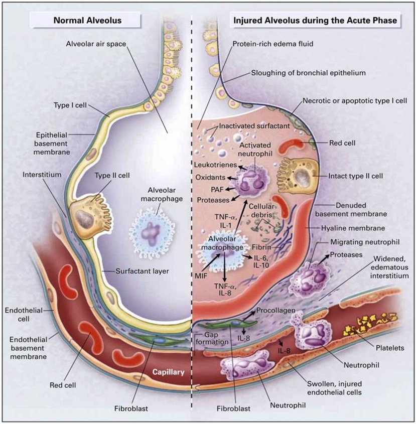
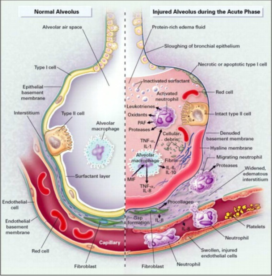
Figure 2. A diagram of the normal alveolus and injured alveolus during the acute ARDS phase (https://www.researchgate.net, CC BY-SA 4.0 via Wikimedia Commons).
Previously, the term "acute lung injury" (ALI) was used to describe a condition that could develop into ARDS. However, the ALI category is now obsolete. Today, we consider a patient to have ARDS if they meet specific clinical criteria. This is a clear, checkbox-based approach to diagnosis. ARDS, although somewhat unpredictable and sometimes discovered randomly, typically manifests after a significant infection or specific physical trauma. Importantly, this trauma does not necessarily need to directly involve the thorax. Figure 2, one of the more widely recognized images depicting ARDS, illustrates this well. On the far left of Figure 2, the key takeaway is that ARDS involves a severe inflammatory cascade. This cascade results in infiltrates and fluid accumulation in the lung acini, accompanied by a leaky pulmonary capillary bed. Platelets begin to aggregate, exacerbating the issues typically associated with an inflammatory response within the lung.
In Figure 2, you can see a comparison between normal alveoli on the left and injured alveoli on the right. The differences are striking. One critical aspect, which may not be clearly depicted in Figure 2, is the deactivation of surfactant. Normally, surfactant reduces surface tension in these tiny lung units. When it is washed out or deactivated, the lung becomes very stiff and non-compliant. Additionally, it is worth mentioning some of the common terminology associated with ARDS or ARDS-like conditions. These terms are often colloquial or clinical slang rather than standardized. However, our focus today is on the standard ARDS criteria.
ARDS stands for acute respiratory distress syndrome. As a side note, the term "baby lung" is often mentioned in the context of ARDS. "Baby lung" refers to the remaining healthy lung tissue that can still be ventilated when ARDS is present. Essentially, it is the functional lung tissue available for breathing. ARDS is considered a syndrome rather than a disease because there is no single known specific cause. it is not black and white. There are patterns and certain indications that a patient may be at risk for developing ARDS, but ultimately, it is a clinical syndrome diagnosed based on clinical features.
Here is a brief overview of those clinical features:
- Progressive dyspnea with increasing O2 requirements
- Alveolar infiltrates on chest x-ray & reduced SaO2 (SpO2) within 6-72h of inciting event
- Lab tests are nonspecific
Alveolar permeability presents a major challenge because it leads to intra-alveolar fluid accumulation, causing a ventilation-perfusion mismatch and right-to-left intrapulmonary shunting. This makes it extremely difficult to oxygenate these patients, who suffer from extremely low lung compliance. It is a vicious cycle: the stiffer the lung, the more creativity is required to ventilate. Otherwise, the cycle of alveolar infiltration continues.
In essence, ARDS can be summarized as sudden respiratory failure unrelated to heart failure or specific fluid overload. Patients present with pulmonary edema, but it is of non-cardiac origin. While there are other aspects to discuss, such as pulmonary capillary wedge pressure monitoring, today, we will focus on the essential items you need to know. Now, back to Figure 2. We know that ARDS pathophysiology is complex. Although it primarily affects the lungs, there is a significant risk of multi-organ involvement. In fact, much of the mortality associated with ARDS is due to multi-organ dysfunction syndrome (MODS). However, that is a topic for another day.
Before we move on, let's focus not only on the alveolar involvement depicted on the right side of Figure 2 but also on the intravascular and extravascular activities, also known as the interstitial processes. Notice the inflammatory mediators, such as interleukin, that are pervasive in both the alveoli and the capillaries. Due to increased capillary permeability, there is a massive fluid shift from inside the capillaries toward the interstitial space and into the gas exchange units. These patients are extremely difficult to oxygenate. Does this sound familiar to something We have encountered en masse over the last few years? The COVID-19 condition often presents similarly, making it challenging to differentiate between the pulmonary presentations of ARDS and COVID-19.
Epidemiology
When discussing any disease, disorder, or syndrome, it is helpful to quickly outline the epidemiology. Here is what we know. A well-done article by Hendrickson et al. (2021), published in the American Journal of Respiratory and Critical Care Medicine, reviewed 50 years of research and publications on ARDS. This makes it a fairly comprehensive source. What is mildly disturbing is the admission from the Canadian group of researchers, despite their bold statement that "No other intensive care syndrome has been as extensively studied as ARDS."
Many basic epidemiologic questions remain unsolved. In other words, the incidence, distribution, control methods, and overall contributing factors of this syndrome are not easily identified. We can define ARDS and occasionally recognize it, but piecing it all together remains challenging. The researchers cite a lack of standardization, and the exact onset of ARDS is not always easy to pinpoint. Even though our understanding of ARDS pathophysiology has certainly improved over the past five decades of published data, gaps remain. Several articles, not just this one, have referred to this extensive timeframe of available information.
Here is what we know: while pharmacologic interventions, such as corticosteroids and diuretics, have their place, the primary target of ARDS management should be the avoidance of ventilator-induced lung injury. We have a lot to consider, including noninvasive ventilation, high-flow nasal cannula, ECMO, and even extracorporeal CO2 removal, which we perform at my local facility. Before diving into these topics, it is important to quickly discuss the famous Lung Safe study by Bellani et al. (2016). Lung Safe stands for "Large Observational Study to Understand the Global Impact of Severe Acute Respiratory Failure." This international, multi-center prospective cohort study included some well-known researchers and investigated patients undergoing invasive or non-invasive ventilation over four consecutive weeks nearly a decade ago.
The study included patients from 459 ICUs across 50 countries on five continents, resulting in a large sample size of just over 29,000 ICU admissions. Remarkably, over 10% of these patients met the criteria for ARDS. This highlights the prevalence of ARDS in ICU settings. If you work in an ICU, you will inevitably encounter ARDS. However, the study also revealed that we are not particularly good at recognizing or treating ARDS, which, unfortunately, remains associated with a high mortality rate. The landscape changed dramatically around 2020 with the onset of COVID-19. It is now clear that differentiating between ARDS and COVID-19 is challenging, as their pulmonary presentations, chest X-rays, lab results, and overall clinical manifestations are often indistinguishable.
Severe cases of COVID-19 are arguably more difficult to treat and require more creative approaches to both ventilator and overall clinical management. Nevertheless, ARDS criteria remain the same. The treatment of the respiratory side of things does not differ dramatically between COVID-19-induced ARDS and classic ARDS. There are over 60 known etiologies of ARDS. Hendrickson et al. (2019) listed some of the most common precursors to the development of this syndrome. If you work at a facility with high trauma cases or a large ICU patient census, you will encounter ARDS frequently. If you have worked in a hospital setting for any significant amount of time, you have likely seen most of these common precursors to ARDS: ARDS in various clinical settings.
- Sepsis
- Aspiration pneumonitis
- Infectious pneumonia (including mycobacterial, viral, fungal, parasitic)
- Severe trauma and/or multiple fractures
- Pulmonary contusion
- Burns and smoke inhalation
- Transfusion-related acute lung injury and massive transfusions
- HSCT
- Pancreatitis
- Inhalation injures other than smoke (e.g., near drowning, gases)
- Thoracic surgery (eg, post-cardiopulmonary bypass or other major surgery
- Drugs (chemotherapeutic agents, amiodarone, radiation)
Understanding these common precursors helps in recognizing and managing. If you work at a trauma center, you are likely to encounter ARDS more frequently than the average clinician. However, ARDS can develop in almost any patient, regardless of age. The official diagnosis is often unclear. There is a great variety of presentations, and no definitive diagnostic test exists to conclusively diagnose ARDS. While we can consider markers like C-reactive protein, no lab value consistently and sensitively alerts us to ARDS. The chest X-ray remains a key feature in the overall assessment of ARDS. At a minimum, a chest X-ray is required, though a CT scan would be ideal. Fluid specifically infiltrates the alveoli without any indication of cardiac failure or typical fluid overload. ARDS is a pulmonary condition, and although it is classified as a syndrome, it manifests as a pulmonary disease.
Stages of ARDS
- Early Exudative Stage (Early)
- Fibroproliferative Stage (Mid)
- Fibrotic Stage (Later)
As with any lung condition, ARDS typically follows an acute phase. While ARDS is characterized by rapid onset and unpredictability, it can be described in stages. These stages help in understanding the progression of the syndrome despite its acute nature. In the early stage, also known as DAD (diffuse alveolar damage), this occurs within the first 10 days of the process. This stage is not dissimilar to various other acute lung conditions. ARDS can also involve a prolonged recovery process. The histological hallmark of this early exudative stage is diffuse alveolar damage, characterized by edema and cellular infiltration, with cells migrating to the site of gas exchange. These cells gather in areas where they did not originate and should not reside.
The next stage, occurring beyond the first 10 days, is known as the fibroproliferative stage. In this stage, pulmonary edema resolves, but cellular infiltration and multiplication continue. Essentially, while the lung fluid decreases, cellular debris continues to accumulate. It is almost like leftover trash in the pulmonary system. The latter stage of ARDS is known as the fibrotic stage. Not all ARDS survivors necessarily progress to this stage, but many do. This stage is characterized by the deterioration of normal lung architecture and the presence of fibrosis or scarring. The extent of scarring varies, with some individuals experiencing minimal scarring while others suffer from severe scarring, requiring home oxygen for the rest of their lives, even after a short period of ARDS in the ICU. According to Dr. Yoshikawa, the subacute phase is a proliferative or organizing phase of ARDS In this phase, polyp-like spindle-shaped fibroblasts infiltrate the alveoli, further complicating the lung's recovery. Of course, anytime the distance between the free surface and the diffusible layer of the alveoli thickens, some degree of refractory hypoxemia is likely to develop. The red arrow in the image points to those spindle-shaped fibroblasts.
ARDS Mortality
To be clear, there is no magic potion or perfect combination of treatments for ARDS. Despite advances in technology and knowledge, the mortality rate remains extremely high. Maca et al. (2017) conducted a systematic review published in the Respiratory Care Journal, categorizing mortality rates by type, such as at 28 days, one month, 60 days, three months, and one year. From nearly 1,100 studies initially evaluated, about 177 papers were included.
Their conclusion was that reported mortality rates are highly variable among institutions, often biased by numerous factors. Without offering an exact number with confidence, we recognize that ARDS-related mortality rates remain high, typically ranging from 30% to 45%, depending on various factors. The highest mortality rate, unsurprisingly, occurs in the ICU. Patients who are critically ill enough to be admitted to the ICU are subject to the most intensive monitoring and diagnostic evaluation. An interesting study by Hendrickson et al. (2020) specifically identified adult patients hospitalized at a single tertiary care center over approximately one year. These patients had acute hypoxic respiratory failure, defined by a PF ratio of less than or equal to 300.
These patients met ARDS criteria while receiving invasive mechanical ventilation for at least 12 hours and ultimately died during hospitalization. The study focused on those who experienced mortality during their hospital stay. ARDS was verified by multiple physicians using the Berlin definition. Ketcham et al. (2020) studied under 400 patients and found that sepsis, pulmonary dysfunction, and neurologic dysfunction were the most commonly identified causes of death. Notably, 70% of those who died exhibited multi-organ failure at the time of death. This underscores the severe impact of ARDS.
Now, let's return to the broader medical community. Although there is rarely complete consensus worldwide, let's review the most updated Berlin criteria for ARDS. We will then quickly move into classification and conclude with some evidence-based ventilator strategies. The overarching summary is that ARDS has a rapid onset, typically less than a week. Chest X-rays will show bilateral infiltrates. Respiratory failure is present and is relatively unexplained by cardiac failure, indicating that the pulmonary edema is of non-cardiac origin. The PF ratio is decreased to 300 or below; normally, this number should be well above 400. In the oxygenation section, it is assumed that all patients are on a minimum PEEP of at least five centimeters of water pressure. This encapsulates ARDS in a nutshell. These criteria allow the ordering provider to code a patient with an ARDS diagnosis.
ARDS Redefined
Very recently, Villar et al. (2023) published a paper in Critical Care suggesting that we should further define or standardize the assessment of ARDS to include both pulmonary and extrapulmonary involvement. They argue that the PF ratio is insufficiently sensitive to guide treatment selection. I cannot disagree with that. They also include a compelling case study of a septic patient with a urinary tract infection. This patient was initially placed on a high-flow nasal cannula, then transitioned to invasive mechanical ventilation, but eventually improved and was discharged after a few days. The key question in their article was whether this patient truly had ARDS.
They contend that their patient initially met the ARDS criteria but quickly fell off after a short period of mechanical ventilation. According to the latest Berlin definition, which includes the PF ratio, their patient had ARDS. However, they question whether the patient truly had ARDS after showing slight improvement in mechanical ventilation. This points to the PF ratio not being the most sensitive indicator of true respiratory status.
Today’s topic is not meant to stir up controversy; we are simply examining the evidence. Villar et al. recommend using new data sets from 2010 and beyond, utilizing more current data. They also suggest that the PF ratio be calculated under more specific standard conditions, such as at predetermined FIO2 and PEEP levels. This approach aims to provide a more accurate assessment of respiratory status in ARDS patients. In other words, leveling the playing field with standardized conditions is not a bad idea. While theoretical concepts do not always easily translate into bedside practice, this one seems feasible. Let's dive into it.
We Are Sure It Is ARDS, Now What?
Let me reiterate that there is no magic potion or absolutely effective treatment for ARDS. We will focus on one of the most important aspects of therapeutic intervention for ARDS, which is mechanical ventilation. ARDS results in respiratory failure with complex oxygenation needs. Sometimes, a less-is-more approach may be acceptable in respiratory failure, but what about with ARDS? I hope to address this moving forward. Multi-organ failure is an unfortunate reality in the wake of ARDS. While not always, it occurs frequently. How do we avoid it? For starters, we seek to ventilate the lungs conservatively and protect the lung tissue as much as possible. A minimalist approach may be best.
Regardless of the journal, it is well established that ARDS and organ failure often go hand in hand, affecting more than just the lungs. Let's look at some strategies to manage this:
- Judicious Ventilation: Employing lung-protective ventilation strategies to minimize further lung injury.
- Minimalist Approach: Using the least amount of intervention necessary to support the patient.
- Organ Protection: Implementing measures to prevent or mitigate multi-organ failure.
By focusing on these aspects, we aim to improve outcomes for ARDS patients and reduce the incidence of multi-organ failure.
Mechanical Ventilation in ARDS
Many recommendations have been made over the years regarding the mechanical ventilation of ARDS patients. Let's focus on the evidence. A team from Vanderbilt (Fuller et al., 2015) published a review in Chest titled "Mechanical Ventilation in ARDS: A State-of-the-Art Review." This article, published over 15 years ago, remains one of the most cited articles of its time. They evaluated only high levels of evidence, specifically randomized control trials, including some of the most renowned trials of the day. In summary, a low tidal volume strategy with the maintenance of plateau pressure below 30 centimeters of water pressure, when possible, is the only method of positive pressure ventilation shown to date that improves survival.
They recommended several useful rescue therapies for severe hypoxemia, including high PEEP, often referred to as "Super PEEP" in some articles, alveolar recruitment maneuvers, and prone positioning. Those of you who regularly work in an ICU have likely seen these techniques integrated over the years for both ARDS and non-ARDS patients when managing severe hypoxemia refractory to usual care.
Do these recommendations, which are approaching 20 years old, still hold today? Let's take a closer look. Before we dive into more recommendations, it is important to note that this is a multi-center clinical trial (Fuller et al., 2015) conducted in collaboration among four academic medical centers, specifically their emergency departments.
Institution-specific influences and other potentially unaccounted-for confounding variables are always at play. Unfortunately, their conclusions may align with what we see at any given time in our emergency departments. Lung-protective ventilation is infrequently used in patients receiving mechanical ventilation in the ED, regardless of ARDS status. It is a concerning finding, especially since this study was conducted among four academic medical centers. We in academic medicine are supposed to be at the cutting edge and evidence-based, right? Yet, this is a reality many of us can relate to. The study also emphasized that the progression to ARDS is very common after admission to the emergency department. The title of the publication says it all.
Spontaneous Breathing
As I mentioned earlier, extensive research on ARDS spans over 50 years. To be clear, a publication by Yoshida et al. (2017) focused on whether ARDS patients, particularly those with severe cases, should be allowed to breathe spontaneously while on positive pressure ventilation. The group presents accumulating evidence that spontaneous breathing may cause or worsen acute lung injury, especially if the breathing is vigorous. We have all seen this type of patient.
In the end, the net impact of allowing ARDS patients to spontaneously breathe while on mechanical ventilation depends on the severity of the lung injury or the ARDS condition. The authors recommend ventilator strategies that either allow spontaneous breathing or use muscle paralysis during mechanical ventilation, depending on the stage and severity of ARDS.
A recent publication by Pelosi et al. (2021) in Critical Care outlines a personalized mechanical ventilation approach. This ongoing recommendation includes a low tidal volume strategy, with volumes as low as four milliliters per kilogram of predicted body weight being acceptable in some patients. There is also a focus on driving pressure, known as Delta P. Additionally, the use of an esophageal balloon to monitor pulmonary pressures, specifically pleural pressures, has well-established merit.
However, most experts argue that there are many barriers to acquiring accurate results and properly interpreting them. Even when the measurements are reliable, their fidelity can sometimes be questionable. While a personalized mechanical ventilation approach is theoretically ideal, implementing it in practice poses significant challenges. How do we truly attain this level of individualized care?
In 2021, a group of Italian physician researchers published what they called the "10 Golden Rules for Mechanically Ventilating ARDS Patients" (Battaglini, 2021). While not groundbreaking, it offers some valuable perspectives. First and foremost, an accurate diagnosis is essential before treating any patient. Classifying the severity of ARDS is also important. Key recommendations include maintaining conservative tidal volumes and minimal plateau pressures, as well as monitoring driving pressures (ΔP). PEEP is crucial, along with recruitment maneuvers and other strategies we have mentioned. Consider prone positioning for patients with refractory hypoxemia and extreme V/Q mismatching, as changing their position can allow gravity to help improve blood flow to the dependent regions of the lung.
- Maintaining conservative tidal volumes: Aim for low tidal volumes to prevent further lung injury.
- Minimal plateau pressures: Keep plateau pressures as low as possible.
- Monitoring driving pressures (Delta P): Focus on maintaining safe driving pressures.
- PEEP: Use appropriate positive end-expiratory pressure to keep the alveoli open.
- Recruitment maneuvers: Implement strategies to reopen collapsed alveoli.
Consider prone positioning for patients with refractory hypoxemia and extreme V/Q mismatching. Changing their position can allow gravity to aid in blood flow to dependent regions of the lung, which can be beneficial. These guidelines reinforce the importance of precise, patient-specific management strategies to optimize outcomes for ARDS patients.
- Classify severity
- Monitor and maintain Vt, Pplateau, ΔP
- Implement/exploit PEEP
- Utilize recruitment maneuvers
- In certain cases, implement paralyzation
- Decrease MV support when possible
- Use prone positioning
- Consider non-traditional rescue therapies as available (i.e., artificial lung, iNO)
- Consider ECMO
- Wean PPV when possible
ARDS-Related Complications
- Barotrauma
- Nosocomial infection (VAP/VAE)
- Delirium
- Thrombi
- GI bleeding
- Sleep disturbances
- Poor nutrition
- Multiorgan failure
If your institution has it, ECMO is a possibility, wean as soon as you can from mechanical ventilation. Almost every breath on positive pressure mechanical ventilation is antiphysiologic. As a side note, there is a relatively short yet well-known list of common mechanical ventilation-related complications encountered by ARDS patients. Many of these pathologies are also present outside the ARDS condition, but they are more commonly seen in ARDS patients, especially those experiencing mechanical ventilation and multi-organ failure. If you have worked with ARDS patients, you have likely seen these complications repeatedly. They almost develop into a pattern, with ARDS-related complications manifesting consistently.
Ventilate This!
Variables of PPV
- Flow
- Pressure
- Volume
- Time
This is a friendly reminder that we are dealing with extremely delicate lung tissue. When a surfactant is suddenly deactivated by the influx of fluid into the gas exchange spaces, you must be creative in avoiding underinflation and the risk of atelectrauma while also being cautious of hyperinflation and volume trauma. Studies suggest that both repeated cyclic opening and closing of lung units, as well as excessive driving pressure, contribute to the already significant pulmonary and systemic inflammation. Therefore, it is crucial to stabilize the lung unit and ventilate minimally.
When considering full inflation, bordering on hyperinflation, versus slight under-inflation, flirting with borderline atelectasis, the sweet spot lies somewhere in the middle. , how do you ventilate fluid-filled, stiff lungs? What is the takeaway? Given the multiplicity of modes, fine-tuned boutique settings, and proprietary options available to some but not to others, what can we do about mechanical ventilation? Clinicians vary in their openness to new methods. Some are evidence-based, while others rely on empirical experience. We have all encountered this variability. As a respiratory therapist, I understand the barriers, but there often comes a point when traditional options are exhausted, and oxygenation remains difficult. In those moments, even the most conservative caregivers become open to trying whatever may work.
I want to leave you with some evidence-based considerations. Ventilating many alveoli with various filling pressures and times, interconnected through the pores of Kohn and alveolar ducts, is a complex task. However, we can simplify it by focusing on the three main variables we can manipulate in real-time mechanical ventilation: flow, pressure, and volume. These variables are all plotted over time.
Flow and Volume
A ventilator is essentially a pressurized flow generator. Oversimplified, it is akin to pouring water into a bag; at its core, it is all fluid dynamics. The process of positive pressure ventilation today is almost identical to its original method, much like auscultation has not fundamentally changed over the years. Understanding these basics helps us navigate the complexities of mechanical ventilation more effectively.
Old Process, New Method
Thanks to technological advancements, the method has indeed evolved. For example, the original stethoscope involved simply placing your head on a patient to listen. The first formal stethoscope, invented in 1816 by French physician Rene Laennec, looks quite different from the modern stethoscope we use today. Similarly, with mechanical ventilation, we are still taking compressed gas and forcing flow under high velocity into a delicate system. While the basic process has not changed much over the decades, our methodologies certainly have.
Equation of Motion
- PVENT + PMUSC = (E x V) + (R x V)
- Pvent – pressure generated by ventilator > PEEP
- Pmusc – pressure generated by inspiratory muscles
- E – respiratory system elastance
- V – Δ lung volume above FRC
- R – respiratory system resistance
- V - flow
Consider Newton's equation of motion without getting too deep into the math. Think of acceleration as the derivative of velocity and velocity as the derivative of position. This understanding of dynamics helps us appreciate the intricate control we now have over mechanical ventilation settings.
Factors to “Overcome”
- Airways resistance (Raw)
- Lung Compliance (CL)
- Elasticity
- Thoracic Wall
In positive pressure ventilation, we must overcome the lung's elastic and resistive forces. The lung resists stretching, especially when the diaphragm is not contracting, and we are controlling each breath. Therefore, the ventilator and the respiratory musculature must work in harmony, particularly when a patient makes spontaneous efforts to encourage adequate ventilation and maintain synchrony.
As the term implies, these are challenges we must address while ventilating patients and each case is unique. According to Poiseuille's law, flow is indirectly proportional to the length of the tube and the viscosity of the gas. This means flow will decrease with a decrease in driving pressure or a decrease in tube radius. Understanding these principles helps us fine-tune our approach to mechanical ventilation for each patient.
Time Constant (T)
- 1 TC = CL x Raw
- How quickly pressure equilibrates between the circuit and alveoli
- 3-to-5-time constants required for complete inspiration/expiration
What is interesting is that even a small change in the cross-sectional area of an airway can dramatically alter the flow received into the lung due to its exponential relationship. Mathematically, if a patient's airway radius decreases by just 16%, the flow rate through that tube is cut in half. This sensitivity underscores the importance of maintaining airway patency.
Now, let’s talk about time constants. A time constant is the time required to fill a particular lung region to 63% of its potential capacity. Understanding this helps in managing ventilation, as different lung regions can have varying time constants, affecting how quickly they fill and empty. The goal is to target three to five-time constants for complete filling. In a normal lung, surface tension is not constant due to a mixture of phospholipids and proteins, known as surfactants, floating atop the alveolar epithelium. This surfactant helps gas flow from larger to smaller alveoli, producing homogeneity in alveolar volume and stabilizing the lung.
In ARDS, however, surfactant is deactivated by the mass infiltration of lung fluid, disrupting this balance. As a result, you can encounter very high airway resistance and, more critically, low compliance. When compliance decreases, as it does in ARDS, the time constant increases because it is a product of airway resistance and compliance. This is why an increase in inspiratory time is sometimes necessary to adequately recruit and oxygenate the lungs. Theoretically, if you allow enough time, even the most noncompliant lung can accept volume within reason. By extending the time, you enable a more equal distribution of gas, allowing the lung to accommodate the necessary volume despite its reduced compliance.
What About Ventilator Mode... Volume-Targeted or Pressure-Tageted?
This question is undoubtedly debated worldwide, let's examine some data. At face value, you could argue either side if you truly know how to manipulate the ventilator. However, many clinicians assume that pressure-targeted modes are more lung-protective, but let’s see what the evidence says. A systematic review and meta-analysis were conducted by Rittayam et al. (2015) to determine whether pressure-control continuous mandatory ventilation (PCCMV) or pressure-control inverse ratio ventilation (PCIRV) has demonstrated advantages over volume-controlled CMV. The Cochrane tool for risk of bias was used to assess methodological quality.The researchers conducted their due diligence and introduced physiological criteria as quality indicators for selecting studies. They identified 34 studies that met the inclusion criteria and compared PCCMV and PCIRV to volume-control CMV. Their results indicated no significant differences in compliance or gas exchange, even with inverse ratio ventilation or when using pressure control with prolonged inspiratory time. They calculated the oxygen index and, after analyzing several patients, observed a poor effect of pressure-controlled inverse ratio ventilation on the oxygenation index in a large portion of patients. Additionally, there was no difference between the modes in terms of hemodynamics, work of breathing, or clinical outcomes. This suggests that the assumed superiority of pressure-targeted modes for lung protection may not hold true, as both modes showed comparable results across various parameters. Simply some things to think about.
Another systematic review by Chacko et al. (2015) evaluated acute lung injury in ARDS, analyzing over 60 years of data from studies conducted between 1950 and 2014. Their conclusions, which you can find in the article, indicate that currently available data from randomized controlled trials are insufficient to confirm or refute whether pressure-controlled or volume-controlled ventilation offers any advantage for patients with acute respiratory failure or ARDS. This suggests that while we may have some insights, the definitive answer remains elusive.
In 2000, the landmark ARDSNet study, a famous randomized controlled trial, demonstrated an approximate 9% absolute mortality reduction using a strategy of low tidal volume. This strategy has been well established since then, with a recommendation of 6 ml/kg of predicted body weight and a limitation of plateau pressure to less than 30 cm H2O as a starting point. This approach is considered ideal, though it represents just the minimum standard for managing ARDS patients.
However, current research suggests that driving pressure might be more crucial than simply focusing on tidal volume and plateau pressure. Driving pressure is the difference between peak inspiratory pressure and plateau pressure, or in pressure-control ventilation, the difference between inspiratory pressure and PEEP. This metric reflects the movement of lung tissue from rest to full inflation.
Gayat et al.'s (2018) article, related to the well-cited FROG study (French and European Outcome Registry study), evaluated 1,570 patients after discharge from the ICU. The term "ICU survivors" highlights that the ICU is indeed a dangerous environment, where patients are often more critically ill than average. The study underscores the importance of considering driving pressure as a significant factor in the management and outcomes of ICU patients, suggesting a need for a more nuanced approach to ventilation strategies beyond traditional metrics.
Ventilator Mismanagement Sign
ICU patients are already at higher risk for mortality upon admission. However, it has also been established that ICU survivors have decreased long-term survival compared to the general population, meaning they often die earlier, even after surviving an ICU stay. This highlights the need for careful management and consultation with pulmonary critical care specialists or respiratory therapists before placing a patient on a ventilator to avoid potential mismanagement.
This situation is sometimes referred to as the "ventilator mismanagement sign," which can occur right after intubation and the initiation of positive pressure ventilation. Always begin conservatively if possible. Where is the so-called sweet spot? The idea is to aim for a balance, finding a middle ground in ventilation strategies. This approach promotes optimal outcomes by avoiding the extremes of under- or over-ventilation and mitigating potential complications.
If you examine a microscopic view of alveoli under hyperinflation, you will see atelectasis and appreciate the dramatic differences between various levels of expansion. These alveoli are delicate, compressible, contractible, and recruitable lung units closely associated with adjacent capillaries that can also become recruited, dilated, constricted, collapsed, or compressed. Pulmonary blood flow is affected in real time as the alveoli expand and contract.
Finding the sweet spot of ideal inflation has been a long-standing concept. This so-called safe window of ventilation aims to avoid lung collapse at the end of expiration while preventing overfilling and overdistension during inspiration. The goal is to maintain this balance, ensuring optimal gas exchange without causing additional injury to the lung tissue.
Safe Window
To ensure this safe window, start with conservative tidal volumes. Typically, this means using 4 to 6 ml/kg of predicted body weight, occasionally up to 7 ml/kg, but preferably on the lower side to minimize lung injury. However, be mindful of hypercarbia, which can develop if you under-ventilate a patient. Permissive hypercapnia might be an option in some cases.
Monitor ventilating pressures carefully, whether you are using volume-targeting or pressure-targeting modes. Evaluate all the available scalars. Modern ventilators often provide a volume-pressure scalar, flow-volume loop, or other compliance curves, which can be evaluated quickly without adjusting settings or stimulating the patient. These tools help ensure you are within the safe window, balancing adequate ventilation and lung protection.
To evaluate a pressure-volume curve in its pure form, the patient should be in a flow-controlled mode with a very slow and prolonged inspiratory time. However, modern technology allows us to approximate these curves more easily. Evaluating lung mechanics in ARDS can be challenging, especially when determining the best PEEP. To generate an accurate pressure-volume curve, the patient must not make spontaneous efforts, and controlled flow delivery is typically achieved in a volume-targeting mode. However, the concept is more important than the specific method.
The goal is to identify two critical points:
- The pressure level at which the lung begins to rapidly fill with volume.
- The pressure level at which the lung begins to collapse on expiration.
By setting the PEEP just above the pressure level where the lung begins to collapse on expiration, you can prevent lung collapse, ensuring a more stable and effective ventilation strategy. To prevent ventilator-induced lung injury, we want to avoid the cyclic closing and reopening of alveoli with each breath. Since their lungs are already injured if they have ARDS, we should strive to minimize additional pulmonary inflammation whenever possible.
Here is a clinical tip: with the ordering provider's approval and supervision, you can occasionally step to the bedside and slightly increase the PEEP while observing the patient's chest rise. If you see an improvement in chest rise, an increase in oxygen saturation, and a sudden increase in expiratory volume (especially if you are in pressure control mode), you have likely recruited additional lung tissue.
There are other strategies to consider as well:
- Using recruitment maneuvers to open collapsed alveoli
- Adjusting inspiratory and expiratory times to optimize ventilation
- Monitoring for signs of overdistension to avoid further lung injury
By carefully adjusting these parameters, you can better manage ARDS patients and improve their respiratory outcomes. You can auscultate while slightly increasing the PEEP, allowing the patient to take a few breaths. If you notice an increase in aeration and air movement, it is likely that you have successfully recruited additional lung tissue. Sometimes, it requires a bit more creativity, such as adjusting inspiratory times and other settings, but this is a good starting point. In mechanical ventilation, we should be particularly concerned with the concepts of stress and strain. These terms, crucial in material science and engineering, are equally important in healthcare and medicine.
- Stress refers to the force applied to a material per unit area
- Strain is the deformation or change in the shape of the material resulting from the applied force
In the context of mechanical ventilation, stress corresponds to the pressure applied to the lung tissues, while strain represents the resulting changes in lung volume. Understanding and managing these concepts is essential to prevent ventilator-induced lung injury and optimize patient outcomes.
Generally Speaking…
- End-inspiratory plateau pressure and Vt/IBW ratio are inadequate surrogates to qualify and/or quantify end-inspiratory stress and strain Gattinoni et al., 2012)
- Large static strains are less harmful than large dynamic strains (Protti et al., 2013)
- An optimal “protective breath” describes one of minimal dynamic strain and energy/power load (Nieman et al., 2016)
- Lung protection > diaphragm protection (Goligher et al., 2020)
Within an alveolus, you can continue to increase pressure, applying stress without seeing any change in shape or recruitment. Only when you achieve a change in shape, through recruitment and increased volume in a particular lung unit, does the lung unit react to the applied force. Some of the most well-known names in clinical research support these concepts. Let's review the key points:
- Lower Tidal Volumes: Using lower tidal volumes is generally better for reducing lung injury.
- Lowest Possible Pressures: Maintaining the lowest pressures possible is crucial to minimize stress and strain on the lung tissues.
- Sustained Inflation: Sustained inflation (such as Super PEEP) is preferable to cyclic closing and reopening of alveoli. This approach reduces the risk of ventilator-induced lung injury.
This concept of sustained inflation aligns closely with the principles of high-frequency oscillatory ventilation (HFOV), which aims to maintain alveolar recruitment and minimize cyclic atelectasis and overdistension. Understanding and applying these principles can help optimize ventilation strategies and improve outcomes for patients with ARDS and other forms of acute lung injury.
The most gentle delivery possible, as tolerated by the patient, is ideal. However, this is often easier said than done. Recently, there has been much debate about whether to allow spontaneous breathing during the more severe stages of ARDS. The consensus seems to be shifting towards prioritizing lung protection over maintaining diaphragm activity, at least in the short term during the most severe phase of ARDS.
If a patient is asynchronous in deteriorating and unable to oxygenate due to issues like clamping down, then removing spontaneous efforts from the equation may be necessary. This approach can help ensure more consistent and controlled ventilation, potentially improving oxygenation and reducing the risk of further lung injury. Ultimately, the goal is to balance lung protection with overall patient stability, adapting strategies as the patient's condition evolves.
Summary
- Lung protective ventilation is a must
- Lower tidal volumes
- Application of optimal (and adequate) PEEP may require higher than traditional settings
- Target low plateau pressure (and low driving pressure)
- Use recruitment maneuvers cautiously
- Consider prone positioning
- Seek to maintain ventilator synchrony – may require neuromuscular blockade
- ***Don’t disregard potential non-traditional intervention as an effective option: NIV, HFOV, APRV, ECMO (Liaqat et al., 2022)
Here are some key points to take away:
- Lung Protective Ventilation: This is essential and begins with using conservative tidal volumes. Even in pressure-control or pressure-targeted modes of ventilation, it is crucial to monitor tidal volumes alongside plateau pressure.
- Adequate PEEP: Finding the optimal PEEP is vital. This "sweet spot" varies for each patient and is dynamic, meaning the PEEP setting at the start of a shift may not be appropriate later on. Regularly reassess and adjust PEEP as needed throughout the day.
- Monitoring and Adaptation: Continuous monitoring and adaptability are key. Pay attention to how the patient's condition evolves and be ready to adjust ventilation settings accordingly to maintain lung protection and optimize oxygenation.
- Balancing Lung Protection and Diaphragm Function: While lung protection is a priority, especially in the severe stages of ARDS, sometimes it may be necessary to eliminate spontaneous breathing efforts if they cause significant asynchrony and impede oxygenation.
By adhering to these principles, you can help minimize ventilator-induced lung injury and improve patient outcomes.
- Recruitment Maneuvers:
- Use Cautiously: Recruitment maneuvers can be beneficial but should be implemented cautiously.
- Open Lung Technique: This technique, which involves applying extremely high pressure for a short period to recruit alveoli, has been around for several years. However, it is less promoted in recent literature, with a preference for simpler recruitment strategies based on scalars and clinical monitoring.
- Prone Positioning:
- Team Effort: Prone positioning requires coordination and buy-in from the ordering provider.
- Quick Transition: When done correctly, it only takes a few moments to move a patient from supine (facing up) to prone (facing down). This position can significantly improve oxygenation and lung mechanics.
By integrating these strategies, you can enhance ventilation management, reduce lung injury, and improve patient outcomes.:
- Driving Pressure: Focus on maintaining a safe driving pressure to minimize lung injury.
- Recruitment Maneuvers: Implement these with caution and base them on clinical monitoring.
- Prone Positioning: Consider this effective strategy, especially for patients with severe ARDS, as it can significantly improve outcomes.
Monitoring for skin breakdown, lines, and other potential issues is crucial when implementing prone positioning, but the benefits can be significant. I recently saw a patient with ARDS who had reached the end of conventional ventilator options, and their oxygenation status improved dramatically upon being placed in the prone position.
- Key Points to Consider:
- Prone Positioning:
- Monitor for Skin Breakdown: Regularly check for skin breakdown, dislodged lines, and other complications.
- Significant Impact: Prone positioning can rapidly improve oxygenation status in patients with severe ARDS.
- Maintaining Synchrony:
- Neuromuscular Blockade is sometimes necessary to eliminate spontaneous breathing efforts and ensure ventilator synchrony. There is supporting literature for this approach.
- Alternative Ventilation Strategies:
- Non-Invasive Ventilation (NIV): Useful in certain scenarios, particularly if the primary issue is oxygenation rather than ventilation.
- High-Frequency Airway Pressure Release Ventilation (HFAPRV): Another option for specific cases.
- ECMO: Considered for severe cases where other interventions fail.
- High-Flow Nasal Cannula (HFNC): Can be effective for oxygenation problems without significant ventilation issues.
- Prone Positioning:
This is indeed a broad but vital topic in respiratory care and clinical medicine. By employing these strategies thoughtfully, we can improve patient outcomes and manage complex cases of ARDS and other respiratory conditions more effectively.
Questions and Answers
Do you find that a high-flow nasal cannula is helpful early on in the weaning process from the ventilator?A high-flow nasal cannula (HFNC) can be very helpful, and I can tell you why. There are two trains of thought here. With HFNC, you can deliver a massive amount of flow with high FIO2s. This can be particularly beneficial in cases of refractory hypoxemia where lung recruitment is not an immediate priority, and the patient is simply in a distressed state. HFNC allows the patient to calm down and may decrease their oxygen consumption, giving you time to perform a better assessment. Not every patient will respond to HFNC, and once they deteriorate and become truly refractory, they will need positive pressure ventilation. However, if you have the option at your institution, trying HFNC early is worthwhile unless you know the patient is in a more severe ARDS condition. With limited information, it is best to try oxygen first. I always recommend trying high levels of oxygen flow before resorting to mechanical ventilation unless you have a chest X-ray and a good clinical history indicating otherwise.
What additional metrics would you find valuable at the bedside to quantify inspiratory stress and strain? For example, if you're closely monitoring driving pressure or dynamic strain based on compliance, what parameters seem easiest to capture and interpret at the bedside?
I wish I had a definitive list, too; it would probably have me giving talks around the world. However, over the years, I have noticed a few key insights beyond what We have discussed today. When you look at the compliance curve or the volume-pressure scalar, you can identify what used to be called the "bird's beak" or the point of overdistention. This is where you see a flattening of the curve after a progressive, almost linear increase in pressure and volume. Suddenly, with each unit increase in pressure, there is very little to no change in volume. This indicates you're overstretching the lung tissue, and it is crucial to avoid this. Not all ventilators provide scalar data with enough detail for precise assessment, but you can still make informed decisions. As you increase the PEEP or other pressure settings, watch for a point where the return volume increases and then suddenly stops. Continuing to increase pressure beyond this point is likely not beneficial. Sometimes, after recruiting lung tissue, you can slightly reduce the pressure while maintaining lung recruitment. Other times, it might not be as straightforward. This is where your assessment skills are essential. Monitor chest rise, the patient’s degree of comfort, the absence of grimacing (if they looked distressed before), oxygen saturation, blood pressure, and heart rate. Simple indicators like these can help you determine if the patient is responding positively. When making changes, observe if they are improving or heading in the right direction. The patient’s response can often guide you better than any scalar or curve alone.
When you're looking at sustained plateau pressures over 30 despite the usual interventions, what other considerations come into play?
One of the first things that come to mind for refractory hypoxemia is permissive hypercapnia. If we are really concerned about their oxygenation level, reducing O2 consumption is key. For instance, using a cooling blanket or even packing the patient in ice in extreme scenarios can help lower their metabolic rate. Administering a paralytic can also reduce O2 consumption in hypermetabolic patients, decreasing the lung's demand for oxygen. Additionally, proning the patient or maintaining sustained pressure for longer periods can help. This technique essentially splints the lung tissue open, promoting more equal distribution of gases and allowing oxygen molecules to diffuse more efficiently across the alveolar membrane. Monitoring fluid status is another critical aspect. Many patients are underhydrated, and ensuring they are adequately hydrated can improve their oxygenation status. In some cases, making the patient hyperdynamic can be beneficial, provided their cardiovascular system is not compromised. Potential pulmonary edema can be managed with diuretics later if necessary. Anti-inflammatories, particularly corticosteroids, are also frequently discussed in the literature. While they are not a cure-all, they can help manage inflammation, although they may not have an immediate impact on oxygenation status. By integrating these strategies—reducing oxygen consumption, optimizing gas distribution, ensuring proper hydration, and managing inflammation—clinicians can better address refractory hypoxemia and improve patient outcomes in severe respiratory distress.
References
ARDS Definition Task Force; Ranieri, V. M., Rubenfeld, G. D., Thompson, B. T., Ferguson, N. D., Caldwell, E., Fan, E., Camporota, L., & Slutsky, A. S. (2012). Acute respiratory distress syndrome: The Berlin Definition. JAMA, 307(23), 2526-2533.
Ashbaugh, D., Boyd Bigelow, D., Petty, T., & Levine, B. (1967). Acute respiratory distress in adults. The Lancet, 290(7511), 319-323.
Battaglini, D., Sottano, M., Ball, L., Robba, C., Rocco, P. R. M., & Pelosi, P. (2021). Ten golden rules for individualized mechanical ventilation in acute respiratory distress syndrome. Journal of Intensive Medicine, 1(1), 42-51.
Bellani, G., Laffey, J. G., Pham, T., Fan, E., Brochard, L., Esteban, A., Gattinoni, L., van Haren, F., Larsson, A., McAuley, D. F., Ranieri, M., Rubenfeld, G., Thompson, B. T., Wrigge, H., Slutsky, A. S., & Pesenti, A.; LUNG SAFE Investigators; ESICM Trials Group. (2016). Epidemiology, patterns of care, and mortality for patients with acute respiratory distress syndrome in intensive care units in 50 countries. JAMA, 315(8), 788-800. doi: 10.1001/jama.2016.0291. Erratum in: JAMA, 316(3), 350.
Fuller, B. M., Mohr, N. M., Miller, C. N., Deitchman, A. R., Levine, B. J., Castagno, N., Hassebroek, E. C., Dhedhi, A., Scott-Wittenborn, N., Grace, E., Lehew, C., & Kollef, M. H. (2015). Mechanical ventilation and ARDS in the ED: A multicenter, observational, prospective, cross-sectional study. Chest, 148(2), 365-374.
Gattinoni, L., Carlesso, E., & Caironi, P. (2012). Stress and strain within the lung. Current Opinion in Critical Care, 18(1), 42-47.
Goligher, E. C., Dres, M., Patel, B. K., Sahetya, S. K., Beitler, J. R., Telias, I., Yoshida, T., Vaporidi, K., Grieco, D. L., Schepens, T., Grasselli, G., Spadaro, S., Dianti, J., Amato, M., Bellani, G., Demoule, A., Fan, E., Ferguson, N. D., Georgopoulos, D., Guérin, C., Khemani, R. G., Laghi, F., Mercat, A., Mojoli, F., Ottenheijm, C. A. C., Jaber, S., Heunks, L., Mancebo, J., Mauri, T., Pesenti, A., & Brochard, L. (2020). Lung- and diaphragm-protective ventilation. American Journal of Respiratory and Critical Care Medicine, 202(7), 950-961.
Hendrickson, K. W., Peltan, I. D., & Brown, S. M. (2021). The epidemiology of acute respiratory distress syndrome before and after Coronavirus Disease 2019. Critical Care Clinics, 37(4), 703-716.
Ketcham, S. W., Sedhai, Y. R., Miller, H. C., Bolig, T. C., Ludwig, A., Co, I., Claar, D., McSparron, J. I., Prescott, H. C., & Sjoding, M. W. (2020). Causes and characteristics of death in patients with acute hypoxemic respiratory failure and acute respiratory distress syndrome: A retrospective cohort study. Critical Care, 24(1), 391.
Liaqat, A., Mason, M., Foster, B. J., Kulkarni, S., Barlas, A., Farooq, A. M., Patak, P., Liaqat, H., Basso, R. G., & Zaman, M. S. (2022). Evidence-based mechanical ventilatory strategies in ARDS. Journal of Clinical Medicine, 11(2), 319.
Máca, J., Jor, O., Holub, M., Sklienka, P., Burša, F., Burda, M., Janout, V., & Ševčík, P. (2017). Past and present ARDS mortality rates: A systematic review. Respiratory Care, 62(1), 113-122.
Matthay, M. A., & Zimmerman, G. A. (2005). American Journal of Respiratory Cell and Molecular Biology, 33, 319-327.
McGurk, K., Riveros, T., Johnson, N., & Dyer, S. (2020). A primer on proning in the emergency department. Journal of the American College of Emergency Physicians Open, 1(6), 1703-1708. doi: 10.1002/emp2.12175.
Nieman, G. F., Satalin, J., Andrews, P., Habashi, N. M., & Gatto, L. A. (2016). Lung stress, strain, and energy load: Engineering concepts to understand the mechanism of ventilator-induced lung injury (VILI). Intensive Care Medicine Experimental, 4(1), 16.
Pelosi, P., Ball, L., Barbas, C. S. V., Bellomo, R., Burns, K. E. A., Einav, S., Gattinoni, L., Laffey, J. G., Marini, J. J., Myatra, S. N., Schultz, M. J., Teboul, J. L., & Rocco, P. R. M. (2021). Personalized mechanical ventilation in acute respiratory distress syndrome. Critical Care, 25(1), 250.
Pham, T., & Rubenfeld, G. D. (2017). Fifty years of research in ARDS. The epidemiology of acute respiratory distress syndrome: A 50th birthday review. American Journal of Respiratory and Critical Care Medicine, 195(7), 860-870.
Protti, A., Andreis, D. T., Monti, M., Santini, A., Sparacino, C. C., Langer, T., Votta, E., Gatti, S., Lombardi, L., Leopardi, O., Masson, S., Cressoni, M., & Gattinoni, L. (2013). Lung stress and strain during mechanical ventilation: Any difference between statics and dynamics? Critical Care Medicine, 41(4), 1046-1055.
Villar, J., Szakmany, T., Grasselli, G., & Camporota, L. (2023). Redefining ARDS: A paradigm shift. Critical Care, 27(1), 416.
Citation
Gilmore, Tim (2023). Acute respiratory distress syndrome (ARDS) and what you should know. Continued.com - Respiratory Therapy, Article 228. Available at www.continued.com/respiratory-therapy
Jun 17, 2024
1 CE HourMechanical Power at a Glance
Keith Lamb, RRT, RRT-ACCS, FAARC, FCCM
Editor’s note: This text-based course is an edited transcript of the webinar, Mechanical Power at a Glance, presented by Keith Lamb, RRT, RRT-ACCS, FAARC, FCCM.
It is recommended that you download the course handout to supplement this text format.
Learning Outcomes
After this course, participants will be able to:
- Define mechanical power its properties
- Describe the literature as it pertains to patient outcomes from excessive mechanical power.
- Explain how to minimize mechanical power applied to the lungs
Ventilator-induced lung injury (VILI), described as such in the literature, stems from our actions or patient-related events. This injury primarily occurs when a patient before you, who is intubated or tracheostomized and under mechanical ventilation, encounters factors often beyond our control—like MVCs, car crashes, viral or bacterial pneumonia, and aspiration incidents. Today, our focus is on areas within our control—ventilator-caused lung injuries.
It is important to understand the differences in those things that you, as a bedside clinician, can do to mitigate or minimize the amount of energy that is applied to the lungs that then, in turn, causes a ventilator-induced lung injury. Once that injury has occurred, no matter what the injury is from, there is an acute release of inflammatory mediators. We will talk more in-depth about that momentarily. Those mediators that are released into the system by that injury circulate systemically. What started in the lungs as a ventilator-induced lung injury then circulates systemically via those inflammatory mediators can cause multi-system organ failure, or those inflammatory mediators go to the liver, the gut, the heart, or the kidney.
What comes next are downstream effects involving other organs. Our prime concern revolves around this subsequent inflammatory reaction, an intensified response to the injury, and the crucial takeaway is that this exacerbation directly worsens outcomes and amplifies mortality rates.
- Ventilator-Induced Lung Injury
- Volutrauma
- Barotrauma
- Atelectrauma
- Biotrauma
- Biotrauma describes a secondary inflammatory response or a biologic response to injury.
- Caused by energy transferred from the ventilator to the lungs.
- Or by the patient P-SILI
For anyone who has responsible for patients under intubation and mechanical ventilation, it is vital to keep these various factors that could harm the patient via the ventilator in mind. The most prevalent concepts we discuss are volutrauma, barotrauma, and atelectrauma. We dive deeper into this shortly, but to offer a general preview, volutrauma denotes the lung stress and strain triggered by excessive tidal volume.
Barotrauma mirrors the lung's exposure to stress and strain under pressure. Atelectrauma signifies the stress and strain the lung undergoes due to the repetitive opening and closing of those alveoli, the delicate single-cell layers. This cycle can ultimately lead to injury or even alveolar fracture.
In the literature, the inflammatory process I previously discussed, should be approached as or leads to biotrauma. This biotrauma constitutes a micro-physiological inflammatory response originating from an injury. We will delve deeper into this shortly, but these are the terms commonly encountered in the literature.
We now explore a deeper understanding of how mechanical power, which is the energy produced by the ventilator, is transferred to the patient. This energy, coupled with the creation of heat, stress, strain, and deformation experienced by the fragile alveoli, eventually culminates in organ failure downstream—a phenomenon referred to as biotrauma in the scientific literature.
Today, we use this term to encapsulate the subsequent inflammatory or biological response to an injury. The patient's DNA and defense mechanisms can partially influence this response. While not entirely equivalent, it bears similarity to someone who gets stung by a bee: one person might feel irritation, while another might face an exaggerated anaphylactic reaction due to their heightened sensitivity to the bee's toxin, potentially leading to a fatal outcome.
A comparable scenario, or something quite akin to it, can be observed in ventilated intubated patients. Some individuals might showcase a substantial reaction to the stress, strain, and energy transmitted by the ventilator, while others may not display such responses. This energy and its corresponding reactions stem from the transfer of energy from the ventilator to the lungs.
As bedside clinicians and educators, we are all aware that positive pressure ventilation, to any degree, is not optimal for patients. It might serve as a temporary life-support measure, but the consequences arising from the energy response or energy transfer to the lungs can be grave. Furthermore, while we will not delve deeply into this aspect today, it is worth noting that the same energy principles—components that explain the details of mechanical power and energy transfer to the lungs—can be encapsulated by a couple of formulas. A relatively newer concept is that the patient's own responses can contribute to this process.
Figure 1. The normal alveolus (Left-Hand Side) and the injured alveolus in the acute phase of acute lung injury and the acute respiratory distress syndrome, Right-Hand Side. (CC BY-SA 4.0 via Wikimedia Commons).
It is fascinating to explore the intricacies of the alveoli and their role in respiratory health. In Figure 1, we have a visual representation that helps us understand the concept of a "sick" alveolus, particularly in the context of Acute Respiratory Distress Syndrome (ARDS). Here is a breakdown of the key features in the illustration:
Healthy Alveolus (Left Side):
- Alveolar Macrophage: This is depicted as a security guard against infections. These cells play a role in the immune response within the alveoli.
- Surfactant Layer: A thin layer of surfactant is shown, responsible for preventing the collapse of the alveolus by reducing surface tension. This is crucial for efficient gas exchange.
- Thin Cellular Structure: The alveolar wall is thin and delicate, enabling efficient gas exchange between the alveolus and the surrounding pulmonary capillaries.
Injured Alveolus (Right Side):
- Injury Response: When the alveolus is injured due to various factors like aspiration, inflammation, or infections, the normal structure undergoes changes.
- Inflammatory Response: Inflammation occurs in response to injury. Alveolar macrophages release pro-inflammatory cytokines, signaling molecules that trigger immune responses.
- Spread of Cytokines: These cytokines cross over from the alveolar capillaries into the bloodstream, affecting various organs and systems throughout the body.
- Neutrophil Infiltration: The cytokines attract neutrophils, a type of white blood cell, into the alveolar interstitium. This response contributes to the inflammation within the lungs.
The concept of biotrauma, a subject we have been delving into extensively, encompasses a secondary inflammatory process triggered by a biological response to injury or a microphysiological reaction to harm. This phenomenon operates at the intricate level of alveoli and within the alveolar-capillary system, intricately avoiding direct visibility. Unlike an evident pneumothorax that can be promptly addressed, biotrauma is not immediately observable. It eludes bedside detection, demanding the deployment of imagination, sophisticated imaging, and clinical insight to effectively manage the intricate transfer of energy from the ventilator to the patient. These inflammatory agents course through the body's intricate systems, implicating other organs, and ultimately culminating in multi-system organ failure. Regrettably, this cascade of events often leads to an amplification of mortality rates. It is worth noting that injuries and inflammation can lead to disruption of the delicate balance within the alveoli, impairing gas exchange and potentially contributing to the development of conditions like ARDS. Understanding these processes is crucial for medical professionals to effectively manage and treat patients with respiratory disorders.
VILI
- Remember that some alveoli are collapsed.
- Some are partially filled with fluid.
- Some are fully functional.
- Alveoli articulate with other alveoli, and what is happening in one alveolus might affect others.
- We refer to this as heterogeneous lung disease.
- One alveolus might “distort” others as they are stretched.
- Some alveoli will be exposed to more or less volume and pressure than others.
To give you a visual, think of the interstitium as the gaps between the alveoli. This is akin to the pleural space. This interstitium serves as a potential area. Typically, nothing interferes with a gas exchange or the permeability of the alveolar-capillary system, allowing for efficient gas exchange. However, when an inflammatory response triggers a release of cytokines – essentially a storm of signaling molecules – the alveolar-capillary membrane system begins to sustain damage. Ultimately, as the integrity of this membrane is compromised, the interstitium and alveoli become flooded with proteinaceous fluid. The once-effective surfactant can no longer maintain alveolar stability.
Consequently, the alveoli collapse due to the absence of surface tension. This dramatic collapse significantly impairs the patient's capacity to exchange gases – a pivotal aspect to consider. Our primary focus lies in understanding the transfer of energy from the mechanical ventilator through the patient's respiratory system.
Then there are those alveoli that remain fully functional. These alveoli untouched by the disease process are impacted directly when volume is introduced. They face the full brunt of stress and strain – terms we will define shortly – from the ventilator. Adding to the complexity, alveoli aren't isolated entities. They are rearranged in clusters, resembling a honeycomb pattern. A fully functional alveolus might neighbor a consolidated one or one that's only partially functioning. it is crucial to grasp this, as it is a reason static measurements on the ventilator might not always be completely reliable. These measurements provide a global pressure, neglecting the interactions within this honeycomb-like network of alveoli. Pressure, volume, stress, and strain experienced by one alveolus can influence another nearby.
The term "heterogeneous lung disease" reflects this complexity, meaning lung pathology is not uniform. Instead, certain areas may differ – dorsal to ventral, apex to base. it is not a consistent picture. Given this, considering that most ARDS cases are heterogeneous, a well-functioning alveolus might inadvertently distort others when overstretched due to pressure, volume, or flow. This might cause it to move away from consolidated alveoli, affecting nearby functional alveoli. This interplay is intricate in microphysiology, but it is essential to bear in mind when caring for these patients.
To put it simply, some alveoli will encounter different amounts of volume and pressure compared to others. Consider this image, which you've likely encountered in your practice. While a standard X-ray might not reveal the full extent of damage, a multidimensional CT scan of the same patient showcases the severity of lung pathology. In comparing the X-ray and the detailed CT slices, you will note that the ventral part of the lungs has some aeration, whereas the dorsal areas seem entirely consolidated. A closer look, however, reveals that certain lung units – individual alveoli – exhibit a mix of both patterns. This is what I mean when I say that numerous alveoli become partially obstructed or consolidated to varying degrees. As they move towards the dorsal regions of the lungs, these alveoli shift into a state of primary consolidation or dysfunction.
The LUNG SAFE Study: A Presentation of the Prevalence of ARDS According to the Berlin Definition!
A few years ago, our team significantly contributed patients to the LUNG SAFE study, conducted by Bellani et al. (2016). This international observational study focused on individuals with ARDS. The study divided participants into two groups: one from the Northern Hemisphere for a few months, and the other from the Southern Hemisphere for a similar duration. Participating healthcare professionals reported their patient data and P/F ratios during this period. The study employed the explicit Berlin definition to assess whether patients met the criteria for ARDS.
We examined a total of 29,144 patients. Upon comprehensive analysis of the collected data, approximately 2,400 of these patients developed ARDS within the initial 48 hours while undergoing invasive mechanical ventilation. When classifying these patients into categories of mild, moderate, and severe ARDS, it is worth mentioning that if you share my perspective – not being the most proficient respiratory therapist, but striving to improve – and if you possess a penchant for literature, you might be aware of the recent redefinition of ARDS.
In contrast to the previous definition, which mandated intubation, a level of PEEP, specific imaging criteria, and a P/F ratio assessment, the new definition takes a different approach. Although that is not our main focus here, I will briefly mention it. The revised definition does not necessarily require intubation, and patients can use a high-flow nasal cannula or non-invasive ventilation. The imaging criteria are more flexible, not exclusively tied to a chest x-ray but extend to other imaging modalities such as ultrasound.
Refocusing on our primary topic, within the cohort of ARDS patients we studied, it was found that 30% had mild ARDS. As a reminder from the Berlin definition, this corresponds to a P/F ratio falling between 200 and 300. Moderate ARDS, delineated by a P/F ratio ranging from 100 to 200, was observed in another group, while severe ARDS, indicated by a P/F ratio below 100, constituted the remaining portion.
The P/F ratio of pO2 to FiO2, offers a straightforward mathematical representation that helps us understand the underlying relationships. To illustrate, consider a scenario where pO2 is 60, and FiO2 is 100%—dividing the pO2 of 60 by the FiO2 of 1.0 results in a P/F ratio of 60.
Delving into the study results, it is noteworthy that 47% of the globally examined ARDS patients fell under moderate ARDS. Among the entire pool of ICU admissions included in the database, 10% of them presented with ARDS. Furthermore, of those patients necessitating mechanical ventilation, 23% were diagnosed with ARDS or fit into its specified classifications.
This presentation aims to convey a vital message from this extensive observational study encompassing 29,000 patients. It emphasizes how sometimes we, as clinicians, may fall short in recognizing evident conditions right before us. Recognition is crucial as it forms the basis for applying the principles of physics and microphysiology in tailoring our mechanical ventilation strategies.
The data underscores this recognition's importance, highlighting that ARDS's clinical awareness stood at 50% for mild cases and 78% for severe instances. This implies that in 20% of cases, severe ARDS went unnoticed. Shockingly, less than two-thirds of all ARDS patients were managed with tidal volumes of eight milliliters per kilogram of predictive body weight or less. Reflecting on the history, we recall the ARDSNet trial 23 years ago, which revealed that employing lower tidal volumes and targeting six milliliters per kilogram of predictive body weight substantially lowered mortality. Yet, even 15 years after this finding, adherence to these practices was lacking.
Exploring another facet of the ARDSNet trial, only 40% of ARDS patients had their plateau pressure measured. Furthermore, a significant 83% of ARDS patients received PEEP levels below 12. Recognizing the impact of PEEP on mechanical power and overall outcomes raises further concerns.
Considering prone positioning, a crucial intervention demonstrated to halve mortality rates in the PROSEVA trial, was astonishingly employed in a mere 16% of severe ARDS cases. Clinician recognition of ARDS correlated with heightened PEEP usage and a greater inclination towards neuromuscular blockade administration. This implies that while our ability to recognize ARDS might be suboptimal, we are more likely to apply appropriate measures once recognized.
Hospital mortality rates for ARDS patients ranged from 35% for mild cases, 40% for moderate cases, and 46% for severe instances. These findings underscore a global phenomenon: irrespective of geographical location, whether it is Charlottesville, Virginia, or diverse regions across the world, the mortality rate for moderate to severe ARDS hovers around 40%. To conclude, ventilator-induced lung injury encapsulates the core theme we have explored.
Biotrauma
The concept of biotrauma, which we have been exploring extensively, encompasses a secondary inflammatory process. It represents a biological response to injury or a microphysiological reaction to such harm. This intricate phenomenon operates within the confines of the alveolar level and the alveolar-capillary system, discreetly eluding direct visibility. It stands in contrast to the more conspicuous pneumothorax, an issue that can be promptly addressed. Biotrauma artfully sidesteps bedside detection, demanding the deployment of imagination, medical imaging, and clinical insight for the adept management of energy transfer from the ventilator to the patient. Within this complex cascade, these inflammatory agents traverse the body's intricate systems, implicating other vital organs. Ultimately, this intricate interplay culminates in multi-system organ failure, a dire outcome that frequently escalates mortality rates.
ARDS Outcomes ARDS (mortality)
Arguably, the most pivotal facet we will delve into pertains to the repercussions stemming from mechanical power or ventilator-induced lung injury. As previously highlighted, regardless of your global location, if a patient grapples with moderate to severe ARDS, their mortality rate typically spans the 40-45% range. Among those afflicted by moderate to severe ARDS, 40-45% do not survive. This juncture underscores its significance: fatalities solely attributed to hypoxemic respiratory failure emerging from severe ARDS are strikingly uncommon.
In fact, only 1 out of 10 ARDS non-survivors succumbs to hypoxemic respiratory failure. This occurrence is remarkably rare. Individuals can tolerate remarkably low pO2 levels. Often, we encounter individuals with severe COPD leisurely traversing malls, their pO2 hovering around 50, yet they persist in their daily routines. it is essential to note that hypoxemia in itself does not bear the sole responsibility for fatality. This uncommon event is so rare that only 1 out of 10 of these patients ultimately succumbs to hypoxemic respiratory failure. A staggering 90%—or 9 out of 10—meet their end due to multi-system organ failure. Grasping the significance of this phenomenon is pivotal, as it unveils a substantial avenue for intervention at our disposal.
Baby Lung
We hold the potential to wield a significant influence in diminishing ventilator-induced lung injury (VILI) by curbing the transmission of mechanical power from the ventilator to the patient. This reduction mitigates the downstream multi-system organ failure, leading to elevated mortality rates. Consider these contrasting chest CT scans of two 5'9" males grappling with considerable respiratory failure. Despite both experiencing severe respiratory failure, the patient on the left showcases less consolidation and alveolar involvement than the patient on the right. Notably, the latter exhibits a pronounced level of alveolar collapse and consolidation.
Now, let's delve into a pivotal question: In a scenario where both patients are 5'9" males, each adhering to a tidal volume of 6 ml/kg of predictive body weight, which patient's lungs would endure more stress? This question spotlights a critical element deserving of attention. When we refer to the predicted tidal volume chart for a 5'9" male and abide by the recommended guideline of employing six milliliters per kilogram of predictive body weight as an initial benchmark, we ascertain a tidal volume of 424 milliliters for each of these patients.
However, it is imperative to grasp that even individuals of the same height can possess varying lung capacities. Take patient A, for instance, who might present diminished alveolar surface area and reduced compliance. Consequently, in this scenario, the operational alveoli in patient A are subjected to elevated stress-strain energy compared to those in patient B, which boasts a more ample capacity.
While I strongly advocate for understanding the significance of the ARDSNet trial and the central role played by tidal volume in patient management, I emphasize that it is not solely the absolute tidal volume that holds influence. Rather, the pivotal factor lies in the interplay between tidal volume and the lung space available.
I hope this rationale resonates clearly. This remains a core principle weaving through today's discussions. This notion forms the essence: individuals do not universally share an identical alveolar surface area to accommodate pressure and volume. Even if both patients receive identical tidal volumes, remember that the chart or electronic medical record—much like the flow sheets employed in our setting—may suggest precise milliliters per kilogram of predictive body weight. While this initial starting point offers value, it is essential to acknowledge that it is not the sole avenue for evaluation.
Ventilation with Lower Tidal Volumes as Compared with Traditional Tidal Volumes for Acute Lung Injury and Acute Respiratory Distress Syndrome (Brower et al. 2000)
Precisely two decades and three years ago, a landmark trial emerged: the Acute Respiratory Distress Syndrome Network's study (Brower et al. 2000) made its debut in the New England Journal of Medicine. In this pivotal randomized controlled trial, patients were divided into two cohorts—one receiving a tidal volume of six milliliters per kilogram of predicted body weight, while the other received twelve milliliters per kilogram. The investigation also diligently tracked static plateau pressures during the course of the study.
Upon scrutinizing the paper, it becomes evident that the patients were randomly assigned to the twelve or six-milliliter groups. There exists an abundance of data here that we won't delve into today. Nonetheless, the findings revealed that the group subjected to lower tidal volumes (6 mLs) exhibited lower mortality rates compared to the 12 mL group, which experienced a mortality rate similar to the average ARDS patient. Even more noteworthy than this observation is the understanding that the individuals in the lower tidal volume group experienced extended periods before succumbing to non-pulmonary organ failures.
A remarkable three days longer, to be precise. This difference bears statistical significance, evident from the extremely low P-value. However, what holds greater importance is that this finding aligns harmoniously with the overarching theme established since the ARDSNet trial: inducing harm to the lungs predisposes patients to the subsequent development of multi-system organ failures. The occurrence of multi-system organ failure exhibits a substantial correlation with mortality. To illustrate this point, consider a patient afflicted by a single-lung failure—an asthmatic serves as a fitting example.
Upon admission, the asthmatic patient struggles with ventilation—intubation ushers in a complex phase of care. The asthmatic often arrives in a hypercarbia state, with CO2 levels potentially nearing 200 and pH levels potentially dropping below seven, say, to 6.9 or even 6.8. Provided we manage to preserve their lungs from ventilator-induced injury, their survival and subsequent discharge from the hospital is feasible. it is not solely the pH levels that pose the predicament; the primary concern is ensuring adequate ventilation. Once ventilation is optimized, pH levels improve, leading to extubation and liberation from interventions like ECMO. This scenario exemplifies a case of single-organ failure. However, the introduction of other organ complications, such as renal or liver failure, inevitably escalates mortality. This phenomenon, we posit, can be attributed, at least in part, to our ventilator management practices.
Another noteworthy aspect of this study underscores that the group subjected to larger tidal volumes—associated with inferior outcomes—exhibited higher mean PaO2 values. This phenomenon highlights that measures implemented to enhance oxygenation do not invariably translate to improved outcomes. A prime illustration of this concept is evident here. The individuals receiving tidal volumes of 12 milliliters per kilogram of predicted body weight demonstrated enhanced oxygenation and inflation. Yet, these markers of improvement were overshadowed by the smaller tidal volume group's superior outcomes and lower mortality.
We hypothesize that this discrepancy can be attributed to the heightened stress-strain energy imposed on the lungs when subjected to larger tidal volumes. Despite potentially improved gas exchange, the deleterious effects of this additional energy ultimately manifest in poorer outcomes.
Driving pressure and survival in the acute respiratory distress syndrome (Amato et al. 2015)
Fast forward to 2015, we encountered the paper titled "Driving Pressure Survival in the Acute Respiratory Distress Syndrome" (Amato et al. 2015). While not a prospective randomized controlled trial, we loosely regard this paper as a landmark due to its initiation of the notion that volume in relation to available space, as opposed to volume alone, might be pivotal. This paper retrospectively examines nine prominent ARDS trials encompassing 3,500 patients. Driving pressure is the focus, defined as the disparity between plateau pressure or static plateau measurement and PEEP.
If your PEEP stands at 10 and your plateau pressure at 20, then your dynamic or static driving pressure is calculated as 10. Upon analyzing these patients, the core of their realization is a discernible correlation: a driving pressure of 15 centimeters of water aligns with increased mortality. When revisiting the diverse ARDS trials—both retrospective and prospective—it becomes evident that a common denominator among the deceased patients is a driving pressure exceeding 15.
- Increasing driving pressure with fixed PEEP leads to higher mortality
- Increasing PEEP with fixed driving pressure has no effect on mortality
- Increasing PEEP leads to a decrease in driving pressure, survival improves
- Significant increase in mortality once driving pressure > 14 cm H2O
Several key insights stem from this trial in terms of interpretation. These insights are gleaned from the cumulative dataset analysis. When dealing with a ventilated and intubated patient, leaving PEEP unchanged but heightening the driving pressure yields unfavorable outcomes. Conversely, elevating PEEP while maintaining a constant driving pressure—unchanged from its initial value—elicits no discernible impact on mortality rates. This equivalence between mortality rates in both scenarios is mirrored within the framework of mechanical power. This brings us to the impending formula explanation.
Additionally, raising PEEP, if it results in a reduction in driving pressure, signifies recruitment of lung capacity. In cases where volume control ventilation is in play, an increase in PEEP coupled with a drop in the difference between plateau pressure and PEEP coincides with enhanced survival rates.
The need to monitor driving pressure emerges as a proxy for assessing mechanical power—essentially, the energy exerted on the lungs. Elevated driving pressure warrants attention. If it surpasses 14 centimeters of water, mortality rates rise, prompting intervention.
Mechanical power, though not delved into with exhaustive formulas, holds significance. The imperative is not universal comprehension of equations but rather an understanding that specific equation components correlate with unfavorable results.
Figure 2. Mechanical Power Equation.
Increasing the power hinges on a quick examination of the equation depicted in Figure 2. The power exerted on the respiratory system can be defined by several factors: the respiratory rate, the alteration in volume (termed delta in volume, corresponding to the tidal volume), the elasticity of the respiratory system, the ratio of inspiration to expiration (I:E ratio), and airway resistance. Notably, airway resistance stands out as a crucial factor—its significance surpasses other elements. Moreover, the change in volume multiplied by PEEP is included, which holds relevance. Any action that heightens or exacerbates these variables invariably results in an increase in power—a concept closely intertwined with ventilator-induced lung injury (VILI). To clarify, the term "power" should immediately evoke thoughts of VILI, and conversely, the term "VILI" should trigger associations with power.
Mechanical power serves as a descriptor for the quantity of energy administered to the lung parenchyma. Another term that might come up is "mechanical energy," often interchangeable with "mechanical work." These equations might be presented in varying formats across different sources—this variability is observable in the equations' structure. However, the takeaway is that within the references provided at the conclusion of this presentation, you'll find these equations delineated diversely. Consulting these references can furnish you with a more detailed grasp of the equation's essence.
The energy disbursed by the respiratory system during a single cycle of inhalation is designated as mechanical power. The terms mechanical power and energy delivered by the respiratory system during a single inspiration cycle are used interchangeably. This is a means of comprehensively portraying the mechanics underlying ventilator-induced lung injury (VILI), capturing its intricacies.
- Stress = Force applied to the lungs (Driving Pressure)
- Strain = Response to applied stress (Distortion)
- Frequency = number of cycles (rate)
- Mechanical Power is the relationship between stress, strain and frequency Joules/time
- If driving pressure of > 16 is injurious, then driving pressure of > 16 X 30 times a minute must be worse (more power applied to the lungs)
- Example = hopping on sprained ankle, once versus all day long
Mechanical Power: A New Concept in Mechanical Ventilation (Paudel et al. 2021)
Fast forward a few years to a couple of years ago, we encountered the emergence of a novel concept in mechanical ventilation (Paudel et al. 2021). To save time, I will not delve into the formulas any further. However, this recent paper essentially outlines our understanding of mechanical power and its constituent elements.
Let's delve deeper into the terminology we have previously mentioned. Stress, in literature, pertains to the actual force exerted on the lungs. Specifically, in this context, we refer to it as driving pressure. This driving pressure stands as the primary stress experienced during a respiratory cycle, defined as the difference between the lowest pressure (PEEP) and the highest pressure (plateau pressure) believed to be exerted on the alveoli.
Strain, on the other hand, represents the reaction to stress. When stress is applied, strain is the subsequent effect on the alveoli, akin to how each alveolus can bear varying degrees of strain. Frequency corresponds to the number of these cycles occurring within a minute—the respiratory rate. This could be either the rate administered by the ventilator or the patient's spontaneous breathing rate. Throughout the literature, you'll encounter descriptions of mechanical power as joules over time.
Mechanical power signifies the interconnectedness of stress, strain, and frequency. As an illustration, consider a patient with a driving pressure of 16, indicating injurious levels. If the rate of cycles increases beyond what's set, let's say 30 times a minute, it logically worsens the situation compared to a lower rate. This scenario should be averted as it results in heightened power applied to the lungs. To elucidate, think of someone repeatedly hopping on a sprained ankle—the analogy holds parallels to an injured lung. The constant, higher frequency of energy delivery exacerbates the situation.
A pertinent analogy involves bending a paperclip. Just as you apply stress to the paperclip by bending it repeatedly, the alveoli undergo a similar process. They experience stress from the cyclic opening and closing, leading to strain and injury. This mirrors the phenomenon of atelectrauma. Much like a sprained ankle, inflammatory responses are triggered—capillaries leak, cytokines are released, and an attempt to heal initiates, causing swelling. The alveoli, illustrated earlier, undergo comparable processes within the surrounding fluid environment.
Understanding these concepts aids in comprehending the interplay of stress, strain, and frequency within mechanical power—a comprehensive lens through which we examine ventilator-induced lung injury.
The Impact of Mechanical Power on Mortality in ARDS Patients in Intensive Care (Coppola et al., 2020)
The impact of mechanical power on mortality in ARDS patients in intensive care (Coppola et al., 2020) study presents intriguing findings. The key finding of this paper suggests that while the anticipation of mechanical power did not exert a significant clinical impact overall, there's an important distinction when comparing lungs with normal and abnormal compliance. Specifically, when applying the same energy to patients with these differing lung compliance levels, those with poorer compliance experienced worse outcomes.
Ventilator-related Causes of Lung Injury: The Mechanical Power (Gattinoni et al., 2016)
Another relevant study by Gattinoni, known for the PROSEVA trial and related literature on proning, provides another example of the mechanical power equation. This equation is essentially an advanced iteration of the equation of motion, incorporating factors such as respiratory rate, and more. You may find it valuable to explore these concepts in depth at a later time. The authors speculate that in the future, ventilators' software algorithms may evolve to report the energy applied to the lung, allowing for prompt reactions based on this information.
Reducing Mechanical Power
- Reduce the number of cycles
- Permissive hypercarbia
- Improve compliance
- Diuresis
- Treat infection
- Effusions
- Redistribute Stress
- Prone positioning
- PEEP
Figure 3. Scalar representation of driving pressure.
Figure 3 illustrates the graphical representation of mechanical power. I encourage you to take your time and familiarize yourself with it when you have the opportunity. Our ultimate goal is to reduce mechanical power. How can we achieve this objective? Let me share a recent patient encounter, though I will provide a concise overview due to time constraints. The trauma case patient required substantial pressure support due to shock and ongoing bleeding. Impressively, he received 90 units of blood products. His condition was characterized by shock and acidemia, resulting in a severe mixed acidosis with a pH below seven. His CO2 levels were also elevated in the 50s, even though his minute volume was as high as 18 liters per minute.
For a patient in such a challenging condition, the options available to mitigate mechanical power are inherently limited, given the intricacies of their case. This complexity underscores the necessity of a multifaceted approach. However, when dealing with a less intricate case, where the degree of acidemia is less pronounced, and the mixed acidosis is milder, the solution might be as straightforward as reducing the respiratory rate – embracing permissive hypercapnia. In this context, it is crucial to prioritize concern over the driving pressure and the quantum of energy being imparted to the lungs, rather than fixating solely on CO2 levels. This introduces a trade-off: if the choice lies between diminishing mechanical power and permitting a moderate CO2 elevation, a careful assessment of the patient is warranted to determine the risk-to-benefit ratio. As we navigate these considerations, one must not overlook avenues to enhance compliance.
Patients can potentially benefit from diuresis in cases where compromised compliance results from an infectious process or pleural effusions. These effusions can be drained if necessary to enhance compliance and minimize energy exerted on the lungs. Additionally, the concept of redistributing stress-strain ratios is pivotal. While an increase in pO2 might not always correlate with improved outcomes, it is plausible that this metric isn't the sole determinant. Take prone positioning as an example; it may not primarily elevate pO2, leading to better outcomes but rather the alteration in transpulmonary pressure. Reducing stress-strain levels could potentially correlate with lower mechanical power expenditure.
PROSEVA Trial: Prone positioning in severe acute respiratory distress syndrome (Guérin et al., 2013)
A valuable technique in enhancing compliance is Positive End-Expiratory Pressure (PEEP), which not only aids in alveolar recruitment but also curbs driving pressure. The PROSEVA study group (2013), a well-known landmark study in critical care, demonstrated the remarkable impact of proning. By proning patients with P/F ratios below 150 for 16-hour intervals, mortality rates were effectively halved.
Case Review
To illustrate the integration of these concepts, let's delve into a concise case study. A previously healthy 31-year-old male encountered a few days of illness, prompting his arrival at the Emergency Department due to fever and chills.
Upon assessment, he exhibited hypotension, tachypnea, and a somewhat elevated white cell count. Additionally, he was experiencing hypoxemia. A glimpse of his vital signs is presented here: marked hypotension, evident tachypnea, and hypoxemia even on room air. His white cell count showed a slight elevation, while other laboratory results remained within acceptable ranges. Notably, his primary acidemia was rooted in metabolic factors, highlighted by a base deficit of nine.
In response, the initial measure involved administering high-flow nasal cannula oxygen; however, this approach yielded no significant improvement. Eventually, the decision was made to proceed with intubation, utilizing an 8.0 oral endotracheal tube. Subsequently, a chest X-ray was conducted, and he was promptly transferred to the Intensive Care Unit. A central line was established, and normal saline infusion was initiated to address his hypotensive state.
The patient displayed significant ARDS, likely of viral origin. He was initiated on volume-assist controlled ventilation with a tidal volume set at six milliliters per kilogram of predictive body weight, amounting to approximately 380 milliliters. The respiratory rate was set at 10 breaths per minute, and the Positive End-Expiratory Pressure (PEEP) was maintained at 10. I have marked these values in red, highlighting what I consider to be the most potentially injurious aspects. Notably, he received 100% FiO2, yielding a plateau pressure of 36 and an abnormally elevated driving pressure of 26, signifying a significant concern.
Conducting a follow-up arterial blood gas analysis, we observe several changes in the patient's condition. His pCO2 has decreased, leading to a slightly improved pH level. However, his pO2 remains relatively unchanged, resulting in a P/F ratio of 67, which is indicative of severe ARDS. This raises a crucial question: What steps should we take next?
Our strategy revolves around addressing the driving pressure, especially since it was notably high at 26 immediately following intubation. Through strategic adjustments, such as reducing his tidal volume and increasing PEEP, we have successfully managed to lower his driving pressure to 16 – a significant improvement, albeit still somewhat elevated. Subsequent blood gas analysis reveals results that, while not ideal, are not causing undue concern.
While our goal is continuous improvement, our primary focus remains on preventing any adverse ventilator-induced effects. Our commitment to pursuing the correct course of action remains unwavering. Consequently, we maintained the same tidal volume while raising PEEP. His persistent high FiO2 requirement and elevated plateau pressure propelled this choice. However, it is noteworthy that the recruitment maneuvers led to a driving pressure below 15, signifying progress in this aspect.
Upon follow-up blood gas analysis, we observe positive trends in the patient's condition. His acidosis is improving, pO2 levels are increasing, and the P/F ratio is showing improvement as well. As a result of these positive changes, we are able to gradually decrease his FiO2 to 50%. Notably, his plateau pressure measures at 29. By subtracting the PEEP value, we calculate a driving pressure of nine, signifying a much safer condition. The application of PEEP for recruitment has played a crucial role in achieving this reduction in driving pressure and, consequently, mechanical power. This shift towards a safer state has also enabled us to lower the FiO2, further contributing to the reduction of driving pressure and overall mechanical power.
This case emphasizes the spectrum of solutions, which can range from simple to complex, as exemplified by the contrast between the patient mentioned earlier and the current case. In this particular scenario, PEEP recruitment has yielded positive outcomes. However, in scenarios where this approach may not prove as effective, alternative interventions could encompass reducing tidal volume while tolerating modest changes in pH, adjusting the respiratory rate with similar pH considerations, and implementing diuresis to maintain drier conditions for compliance, among other strategies. Furthermore, considering culture-specific antibiotics, when suitable, might enhance compliance and overall progress.
The concept of mechanical power revolves around the energy transmitted from a mechanical ventilator to the lungs during the breathing process. Acquiring a grasp of mechanical power offers valuable insights into the components of a mechanically-assisted breath that potentially contribute to ventilator-induced lung injury. Mitigating this energy burden on the lungs leads to improved patient outcomes. While reducing mechanical power is often straightforward, certain cases require a more intricate approach, demanding close collaboration among healthcare professionals and a multidisciplinary team. This collaborative effort involves exploring a range of strategies to optimize patient outcomes and minimize ventilator-induced complications.
References
Acute Respiratory Distress Syndrome Network, Brower, R. G., Matthay , M. A., Morris, A., Schoenfeld, D., Thompson, B. T., & Wheeler, A. (2000). Ventilation with lower tidal volumes as compared with traditional tidal volumes for acute lung injury and th e acute respiratory distress syndrome. The New England journal of medicine, 342(18), 1301 – 1308.
Amato, M. B., Meade, M. O., Slutsky, A. S., Brochard , L., Costa, E. L., Schoenfeld, D. A., Stewart, T. E., Briel , M., Talmor, D., Mercat , A., Richard, J. C., Carvalho, C. R., & Brower, R. G. (2015). Driving pressure and survival in the acute respiratory distress syndrome. The New England journal of medicine, 372(8), 747 – 755.
Bellani , G., Laffey, J. G., Pham, T., Fan, E., & LUNG SAFE Investigators and the ESICM Trials Group (2016). The LUNG SAFE study: a presentation of the prevalence of ARDS according to the Berlin Definition!. Critical care (London, England), 20(1), 268.
Coppola, S., Caccioppola , A., Froio , S., Formenti , P., De Giorgis , V., Galanti , V., Consonni , D., & Chiumello , D. (2020). Effect of mechanical power on intensive care mortality in ARDS patients. Critical care (London, England), 24(1), 246.
Gattinoni , L., Tonetti , T., Cressoni , M., Cadringher , P., Herrmann, P., Moerer , O., Protti , A., Gotti , M., Chiurazzi , C., Carlesso , E., Chiumello , D., & Quintel , M. (2016). Ventilator - related causes of lung injury: the mechanical power. Intensive care medicine, 42(10), 1567 – 1575.
Guérin , C., Reignier , J., Richard, J. C., Beuret , P., Gacouin , A., Boulain , T., Mercier, E., Badet, M., Mercat , A., Baudin , O., Clavel , M., Chatellier , D., Jaber, S., Rosselli , S., Mancebo , J., Sirodot , M., Hilbert, G., Bengler , C., Richecoeur , J., Gainnier , M., … PROSEVA Study Group (2013). Prone positioning in severe acute respiratory distress syndrome. The New England journal of medicine, 368(23), 2159 – 2168.
Paudel , R., Trinkle, C. A., Waters, C. M., Robinson, L. E., Cassity , E., Sturgill, J. L., Broaddus, R., & Morris, P. E. (2021). Mechanical Power: A New Concept in Mechanical Ventilation. The American journal of the medical sciences, 362(6), 537 – 545.
Ware, L. B., & Matthay , M. A. (2000). The acute respiratory distress syndrome. The New England journal of medicine, 342(18), 1334–1349.
Citation
Lamb, K (2023). Mechanical power at a glance. Continued.com - Respiratory Therapy, Article 196. Available at www.continued.com/respiratory-therapy
Jul 27, 2023
1 CE HourWhat’s Anesthesia Got To Do With It?
Steven Vela, CRNA, MSNA
Editor’s note: This text-based course is an edited transcript of the webinar, What’s Anesthesia Got To Do With It, presented by Steven Vela, MSNA, CRNA.
Learning Outcomes
After this course, participants will be able to:
- Recognize the role of the anesthetist in the hospital setting
- Identify airway equipment and strategies for intubation
- Determine induction agents as a tool for optimization of intubation
- Describe the differences in sedation strategies for the ventilated patient.
- Recognize the function of neuromuscular blocking agents
- Explain the importance of the cuff leak during planned extubation
Introduction
Nurse anesthetists, the first health providers dedicated to the specialty of anesthesia, have their roots in the 1800s when nurses first gave anesthesia to wounded soldiers on the civil war battlefields. Today, certified registered nurse anesthetists (CRNAs) are advanced practice registered nurses with graduate-level education who enjoy two high degrees of autonomy. CRNAs provide anesthetics to patients in every practice setting and for every type of surgery or procedure. They are the sole anesthesia providers in nearly all rural hospitals and the primary anesthesia provider to the men and women in the US armed forces. On average days, we might be on code pager duty, meaning we respond to hospital-wide airway emergencies, code blues, or consult for airway management. Sometimes, we are consulted for planned extubations with patients who are at higher risk for post-extubation stridor in intensive care units. We are in the operating rooms providing anesthesia for various surgeries or assigned to the obstetrician-gynecologist department where regional anesthesia is administered for labor and deliveries.
Anesthesia History
I want to get started by talking about anesthesia history. When people think about historical anesthesia, it is easy to conjure up images of the medicine man pouring a cocktail of some sort that would have mind-altering effects or even someone taking a shot of whiskey or some ancient form of alcohol and told to bite hard on a stick as the doctor worked on a wounded limb. Well, those theatricals are not too far from historical truth.
Early anesthesia is traced back to the true ancient times. The Babylonians, Greeks, Chinese, and Incas are ancient civilizations and all practiced anesthesia. The Egyptians used the combination of opium poppy and Hyoscyamus. A similar combination of morphine and scopolamine is still used for pre-medication today. Incan surgeons were known for chewing cocoa leaves and spitting saliva, presumably containing cocaine, which by the way, is a local anesthetic into the surgical site or wound. Around 1250, Theodoric Borgognoni, an Italian physician and Bishop, soaked sponges with opium and mandragora for surgical pain relief. In 1540 Valerius Cordus synthesized Sweet Oil of Vitriol in Germany by distilling ethanol and sulfuric acid, discovering that this oil had anesthetic properties.
It is important to note that most early anesthetics were inhalational, followed by local, regional, and finally intravenous anesthesia. Intravenous anesthesia was late to the scene because the hypodermic syringe and needle were not invented until 1855. It was destiny that the first eight anesthetics were inhalational.
As mentioned earlier, the Sweet Oil of Vitriol was later called Ether. At the time of its early discovery, its true potential was largely untapped. Ether was used by the medical community mainly for frivolous purposes. Traveling lecturers would dispense ether to a volunteer from the audience to test its mind-altering effects. These early frivolous uses are known in history as ether frolics. In 1842, William E. Clark and Crawford Long used ether independently on patients for various surgeries. However, they did not publicize this discovery. Such a shame is that most sources do not credit these two surgeons.
It was not until four years later that William TG Morton conducted the first publicized demonstration of general anesthesia using ether. The success of that exhibition led the operating surgeon to exclaim to an audience of skeptics, "Gentlemen, this is no humbug." Despite its early use, due to underutilization and lack of understanding, it only truly evolved in the mid 19th century and became firmly established less than six decades ago. The development of surgical anesthesia is considered one of the most important discoveries in human history.
Does anyone know who discovered oxygen? Historically, English chemist Joseph Priestley is credited with independently discovering oxygen in 1774 by the thermal decomposition of mercuric oxide in isolation. However, it is a trick question because Swedish chemist Carl Scheele likewise made the same discovery in 1772, two years prior, but Priestly publicized his findings first. Maybe this would be the equivalent of today, who posted on social media first. Thus a brief history of anesthesia is explained.
You might be asking, What's anesthesia got to do with it? Truthfully, a lot. My realization that there needed to be some content that bridges the gap between anesthesia and respiratory therapy did not come from my three years working as an ICU nurse alongside respiratory therapists (RT). It came from recently working with the courageous respiratory therapists who volunteered to work in our hospitals' COVID unit during this historical pandemic. As a young ICU nurse, I was too inexperienced to understand the actual value that respiratory therapists bought to the table. Sure, I knew the profession was knowledgeable and an essential part of the critical care team, but working side by side over the past year, I have grown a new appreciation for the ingenuity of the RT, the adaptability, the vast knowledge base concerning ventilator management of incredibly sick patients. I cannot tell you how many times our RTs would find ways to make equipment work or sort of “McGyver” things to function in times of shortages. When the system became stressed, the RT was not. The world of critical care is lucky to have such a valuable profession at its side.
Now begins our journey into anesthesia. We will start by talking about airway equipment, walk down the path of airway management techniques, discuss how pharmacology works and how it is used to help optimize the patient for intubation and ventilation. I will try to paint a picture of certain pitfalls you might encounter and discuss ways to navigate your way out of them, with preparation being the most crucial tool in your toolbox.
Airway Equipment
What do we need in our toolbox? In other words, if you are "it" at your hospital and you are called to an airway emergency for intubation, what are your essentials? I understand every person's different and how they practice or their approach. Some hospitals or institutions have certain medical personnel who are designated to intubate patients. However, for this presentation, I will assume you are intubating either alone or with me. Let's talk about the essentials we see in Figure 1 on airway equipment.
Figure 1. Airway Equipment. Source: Continued (licensed from Getty Images); Dr. Lorimer, CC BY-SA 4.0 via Wikimedia Commons.
A bag valve mask or Ambu bag is first. This fundamental piece of equipment. The ability to ventilate a patient should never be undervalued because an experienced provider can properly oxygenate and ventilate a patient until a secure airway device is placed into the trachea. Depending on where you are called, I always hope a suction setup is available, but sometimes this can be a luxury item if the patient needs assistance in an area where suction is not available, like a hallway, if they collapse outside of their room, or even in route to a procedure. For this scenario, let's assume we are needed in a patient's room, and we have the luxury of a suction apparatus. If we have difficulty bagging, always have an oral and or nasal airway available. If there is any doubt that we are not achieving adequate oxygenation and ventilation, including oxygen saturation is 70%, minimal condensation in the mask, chest rise is less than optimal, and suspect obstruction, always place an oral or nasal airway.
Genioglossus Muscle
Most times, in the anesthetized or unconscious patient, there is a degree of loss of airway muscle tone or weakness of the genioglossus muscle (as shown in Figure 2).
Figure 2. Illustration of Genioglossus muscle. Source: Primal Pictures.
This weakness allows for airway collapse, and you can see the picture in Figure 2. More specifically, the tongue and epiglottitis fall back against the posterior wall of the pharynx, rendering the patient unable to oxygenate or ventilate. The preferred method to offset this obstruction is repositioning the head or adding a jaw thrust. However, the placement of an oral or nasal airway and repositioning can help maximize airway patency and make life easier while other airway equipment is being prepared for actual intubation.
It is crucial to remember that a patient who has intact laryngeal reflexes may cough or even develop laryngospasm during oral or nasal airway insertion. If the decision to intubate has already been made, medication to help blunt this reflex is recommended. Because of the risk of epistaxis, a nasal airway should not be used in anticoagulated patients or patients with baseline skull fractures. The nasal airway should be lubricated and advanced long the floor of the nasal passage. Frequently we will see someone aim towards the apex of the nasal passage. This approach can lead to traumatizing the turbinates or roof of the nose and causing a bloody airway that is going to make things more difficult.
It is critical to place an airway device in patients smoothly. The placement of an oral airway can be facilitated by using a tongue depressor and selecting the correct size. You can "guesstimate" the size, but there is some evidence that reveals an easy way to determine the appropriate size for patients, and it does not require using a ruler measuring tape.
Oropharyngeal Airway Size in Adults
Research by Kim et al. (2016) supports a quick measure for the maxillary incisors (as shown in Figure 3). The angle of the mandible can help in your choice of size.
Figure 3. Oropharyngeal Airway Size in Adults. Source: Primal Picture; Continued (licensed from Getty Images).
If you are too short, you may not be behind the tongue, and if you choose a size that is too large, you may further worsen obstruction as it will bunch the tongue back into the oral pharynx. The nasal airway length is determined by measuring the distance from the nares to the meatus of the ear. It should be approximately 2 to 4 centimeters longer than oral airways because you have more ground to cover when approaching the nasal route.
Bag-Mask Ventilation (BVM)
In our scenario, we have taken our quick measurement and placed an oral airway—let's get to ventilating more effectively. The technique is critical. Often this essential step is lost in the chaos of the situation. The patient is coding, and chest compressions move the head and body; a room with multiple people can be overwhelming, including sensory overload, and focus can be easily lost. It is important to remember that your focus should remain on the task at hand, airway support. We always need a tight mask fit. The mask is held against the face by exerting downward pressure on the mask body by the left thumb and index finger (as shown in Figure 4).
Figure 4. Bag-mask ventilation (BVM). Source: Continued (licensed from Getty Images).
The middle finger and ring fingers should be grasping the mandible to help extend the atlantooccipital joint, a key point. Keep your fingers on the bone and avoid the soft tissue. This subtle technique maneuver can help to avoid further airway obstruction. Let's not forget the pinky. Place it under the angle of the jaw and thrust it anteriorly towards the ceiling. Never will your pinky feel more important. In certain situations, a two-hand mask technique is warranted for the morbidly obese patient, the patient with a beard, or with a large head.
Airway Management
Another device aiding ventilation is a laryngeal mask airway (LMA). The LMA is not an airway security device. These are not popular devices in a code situation or any situation that requires proper airway security. Think of it as more of an invasive way to mask-ventilate. It also carries with it the increased risk of gastrointestinal aspiration. Regardless, it is certainly something I keep in my toolbox when responding to airway emergencies or in preparation for a possible difficult airway. Each operating room has an abundant supply of various sizes in the hospital I work in because you never know when you might need it. Even some surgeries requiring general anesthesia can be done with an LMA, but of course, these are on patients that have been NPO (nothing by mouth) for at least eight hours.
If you ever see an LMA used in an airway emergency setting, things are likely not going as planned. Moreover, the placement of the LMA indicates a pathway for ventilation for a patient, which is not easy to intubate. It serves as a temporizing measure in patients with difficult airways. The advantages of the LMA are:
- Being easy to insert and has a relatively high success rate of 95- 99%
- Being used as a conduit for an intubating stylet - bougie, flexible bronch, or an endotracheal tube
- Available in both infant and adult sizes
In our scenario, we are proving we are effectively ventilating and oxygenating our patient. Our oxygen saturation is acceptable. We see chest rise. We have end-tidal CO2 monitoring connected and see an acceptable range of CO2 exchange. The patient remains obtunded. There is some degree of hemodynamic instability. We can occasionally see an effort to breathe, but the decision is made to intubate.
Endotracheal tube
What else do we have in our toolbox? The pinnacle of airway security is the endotracheal tube. As a CRNA, this is the airway device I place most often. A fundamental piece of equipment that should not be taken for granted. Known as the "breathing tube," the windpipe that goes into the windpipe. In my practice, various tube sizes are available in the operating room or when responding to emergencies.
Endotracheal tubes are not a "one size fits all tool." The standard endotracheal tube has a beveled tip, which you can see here, to aid in visualization and insertion through the vocal cords. Also, you notice that the side hole called Murphy's eye, located at the distal end of the tube. The Murphy's eye helps decrease the risk of occlusion if the distal tube abuts the carina or trachea.
Some formulas help determine internal diameter (ID) tracheal tube size, but 7 to 7.5 ID is generally acceptable for most adult females. Adult males usually require 7.5 to 8 ID, and even up to 9 ID. Usually, I see around 7.5 to 8 ID. If you examine the endotracheal tube, you will most likely notice a radiopaque line, which is when you want to confirm tube position on a chest X-ray—another endotracheal tube fun fact. You will also notice it as a natural curve to it. The Magill curve was a happy accident. When Sir Ivan Magill first used the single lumen rubber tubes for nasal tracheal intubation in the 1920s, he cut them from a coil of tubing; hence they were curved. He found the curve helpful. This natural curve allows for the omission of an intubating stylet in most patients.
In a highly controlled environment during training, most anesthesiologists and CRNAs do not routinely use a stylet for intubation in the operating room (OR) setting. Some training facilities prefer individuals training for intubations not to use a stylet as it creates a more rigid distal end, and the novice tends to "saw" or move back and forth the endotracheal tube as they are trying to get it into the trachea, causing airway trauma. I highly recommend using a stylet for any airway situation outside of the OR setting, precisely any airway emergency or code situation, because it is in the endotracheal tube if you need it, locked and ready to go. These patients are already compromised and usually have little oxygen reserve. The loss of valuable seconds because the stylet is not within the tube means further patient compromise. I am not willing to bend that rule. No pun intended.
Laryngoscopes
Back to our scenario, we decide to intubate our patient. We have a male patient of average size. We have our endotracheal tube ready with a stylet in place and the cuff intact, checked for integrity. What else do we need? We need the laryngoscope (as shown in Figure 6).
Figure 6. Types of Laryngoscopes: Mac and Miller blades. Source: DiverDave, CC BY 3.0, via Wikimedia Commons.
As we prepared, we have the most commonly used one. Herein lies an age-old question, Miller or Mac? This debate always makes me laugh a little bit. If you have ever listened to the argument by two seasoned laryngoscopists, each person will sound very passionate about why their blade is superior to the other. At the end of this debate, you will be convinced that both are superior blades. I will not get into this one too much, but I have discovered that the best blade is the one you are most comfortable with. Whoever is intubating, the RT, anesthesiologist, CRNA, pulmonologist, EMT, paramedic, should learn how to use and be proficient with both and then determine which they like best.
It is very subjective. The main difference, the Miller, is straight, and the Mac is curved. The Miller is placed underneath the epiglottitis. In contrast, the Mac is placed in the vallecular space to visualize the airway. In the last decade, Mac's direct laryngoscopy methods, e.g., videoscopes, have become popular, and some circles argue it will become the intubation standard in the future.
Glidescope
At the institution I work in, we have the GlideScope (as shown in Figure 7).
Figure 7. Example of a Glidescope. Source: DiverDave, CC BY-SA 3.0 via Wikimedia Commons.
The GlideScope is one variation of a group of videoscopes available, but this is the one that I am most familiar with. A videoscope is an excellent adjunct tool to have when there is little time to assess the patient's airway, such as a code situation or emergency. Evidence suggests that using the GlideScope yields a success rate of 90% within 120 seconds, within the first attempt, even for untrained personnel. In this particular study (Nouruzi-Sedeh, Schumann, & Groeben, 2009), these personnel only had mannequin training before human attempts. The evidence for the use of the GlideScope is compelling and certainly supports its use. In my practice, we always have a GlideScope available or within reasonable reach in the OR area, and we always take one with us to emergency airway calls. It is one of those things where it is better to have it and not need it than to need it and not have it.
Something I have found to be very profound as well, as mentioned, the anesthesia department where I work is responsible for airway calls throughout the hospital, including the emergency department. Before getting the GlideScope, the emergency department would call us at least once or twice a week for help with intubations. When they finally purchased the GlideScope, these calls dropped significantly. We might get a call from these guys, maybe once every two to three months. We know if they are calling us, the airway is genuinely difficult.
Pharmacology: Induction Agents
Let's say we have done all the things. Our patient is safely intubated, and for the RTs, security of the airway device and ventilator management becomes paramount. Thank goodness you have all the tips and tricks in airway security devices. In the operating room setting, CRNAs do a great job securing the endotracheal tube, but let's be honest, RTs do the best job. Our patients are under general anesthesia in the operating room, and patient movement is not an issue until it is time to extubate. There is less time or somewhat less of a chance of inadvertent extubation. In the ICU, it is an entirely different ball game. There are spontaneous breathing trials, prolonged weaning from the ventilator, awake intubated patients who may be uncomfortable, delirious, or agitated. The airway security device is critical and leads us to the next topic.
Our patient is safely intubated, an airway security device is in place. What is the best way to keep our patient comfortable on the ventilator? The answer is quite simple- pharmacological agents, anesthetic agents. Coming from an anesthesia background, in the OR, we have the benefit of inhalational agents. No, not ether but more modern and safer agents. I will talk a little bit about them later, but since you will not be seeing them, we will talk about the everyday things you will see outside of a surgical area. I recommend going over pharmacology and figuring out what agents are best for patient comfort while on the vent.
Back to our patient that we just intubated, first ask, what is still lingering from the intubation? Yes, we gave some medication for the intubation, but I wanted to keep the discussion of pharmacology in one area and keep the dialogue less convoluted. What is still lingering from the intubation? It will likely be a hypnotic sedative agent, specifically Etomidate, Propofol, or Ketamine combined with a paralytic agent. In most cases, the paralytic will either be Succinylcholine or Rocuronium.
Why are we using these agents? Our patient was semi-conscious or obtunded at worst. Will the patient remember anything? Well, we are not sure. Recall memory is hard to assess at that point, and we have to ensure safety and comfort for the patient before intubation. In this scenario, we used medication for intubation. Keep in mind that there will be times where medication is not necessary. Such as in the patient who is actively coding, but each situation is different. Again, in our case, we chose to medicate. Medication will aid in the optimization of the airway for intubation, and the choice of medication has to be tailored specifically to each patient. It should not be a cookbook or recipe-type thing. Ideally, you will need to ensure three things before intubation:
- Amnesia
- Analgesia
- Muscle relaxation
Amnesia will come by rendering and guaranteeing that your patient is unconscious with Etomidate, Propofol, or Ketamine agents. These are your brain scramblers, and we will talk about them in-depth shortly.
What is GABA?
Before we delve into it, I would like to mention one crucial neurotransmitter and receptor. You will hear it mentioned from time to time, and I want to ensure you have some detailed knowledge of it so what I am saying makes a little more sense. The important neurotransmitter and receptor is called the Gamma-aminobutyric acid or GABA (as shown in Figure 8).
Figure 8. GABA Receptor. Source: BruceBlaus, CC BY-SA 4.0 via Wikimedia Commons.
GABA is the primary inhibitory neurotransmitter in the brain and is a major inhibitory neurotransmitter in the spinal cord. Many anesthetics enhance GABA inhibition of the central nervous system. GABA receptor agonists appear to enhance anesthesia, whereas Gabba antagonists reverse some anesthetic effects. When you hear GABA, understand things are slowing down, and your neurons are less likely to fire on the action potential. For example, ever hear someone talk slower after a few glasses of wine? You can thank GABA for that.
Etomidate
Etomidate depresses the reticular activating system and mimics the inhibitory effects of GABA. It also may have disinhibitory effects on parts of the nervous system that control extrapyramidal motor activity. This inhibition offers a potential explanation for the 30-60% incidents of myoclonus with its use. What you might see is mild to profound muscle jerks or tightening shortly after Etomidate administration. The recommended dose for intubation is 0.2 to 0.5 milligrams per kilogram IV.
The benefit of Etomidate is it has minimal effects on the cardiovascular system. A mild reduction in peripheral vascular resistance is sometimes responsible for a slight drop in blood pressure, but I have rarely seen that.
Cardiac output and myocardial contractility are usually unchanged, and it does not release histamine. In terms of respiratory effects, even induction doses do not usually result in apnea. Some intensivists are concerned because it can cause adrenal suppression, but it is transient with a single dose. This medication is ideal for your cardiac patients who have heart failure or hemodynamically unstable, or both. Although you may not see apnea with a second dose, it can sometimes cause respiratory depression.
Propofol
Our second agent, Propofol, is probably the most widely used induction agent for intubation in the OR setting. Outside of the OR critical care areas, these drugs have to be used judiciously. It will induce a state of general anesthesia, and in my world, it is sometimes called Milk of Amnesia because of its milky white appearance. It works by facilitation of inhibitory neurotransmission mediated by GABA.
Usual induction doses range from one to two and a half milligrams per kilogram IV. It has a rapid onset and relatively rapid recovery time. The significant cardiovascular effect of Propofol is a decrease in arterial blood pressure due to a drop in systemic vascular resistance, cardiac contractility, and preload. May see hypotension after its use, after induction for intubation. Factors exacerbating the hypotension include large doses, rapid injection, and administration to the elderly. That is why the dose range mentioned previously is so broad. The older you are, the less you get, the sicker your heart is, the less you get, especially if you have heart failure and compromised heart function.
Propofol's respiratory effects are profound, and apnea occurs after an induction dose. Even when used for conscious sedation and subanesthetic doses, a Propofol infusion inhibits hypoxic ventilatory drive and depresses the typical response to hypercarbia. Although Propofol does not induce a histamine release, it is not contraindicated in patients with asthma as the histamine release is pretty insignificant. Of course, these side effects make Propofol sound dangerous, but with proper dosing, its safety profile is excellent, and it is ideal for most patients, especially when you are trying to achieve an apneic state before intubation.
Ketamine
Finally, we have Ketamine. Ketamine has multiple effects throughout the central nervous system, including blocking polysynaptic reflexes in the spinal cord and inhibiting excitatory neurotransmitter effects in selected areas of the brain. Ketamine functionally dissociates the thalamus from the limbic cortex, which is involved with the awareness of sensation. Although some brain neurons are inhibited, others are tonically excited.
What might you see clinically? Most patients appear conscious, have open eyes, can swallow, and have muscle tightness but cannot process or respond to sensory input. After administration of Ketamine for intubation, a glazed look on the patient appears as it takes effect. They may not close their eyes like they do with the Etomidate or Propofol. They may continue to stare out into space until the paralytic hits their system.
A quick anesthesia fun fact, Ketamine is a structural analog of phencyclidine. Does anyone know what phencyclidine is? Well, this is also known as angel dust. It is 1/10 as potent yet retains many of phencyclidine’s psychotic-mimetic effects. Even sub-therapeutic doses of Ketamine can cause hallucinations. Cardiovascular effects are quite the opposite of what we see with typical induction agents. We usually expect some drop or at least minimal change in hemodynamics, as discussed earlier with Propofol and Etomidate, but Ketamine is unique and sometimes casually referred to as Special K in the anesthesia world.
It increases arterial blood pressure, heart rate, and cardiac output. These indirect cardiovascular effects are due to central stimulation of the sympathetic nervous system and inhibition of the reuptake of norepinephrine. It is going to make the patient's heart work a little more. For these reasons, we try to avoid it in patients with coronary artery disease, uncontrolled hypertension, congestive heart failure, and unknown arterial aneurysms. Indirect stimulatory effects of Ketamine can prove beneficial to patients with acute hypovolemic shock.
From a respiratory standpoint, Ketamine's effects are well-established. The inhibitory drive is minimally affected by the usual induction doses of Ketamine. That induction dose is 1-2 milligrams per kilogram. A quick side note, doses needed for conscious sedation can be a great adjunct to the anesthetic because it minimally affects ventilation. Another thing that makes Ketamine unique is its known bronchodilator effects, making it the ideal induction agent for asthmatic patients. We might give it to patients who exhibit signs of bronchospasm post-intubation in the OR, and I have seen it work nicely. Another advantage is that Ketamine has analgesic properties as well. One of the drawbacks of this particular anesthetic is its ability to increase patient salivation. You might see increased secretions if this drug is used anytime during a patient's care.
Sedation Strategies
We have discussed some common induction agents, simply put, a stabilizer, a potential downer, and a stimulator, Etomidate, Propofol, and Ketamine, respectively, each with their unique characteristics can help provide optimal intubating conditions for the patient. Of course, we will add a muscle relaxant for our intubation. We will talk about that more in-depth later.
Let's get back to that guy. Remember the one you intubated smoothly. His heart was healthy, but because he had hemodynamic instability early on, you chose Etomidate, which worked well, but the Etomidate will not last forever. It is only a matter of time before he starts coughing and "bucking" the vent. How will we make him "like being on the vent" and keep him comfortable for his condition to be worked up and resolved? Again, more pharmacology.
Benzodiazepines
Let's talk amnesia. Breathing through a straw can be very stressful, an event most individuals do not want to remember. That leads us to a commonly used drug in the ICU, the group known as Benzodiazepines that interact with specific receptors in a central nervous system, particularly in the cerebral cortex. Benzodiazepine receptor binding enhances the inhibitory effects of various neurotransmitters. For example, the Benzodiazepine receptor binding facilitates GABA receptor binding, increasing chloride ion's memory conductance.
What does this all mean? It means that, again, things slow down, and it is harder for neurons to fire. Common Benzodiazepines include Versed (Midazolam), Ativan (Lorazepam), and Valium (Diazepam), and you will commonly see Versed, or Ativan drips in the ICU setting post-intubation. Effects on the cardiovascular system are minimal. Although it is not uncommon to see a slight decrease in blood pressure, a patient is more relaxed and less anxious, making sense. Effects on heart rate are variable but will often see the heart rate unchanged or slow down a little. Respiratory effects from Benzodiazepines, we know they depress the ventilatory response to CO2.
Your patient gets a big bolus of Versed, guess what? They may not breathe faster if their PCO2 rises. It could be good or bad, depending on what you are trying to achieve in the ICU. IV Benzodiazepines, in particular, are known respiratory depressants, and patients on Versed can even become apneic. This is great if you are trying to breathe for someone with sick lungs. Remember, we want the patient to “like” our vent settings because they were most likely intubated for a significant reason after all. We want to maximize comfort and minimize ventilator-associated lung damage.
Opioids
What other drugs will you see in combination with Benzodiazepines? Well, I did bring up comfort. How are we going to reduce the discomfort and/or pain of ventilatory support? Well, we need pain medicine. That brings us to the class of drugs known as Opioids. Fentanyl, Dilaudid, or Morphine infusions are pretty standard in the ICU. in particular, at my hospital, Fentanyl is usually the go-to. It is important to keep in mind that all Opioids have some sedation effects. They are most effective at producing analgesia. There are four major types of opioid receptors, Mu, Kappa, Delta, and Sigma. The pharmacodynamic properties of specific Opioids depend on which receptor is bound, the binding affinity, and which receptors are activated.
In general, Opioids do not seriously impair cardiovascular function. High doses of morphine and Fentanyl are associated with vagus-mediated bradycardia. That is why you might see the patient get bradycardic after a Fentanyl bolus, and it is producing its analgesic effects. It helps to attenuate the adrenergic response to stimulation. In our case, that stimulation is the vent in the endotracheal tube. Picture your patient in ICU on Versed and Fentanyl drips, the combination working synergistically, and with the addition of the Fentanyl, you see some bradycardia, venodilation, and decreased sympathetic reflexes. It is not uncommon to see blood pressure fall a little bit in patients supported on low-dose vasopressors. At least at my facility, there has been a trend of seeing fewer morphine infusions being used. I have not seen it used in years.
Fentanyl and Dilaudid have a reputation for being cleaner drugs in terms of clearance and side effects. Morphine also evokes histamine release, and it can lead to profound drops and systemic vascular resistance and arterial pressure, decreasing its popularity because its ability to depress ventilation is excellent for patients on the ventilator, particularly in patients who need full ventilatory support for sick lungs. The drive to breathe despite increased PCO2 is blunted with Opioids—also, the hypoxic drive decreases. The respiratory centers in the brainstem mediate these effects. It is also important to note that Opioids can induce chest wall rigidity severe enough to prevent adequate ventilation; this usually occurs with substantial doses not commonly given in the ICU. I have never seen it, but if we ever did see it, the treatment is a paralytic agent, which, again, we will talk about shortly. Finally, I would like to mention that Opioids can also blunt the bronchoconstrictive response to airway stimulation during intubation, making Opioids ideal drugs for pre-intubation or even "bolusing" for bronchoscopy in the ICU. You will notice lower peak pressures as bronchoconstriction resolves.
Dexmedetomidine (Precedex)
Another anesthetic agent you might see for sedation that is not used for standard and intubation but will undoubtedly be used to help with mechanically ventilated patients is Dexmedetomidine or Precedex. Precedex is a centrally acting highly selective Alpha2 adrenergic agonists properties that proved very useful in the intubated patient.
First, it has potent sedative and analgesic expiring properties. In therapeutic doses, it does not cause respiratory depression, making it extremely attractive for infusion sedation. However, it causes reduced sympathetic outflow. Blood pressure and heart rate drops can be seen. Because of its properties, it is a valuable agent in the ICU. Patients can even be weaned from other sedatives and mechanical ventilation faster with it on and be extubated without discontinuing this infusion. I mentioned the Opioid. We can reduce Opioid use in a patient on Precedex, subsequently avoiding unneeded respiratory depression when we need it from the ventilator.
Back to our patient, we now have a few drugs in our arsenal working for him, Versed, Fentanyl, maybe Precedex if he needs another. You can try a combination of all the medications we discussed, in particular Propofol and or Ketamine. Sometimes Propofol can be used alone if you anticipate a short intubation period due to relatively rapid recovery. However, our patient is sick. His lung compliance is worsening, showing signs of acute respiratory distress syndrome (ARDS). Our vent settings are unnatural with lung protective strategies in place.
Neuromuscular Junction
Despite all we are doing with pharmacology, he is dyssynchronous on the event, he is “desatting” despite sedation boluses your ABG is looking worse, you notice occasional bucking on the vent, and with each cough, there is significant desaturation. He is not happy with what we require on the vent for him. Along with fewer collaborations, the team decides he is receiving enough sedation and pain control. At a certain point, you and your ICU team need to consider neuromuscular blocking agents or paralytics—first, a little background on how these agents work and where they work with regular neuromuscular transmission. The neuromuscular junction is a region between a motor neuron and a muscle cell (as shown in Figure 9).
Figure 9. Illustration of the Neuromuscular Junction. Source: Paul Hege, CC BY-SA via Wikimedia Commons.
Cell membranes of the neurons and muscle fiber are separated by a narrow gap called the synaptic cleft, which you will see right here. The nerve action potential and various ion release, acetylcholine molecules, a neuro-transmitter, diffuse across the synaptic cleft to bind specific areas on the muscle membrane, the motor endplate. Right here, you'll see these acetylcholine receptors. Once an inflate potential is generated, the muscle here contracts, the breakdown of acetylcholine by an enzyme, acetylcholinesterase, eventually causes the muscle to relax. So our neuromuscular blocking agents are divided into two classes, the polarizing and nondepolarizing. The difference lies in the mechanism of action.
Succinylcholine
Let's go back in time to the guy we intubated. Once we decided to intubate, someone in the crowded room shouted, Can someone grab the succs? Succinylcholine is our only depolarizing muscle relaxant, similar to a seat of acetylcholine which causes the muscle to contract or depolarize. Succinylcholine will occupy the receptor constantly stimulated, supercharge the postsynaptic membrane and cause action potentials to fire over and over again. By doing this, they essentially tire out the postsynaptic membrane. This excitement to rephase is going to present as muscle contraction to you. We call these contractions fasciculations and will present as mild to intense depending on your patient's size or muscle mass.
I have had some elderly patients who may show little to no fasciculations and young adult females or males with strong ones. When I say strong, I mean pretty impressive, almost looking like they are trying to wiggle their way off the bed. Once after Succinylcholine administration, I had a physician at the foot of the bed shout, Hey, this guy tried to kick me. The popularity of Succinylcholine is due to its rapid onset of action, about 30 to 60 seconds, and its short duration of action, around 10 minutes, making it ideal for intubation. Despite its many advantages, it has some drawbacks to keep in mind because normal muscle releases enough potassium during Succinylcholine-induced depolarization to raise serum potassium levels by about 0. 5 milliequivalents per liter. It can be life-threatening and patients with hyperkalemia. We see this often in patients with renal disease, burn injury, or massive trauma, among other conditions. If you are ever concerned about a patient's potassium level, you are better off not using Succinylcholine for intubation. Aside from increasing potassium level, it can induce muscle pain, increase intraocular pressure, intracranial pressure, or even induce a life-threatening condition called malignant hyperthermia.
Nondepolarizing Relaxants
Let's say our guy, for whatever reason, had a potassium level of 5.8, and we were alerted he had a history of renal disease. For us, Succinylcholine is ruled out. What do we use? What was our paralytic of choice for our intubation earlier? Well, we played it safe and went with a class of paralytics known as the nondepolarizers. In contrast to our Succinylcholine (depolarizer), there is a wide selection of non-depolarizers, Rocuronium, Vecuronium, Cisatracurium, to name a few. Due to the drug agents' longer duration of action, they are ideal for muscle relaxation during surgery and, in our patient's case, for intubation who has a high potassium level and the need for paralysis to permit de-recruitment from asynchrony on the ventilator.
Why do we even need paralysis for intubation? Paralysis will drastically improve intubating conditions. Your patient will be flaccid, making it easier to ventilate them, their vocal cords will be relaxed, and paralysis guarantees no diaphragmatic movement. The literature on this is convincing. Studies have shown that emergent intubations without the use of paralytic agents lead to an increase in adverse events. More recently, Walls et al. (2011) looked at 8,937 emergency department intubations and found that the likelihood of any associated event was 1.7 times higher with sedation without paralytics comparing to using sedation with paralytics. It is important to note the paralytics do just that. They only paralyze. When administrating a sedative before a paralytic, make sure the onset of your sedative has the same onset of action as your paralytics as there will be no awareness.
That takes us back to our guy. We did that. We gave Etomidate and chose our nondepolarizing agent, Rocuronium, because you will often see it used for rapid sequence induction with the comparable onset time to Succinylcholine, but it will last a bit longer. A rapid sequence induction dose usually lasts about 45 minutes to an hour, depending on the patient's metabolism, but how is it working differently from Succinylcholine? Our nondepolarizers bind acetylcholine receptors and sit there, blocking all activity. To compensate for its longer onset time, we have to increase the standard dose for the rapid sequence induction, at least 1.2 milligrams per kilogram IV, and about 45 to 60 seconds for adequate intubation conditions is the waiting period.
Let's recap. We have done a lot for this patient in a short time. We have opted to intubate. We used our sedative Etomidate then administered Rocuronium, avoiding Succinylcholine due to its elevated potassium level. We knew the Etomidate would wear off within a few minutes, our nursing staff was ready with Versed and Fentanyl drips to maximize comfort, given he was on the ventilator as the Rocuronium that we used for intubation was wearing off. He began to cough, buck the ventilator, and sedation boluses worked until his lungs became challenging to ventilate. As our ventilator settings changed, he was not happy. We opted to maintain sedation and added a paralytic to avoid the recruitment, increased oxygen consumption, and dyssynchrony on the vent. Nice work.
Ventilator Weaning
Days pass and the patient's lungs begin to heal. The paralytic is no longer necessary. We begin to show signs of improvement overall. The set requirements are minimum, so how will we smoothly wean this guy off sedation for his extubation? Well, truthfully, weaning sedation should be tailored to the individual patient. As patient conditions improve, this is something that should be worked on daily. If you were to go on your computer and do a Google search, the yield would be high for various methods. Indeed, an arena of critical care medicine where the opinion-based practice is currently hard to avoid because robust evidence is lacking. I can give you some general guidelines, and my references will point you to a few areas for help, but this entire topic could easily be worthy of another separate presentation.
However, I will say that long-acting medications like benzos should be weaned first, and literature supports that. Remember also that there are medications that you can keep on while working on extubation. In my experience, low dose Opioids and or Precedex are preferable and perfectly acceptable to keep on as long as they do not affect your patient's respiratory status. Another thing to remember is that rapid and abrupt termination of an established sedative regimen should be avoided, primarily when longer-acting sedatives have been used. It could lead to withdrawal symptoms. Frequent signs of withdrawal include agitation, diarrhea, fever, tachypnea lacrimation, and hyperactive delirium. According to one recent study, patients who developed a withdrawal syndrome spent almost double the amount of time receiving mechanical ventilation. It is important to note that some of these symptoms may mimic respiratory distress on the vent that may lead you to think the patient is not ready for extubation. Imperative to use drug strategies to help for a smooth and uneventful wean.
Let's say, for our purposes, our patient is doing fine and as we wean, and his vent settings are minimal. He is doing great with spontaneous breathing trials, his Benzodiazepine has off, and he is following commands. He is on low-dose Precedex, and you alert the nurse when he gets tachypnea mildly. When the nurse gives a small dose of Opioids, in this case, Fentanyl, the patient calms down and continues to pull adequate volumes. Everything points to, hey, this guy is ready to extubate.
What About a Cuff Leak?
Kuriyama & Kamei (2020) concluded laryngeal edema and airway obstruction following extubation is a major cause of extubation failure. Post-extubation stridor, its clinical sign, has a reported incidence of 2–26% and frequently results in reintubation. Reintubation is associated with an increase in morbidity, duration of mechanical ventilation, and ICU stay. The cuff leak test has excellent specificity but low to moderate sensitivity for post-extubation airway obstruction. Your input and the Intensivist both decide the patient is ready, but the nurse says there is no cuff leak (see Figure 10).
Figure 10. Illustration of an endotracheal tube and inflated cuff. Source: Continued (licensed from Getty Images).
What are your thoughts? Should you hold off? Well, honestly, opinions vary on this topic. Most evidence suggests that the cuff leak test has excellent specificity but low to moderate sensitivity for post-extubation airway obstruction. It can be a valuable tool in deciding whether or not to extubate a patient, but the low sensitivity suggests that a negative test, meaning the presence of a leak, cannot wholly exclude post-extubation airway obstruction.
Therefore, this test should not be the sole variable in deciding to extubate if all other factors suggest you should. The team and their collaborative efforts should decide if it is safe and factor in other things such as, was the patient a difficult airway upon initial intubation? Does there continue to be airway swelling? Do you think there may be encrusted secretions accounting for no-leak rather than upper airway swelling? My point is here, what your team deems the higher-risk patient, it is probably safer to give steroids and wait, but if they are not, it might be perfectly acceptable to extubate. The global picture and context of your patient's history and condition should be factored in as well.
Clinical Pearls of Wisdom
Finally, to wrap it up, some clinical anesthesia pearls. When we, the CRNAs, come from the OR and you are in the ICU ready for us to transfer care, the detailed report should paint a clear picture of what is coming to you. Sometimes we bring intubated patients, extubated ones, wide-awake ones, sleepy ones, and some unpredictable. Ideally, the transfer of care is smooth and uneventful, but sometimes anesthetics can be unpredictable. If the surgery was long, then it is possible that the inhalational agent or anesthetic gas is not washed out of the patient's system. Quite commonly, you will see some mild tachypnea that is directly related to the inhalational agent. But this should resolve within a few minutes, something you to be aware of. So back to the toolbox. I mentioned a few things I keep handy, but there is so much more, keep in mind for this presentation. In our airway scenario, I talked about what we used and the must-haves, but we are never limited to those. Fiber optic scopes, bougies, colorimetric detectors are some other things I know are immediately available to me. You need to become familiar with what you have available at your facility. The last thing, in any clinical situation, do not to forget to look at your patient. With today's technology, we can sometimes become lost in the monitors, the numbers, labs, vent settings, and those things are plentiful in the holistic picture, but looking at and observing your patient should not be lost in the shuffle, after all, that is whom we are taking care of.
Question and Answers
Are there ever instances where you would absolutely not use a neuromuscular blocking agent for intubation?
That is a great question. Well, I do not like to ever think of absolutes, but I will say that I certainly do my best to avoid them in patients who have a history of a difficult airway. For these patients, I might try to intubate with higher doses of hypnotic sedative agents like higher dose Propofol and avoid paralytic and take a look with a video scope or depending on the history, what do you mean to consider awake fiberoptic intubation, where I would use local anesthetics to numb up the airway and keep them breathing on their own. At the same time, I enter the trachea with a scope first. Another situation that I would avoid paralytics is in the case of an anterior mediastinal mass. Why? There's a potential for major airway and vascular or compression with these patients, and the paralytic dose could be fatal to them. Useful strategies to consider for these types of patients include awake fiberoptic intubation, maintenance of spontaneous ventilation as mentioned avoidance of muscle relaxants, intubation distal to the airway compression with the ENT team presence, positioning changes, immediate availability of rigid bronchoscopy, and you might even consider elective cardiopulmonary bypass in extreme cases. And then, of course, as mentioned in my talk, sometimes a patient is actively coding, and for those patients, they are usually already flaccid. There is no need for paralytics or drugs, period.
Are the muscle relaxants reversible and how often do you use these reverse agents?
I did not talk about the reversal muscle relaxants. It is a good question. The classic paralytics known as the nondepolarizers that I talked about are reversible. We use a drug called neostigmine, which is in the drug class known as cholinesterase inhibitors. These drugs indirectly increase the acetylcholine available to compete with whatever nondepolarizing agent we use for paralysis. If timed right, the reversal process optimizes within about seven to 12 minutes. Because of the side effects and neostigmine, one being bradycardia, we always couple it with a drug that counteracts this bradycardia, known as glycopyrrolate or ropinirole. There is some art form to all this, and I will not go into detail, but these drugs have to be adequately timed and given to optimize the patient's condition for extubation. We usually give this combination of reversal agents at the end of an operation where we are getting ready for the emergence of anesthesia. Another more recently introduced agent is Sugammadex, and this is a reversal more specific for rocuronium and vecuronium, both of which are nondepolarizers. Its mechanism of action works differently, but its popularity is definitely on the rise, and you do not have to couple it with another medication. As far as the reversible agents for Succinylcholine, I have been asked that question specifically, is there a reversal for Succinylcholine? All I can tell you is if the only thing that reverses it is time, and luckily, because it is a short duration of action, that time is not too long, about 8 to 10 minutes. This is a topic that we can discuss more with RTs and anesthesia collaborating specifically in critical care. I mean, there is still so much more information out there, and the gap between anesthesia and respiratory therapy in critical care is narrowing, and there must be more overlap of the knowledge base.
References
Arroyo-Novoa, C. M., Figueroa-Ramos, M. I., Balas, M., Rodríguez, P., Puntillo, K. A. (2020). Opioid and Benzodiazepine Withdrawal Syndromes in Trauma ICU Patients: A Prospective Exploratory Study. Critical care explorations, 2(4), e0089.
Conti, G., Mantz, J., Longrois, D., Tonner, P. (2014). Sedation and weaning from mechanical ventilation: time for 'best practice' to catch up with new realities?. Multidisciplinary respiratory medicine, 9(1), 45.
Kim, H. J., Kim, S. H., Min, N. H., Park, W. K. (2016). Determination of the appropriate sizes of oropharyngeal airways in adults: correlation with external facial measurements: A randomised crossover study. European journal of anaesthesiology, 33(12), 936–942.
Kuriyama, A., Jackson, J.L., Kamei, J. Performance of the cuff leak test in adults in predicting post-extubation airway complications: a systematic review and meta-analysis.Crit Care 24, 640 (2020).
Lafferty, MD, K., Windle, PharmD, M., & Dillinger, R. (2019). What is the benefit of paralytic agents during tracheal intubation? Medscape. Published.
Morgan, G., Mikhail, M., Murray, M. (2005). Clinical Anesthesiology, 4th Edition (4th ed.). McGraw-Hill Medical.
Nouruzi-Sedeh, P., Schumann, M., Groeben, H. (2009). Laryngoscopy via Macintosh blade versus GlideScope: success rate and time for endotracheal intubation in untrained medical personnel. Anesthesiology, 110(1), 32–37.
Walls, R. M., Brown, C. A., 3rd, Bair, A. E., Pallin, D. J., NEAR II Investigators (2011). Emergency airway management: a multi-center report of 8937 emergency department intubations. The Journal of emergency medicine, 41(4), 347–354.
Citation
Vela, S. (2021). What’s anesthesia got to do with it? Continued.com - Respiratory Therapy, Article 94. Available at www.continued.com/respiratory-therapy
Jun 29, 2021
1 CE HourAspiration Pneumonia: Who Gets Sick and How Can We Help?
Angela Mansolillo, CCC-SLP, BCS-S, MA
Editor’s note: This text-based course is a transcript of the webinar, Aspiration Pneumonia: Who Gets Sick and How Can We Help?, presented by Angela Mansolillo, MA/CCC-SLP, BCS-S, Board Certified Specialist, Swallowing and Swallowing Disorders.
Learning Outcomes
After this course, participants will be able to:
- Describe the role of aspiration in the development of pneumonia.
- Identify three modifiable risk factors for pneumonia.
- Explain three strategies to reduce the risk of pneumonia.
Introduction/Pneumonia Costs
Let's start by talking a bit about the cost of pneumonia.
Dollars
Mean cost per pneumonia episodes:
- $429.1 for office/outpatient visits
- $1126.9 for ED/urgent care visits
- $10,962.5 for hospitalization
(Tong et al., 2018)
This is some data from 2018 in terms of healthcare dollars. Data two or three years old is considered pretty up-to-date as it takes some time to gather it. The mean costs for pneumonia episodes are hundreds of dollars for outpatient visits, outpatient care, emergency care, and hospitalizations.
- Medicare cost for those with pneumonia during an episode and 1 year after $15,682 higher than age-matched peers without pneumonia; >87 billion dollars cost to Medicare annually (Parks Thomas et al., 2012)
- Annual VHA medical expenditure related to pneumonia = $750 million (>half of which was spent on patients over 65 years of age (McLaughlin et al., 2015)
This is some data from Medicare. They calculated costs not only for the care during the hospitalization for pneumonia but also the accumulating healthcare costs to take care of these folks as they recover. They calculated costs for up to a year. You can see that we are talking about billions of dollars in healthcare costs. We also have some data from the Veterans Health Administration (VHA). They spend millions of dollars in caring for vets with pneumonia.
Mortality
- VHA: Mortality rate = 36% for high-risk patients >65 years of age (McLaughlin et al., 2015)
- 102821 annual deaths in the United States. Mortality at 30 days, 6 months, and 1 year was 13.0%, 23.4%, and 30.6%, respectively
- >1.5 million unique adults being hospitalized annually, 100000 deaths occurring during hospitalization, and approximately 1 of 3 patients hospitalized with CAP dying within 1 year
(Ramirez et al., 2017)
It is not a cost in dollars. We also have to think about cost in terms of lives and mortality rates. This is some data from the VHA. Their mortality numbers are very high, with thousands of annual deaths in the United States and thousands of hospitalizations.
A Serious Problem...But
- Not all pneumonias are aspiration pneumonias, and,
- Pneumonia isn’t the only possible consequence of aspiration
This is a very serious disease and affects a lot of people. We recognize that pneumonia is a serious problem and has a high cost in dollars and lives lost. However, we need to distinguish between the types of pneumonia as not all are caused by aspiration. And, pneumonia is not the only possible consequence of aspiration. Every aspiration event does not inevitably lead to aspiration pneumonia. As a speech pathologist, I often get consulted when people have pneumonia. It is important to have this distinction.
Some different terms are used in terms used when identifying pneumonia. It is important to understand that not everybody uses these terms in the same way. We have to have conversations with the team (physicians, respiratory therapists, pulmonologists, nursing, et cetera) to build some consensus on defining these terms and using them.
Community-acquired Pneumonia
- Nosocomial
- (Healthcare-associated pneumonia, Hospital associated pneumonia)
- Contracted while in hospital, nursing facility
- Bacterial – often staph
- Recent intubation, ventilation
- Weakness, debility, change in mental status increase the risk
One of those terms is community-acquired pneumonia. These are generally defined as pneumonia following a viral illness that turns into a chest infection. The chest x-ray results are fairly focal, but that is not a necessarily defining characteristic. Certainly, people who are cigarette smokers with underlying lung disease are at higher risk for community-acquired pneumonia. Additionally, individuals who use proton pump inhibitors to manage their reflux are also at higher risk. This is related to the medication decreasing the acidity in the stomach, which has the potential to lead to bacterial overgrowth. These folks are certainly at higher risk than our elderly. There are certain risk characteristics, but really anyone could get community-acquired pneumonia from a viral illness that has progressed.
I want to come back to my earlier point. Not everybody uses the term "community-acquired" in the same way. You can have someone living in the community being treated for a swallowing impairment post-stroke. You may wonder if this is aspiration-related pneumonia. Another person may say it is community-acquired because they were living in the community. Again, we need to know how our team members are using these terms to build consensus.
Nosocomial Pneumonia
Another term we commonly hear is nosocomial pneumonia. You might also hear it referred to as healthcare-acquired pneumonia, healthcare-associated pneumonia, or hospital-associated pneumonia. These are all variations on the same theme. This type of pneumonia is contracted while a patient in the hospital or a skilled nursing facility. Sometimes the definition is expanded to include those folks living in the community but are in health care environments regularly. Examples may be those on renal dialysis and are in the dialysis center three times a week or your head and neck cancer patients who are getting radiation therapy five days a week in the oncology center. These folks are sometimes considered to have nosocomial pneumonia because of their time in the healthcare environment.
If you think about the pathogen, it is typically bacterial rather than viral. And, if an individual goes into the healthcare environment with some weakness or durability or a change in mental status, their susceptibility to nosocomial pneumonia is higher. For example, someone in your hospital who has a urinary tract infection and known dysphagia. They may get pneumonia in the hospital. You may wonder if it is aspiration-related or nosocomial pneumonia. This is why it is important to clarify these terms, at least amongst our own teams.
Ventilator-Associated Risk Factors
- Duration of ventilation
- Tube feedings
- GERD
- GCS<9
Ventilator-associated pneumonia is a subset of nosocomial pneumonia. People who are mechanically ventilated are at higher risk for illness. Risk factors include the duration of ventilation. The longer a person is on the vent, the higher their risk. The risk of pneumonia is higher if they are on tube feeding or have known reflux disease (GERD). Lastly, alertness is another risk with a Glasgow Coma Scale (GCS) score of less than nine.
Infectious Disease Society of America and the American Thoracic Society
- Infectious Diseases Society of America and American Thoracic Society – 2016
- Recommended removal of the concept of healthcare-associated pneumonia
- HAP, VAP caused by different organisms and require different antibiotics
(Kalil et al., 2016)
A few years ago, the Infectious Disease Society of America and the American Thoracic Society recommended removing the blanket term healthcare-associated pneumonia. Their feeling is that what is technically healthcare-associated pneumonia and what is ventilator-associated pneumonia are not the same. They are caused by different organisms that require different types of treatment. As such, lumping them all together under healthcare-acquired or healthcare-associated pneumonia does not make a lot of sense. We still see this terminology being used in facilities, however. Again, you have to know who is using the terms and how they are using them.
"Stroke-associated Pneumonia"
- High incidence of pneumonia in stroke patients…generally thought to be aspiration-related. However:
- Higher incidence of pneumonia in dysphagic stroke patients than in dysphagic patients overall
- Most pneumonia occurs in the acute stroke phase in the presence of a maximum neurological deficit
- So, is there another mechanism involved?
Another term to be aware of is stroke-associated pneumonia. The first few days after a stroke are a very high risk for pneumonia. It is generally thought to be aspiration-related, as many people post-stroke have dysphagia. However, dysphagia does not really explain the whole picture. There is a higher incidence of pneumonia in dysphagia patients after a stroke than in dysphagia patients overall. Thus, there is something about the stroke that puts these folks at higher risk. Most stroke-associated pneumonia occurs in those first few days. There seems to be something related to the general inflammatory response after a stroke that may be a factor.
- Stroke associated immunodepression
- Infection (particularly fever) leads to neuronal excitotoxicity and electrolyte imbalances which worsen stroke
- Most pneumonia occurs in the setting with the highest degree of disability (within the first 7 days)
- Also…
- Oral flora is altered after stroke – increased colonization overall; changes in bacteria type
(Hannawi et al., 2013; Eltringham et al., 2020
There is also a period of significant immunodepression after a stroke. All of the systems are in an intense inflammatory state. There is also a relation between pneumonia and the degree of disability as most stroke-associated pneumonia occurs in those with the most disability and those first few days. Interestingly, we also know that some important oral bacteria/flora changes occur right after a stroke. This is most likely related to the immunodepression and generalized inflammatory response. There is more colonization of bad bacteria, if you will that could be contributing. It is complicated with several things going on there.
COVID Pneumonia
- More likely to be bilateral
- Significant ground-glass opacities on CT
- “Silent hypoxia”
We are now seeing COVID pneumonia. This type is more likely to be bilateral. It is related to the COVID infection, and on imaging, we see significant ground-glass opacities. The important characteristic of COVID pneumonia is what has been termed silent hypoxia. These are folks who have very low oxygen saturation but are not necessarily perceiving significant dyspnea. People with COVID pneumonia are not always the best judge of their saturation status.
Aspiration Pneumonia
- Aspiration Pneumonia
- Can be Nosocomial or Community Acquired
- No consensus re: diagnostic criteria
- No “gold standard” for assessment and diagnosis
- Aspiration of colonized oropharyngeal material (food, gastric contents, secretions)
- Pulmonary inflammation in response to bacteria
- CXR: Opacities in dependent portions of the lung (LL, posterior UL)
- Dysphagia, gastric dysmotility are risk factors
The next is aspiration pneumonia, a common one with SLPs. Aspiration pneumonia can be either nosocomial or community-acquired. There is no consensus or gold standard for assessment. And it is challenging to determine specifically what causes pneumonia. When we think about aspiration pneumonia, this is caused by aspiration of secretions, food, or gastric contents.
Reflux is an important risk factor for aspiration pneumonia. On a chest x-ray, you are likely to see opacities in the dependent portions of the lungs. This means that gravity is taking that aspirated material as low as it will go. As an example, lower lobe pneumonia is most likely a result of aspiration. Every lower lobe pneumonia is not aspiration-related, but that certainly is part of the process that physicians go through as they are ruling out the cause.
You also have to think about how the patient is positioned. Suppose this is someone who is bed-bound and spends a lot of time and recline. Where is gravity going to take that aspirated material? It is more likely to be in the posterior upper lobe. In contrast, people who have dysphagia are at higher risk for aspiration pneumonia, as are individuals with gastric motility or reflux problem.
- Associated with Acid-Suppressive Medication
- PPI use results in higher bacterial load in gastric contents
- Aspiration of gastric contents, then, results in aspiration of large amounts of bacteria
There is a relationship between acid-suppressive medications like proton pump inhibitors (PPI) and aspiration pneumonia. Individuals taking proton pump inhibitors usually have some known gastric dysmotility or a known reflux problem. These medications make the reflux less acidic, but they do not stop the backflow of the reflux site. They just make it less acidic, so there is less damage to the esophagus. This backflow of stomach contents, although less acidic, has bacterial overgrowth in it and can be aspirated. With this, you are likely to get sick.
Pneumonitis
- Chemical Pneumonitis/Aspiration Pneumonitis
- Lung inflammation resulting from aspiration of acidic and/or particulate gastric contents (20-25 ml of material of pH>2.5)
- LOC (including post-surgical, seizure, CVA
- Respiratory distress within minutes/hours
- Subsequent bacterial infection in some
There is a distinction between pneumonia and pneumonitis. You may see the term chemical pneumonitis or aspiration pneumonitis. In fact, chemical pneumonitis seems to be replacing aspiration pneumonitis. Pneumonitis is an inflammatory process as the result of aspiration of large amounts of acidic stomach contents. The person is usually not awake or very alert. This may occur with the college student who drinks until he passes out, vomits, and aspirates. Or, this could be the patient that has a seizure, loses consciousness, and aspirates vomit. This could occur if a person does not fast due to emergency surgery and vomits during the procedure. Pneumonia can take a while to percolate, but these folks with pneumonitis are in trouble right away. They are short of breath and in respiratory distress minutes to hours after the aspiration event. In some people, this progresses to a more diffuse bacterial infection, but initially, it is more of an inflammatory process.
Is It All About The Coding?
- Much variability in how aspiration pneumonia is diagnosed and coded:
- Physicians often have difficulty diagnosing pneumonia etiology
- Aspiration pneumonia and pneumonitis can be difficult to differentiate clinically
- Regional differences in practice observed
- And…
- Aspiration pneumonia typically associated with higher payment rates
(Lindenauer et al., 2018)
We do not really have a good handle on pneumonia types because of all of the variability of how these terms are used and coded. It is not an easy thing to determine exactly what is causing pneumonia, and this contributes to some of the variability. There are some regional differences as well as differences in reimbursement. Community-acquired pneumonia and aspiration pneumonia have different reimbursement levels, as one example, and aspiration pneumonia is typically billed higher. As such, there may be a tendency to err on that side of caution/profit and get paid the higher rate. As you can see, many external factors can confuse diagnosing and billing for pneumonia.
GERD and Aspiration
- Pulmonary Consequences of Aspiration of Gastric contents:
- Asthma exacerbations
- COPD exacerbations
- Chronic cough
- Bronchiolitis
- Respiratory difficulties
- And…
- Pneumonia
I want to come back to this idea of reflux disease and aspiration. The consequences of aspirating stomach contents are significant. This can result in pneumonia, as we have been discussing. We also know now that even microaspiration of acidic stomach contents can exacerbate asthma, making it harder to control. It can also result in exacerbations of COPD and hospitalizations related to COPD exacerbations. It can cause chronic cough, respiratory, and bronchiolitis. The point is that many physicians and healthcare teams tend to focus on swallowing and the aspiration of food and liquid. I am not saying that is not important. Still, we need to broaden our perspective a bit and think not just about prandial aspiration but also about the aspiration of stomach contents. Getting reflux under control is going to be an important treatment consideration.
Something Else To Consider
- 25 patients with occult aspiration as diagnosed via lung biopsy
- 96% had GERD
- 40% had esophageal dysfunction
- 40% had oropharyngeal/laryngeal dysfunction
- 32% had HH
- 32% Obstructive sleep apnea
- Radiological presentation = multilobar/centrilobular nodularity, tree in bud, airway thickening, and fibrosis (less commonly)
(Cardasis et al., 2014)
This study is from a few years ago. The subjects had undergone lung biopsies and were found to have aspirated materials, both food and liquid. They did a retroactive study to see which individuals were not known to be aspirating. They found that 96% of them had reflux disease, 40% had some esophageal dysfunction, 32% had a hiatal hernia, and 40% had oropharyngeal dysfunction. Again, I am not saying that we do not have to worry about dysphagia. I am only saying it is not the only thing we have to worry about. Obviously, many of these folks had more than one thing going on, which was probably a contributing factor to their difficulties as well.
Aspiration-Clearance Mechanisms
- Cough
- Mucociliary escalator
- Cellular defenses
What happens in your lungs when you aspirate, and what are the pulmonary clearance mechanisms. Cough is the primary protective mechanism against aspiration. We use a cough in two ways. We use cough to keep material out of our airway and to get material out of our airway. If we aspirate and the material is in the trachea or even in the first few bifurcations on the bronchi, we have the potential to use a cough to clear the airway. And, you are going to cough regardless of what it is you are aspirating. It does not matter to your airway if the material is coming down from your mouth or up from your stomach. The healthy airway is going to respond.
Once we have aspirated and the material is deeper in the lungs beyond where the cough response can respond, the mucociliary escalator and cellular defenses. These two systems work simultaneously. The cilia are coated with mucus and have the potential to catch and trap the aspirated material. Then, the wave-like motion of the cilia and the lung fluid brushes that mucus impregnated aspirate up to the point where you can then cough it out. At the same time, we also have cellular defenses working. These cellular defenses are mediated by your immune system, which sends white blood cells. There are several different types and subtypes of white blood cells, but some of them can complete phagocytosis on the aspirated material and even bacteria.
If these three systems fail and the aspirated material is not cleared, then you get sick. A patient with aspiration pneumonia has failed pulmonary clearance mechanisms.
Aspiration and the Airway
- Laryngeal Penetration:
- Frequent occurrence in normals
- Increases in frequency for larger liquid boluses
- Frequency increases with age (>50 yrs)
- (Daggett et al., 2006)
- AND…
- As many as half of the healthy adults aspirate small amounts of their secretions...usually in our sleep (between 0.1 and 2 ml) (Gleeson et al., 1997)
I want to make a distinction between laryngeal penetration and actual aspiration. We use the term penetration to refer to that material that gets into the laryngeal vestibule but does not get past the vocal folds or enter the trachea proper. We know that laryngeal penetration material that enters the laryngeal vestibule and eventually gets cleared is a frequent occurrence in normal swallowers. It happens a lot, especially with larger bites and sips and straw drinking. This is also more common in older people. We also know that aspiration is a fairly normal occurrence. I am not suggesting that everybody is constantly aspirating but everybody aspirates once in a while. I am sure you have experienced this while talking or laughing when eating and not paying attention to what you were doing. Most of us have also aspirated some of our saliva in our sleep. However, we are not all walking around with aspiration pneumonia because of those pulmonary clearance mechanisms that we just discussed.
How Much Aspiration Is Too Much?
If everybody aspirates some and we have the potential to clear aspirated material, this begs the question, "How much aspiration is okay, and how much aspiration is just too much?"
- What was aspirated?
- Acidity
- Fat molecules vs. Water molecules
- Weight
- Bacteria
As we have said, aspiration of acidic stomach contents is very dangerous in the lungs and very hard to clear. Thus, getting the reflux under control is going to be an important strategy to reducing pneumonia risk. What about the aspiration of food and liquid? Everybody is always concerned about this, and certainly, as a speech-language pathologist, I spent a lot of time thinking about it. It matters what you aspirated. You can think about this as a continuum of safety. There are fairly safe things at one end of the continuum and others that are more dangerous at the higher end of the continuum. One thing that is fairly benign in your lungs is water. Water is pH neutral, and some of it seems to be absorbed by the lung fluid. There also are molecular channels called aquaporins that have been identified as having a role in channeling water out of the lungs and back into the bloodstream. I am not saying you can pour water into the lungs, but the amount you are likely to aspirate while you were having a drink will be fairly well tolerated.
In terms of risk continuum, up from that would be those foods and liquids that have water-based molecules. These would be things like fruits and most vegetables. They are not as safe as water but safer than those foods and liquids at the higher end of the continuum.
Fatty-based and protein-based molecules are higher on the continuum as they are not very well cleared. This is about phagocytosis and those white blood cells scavenging through the lungs. If the aspirated material is water-based, the absorption occurs much more efficiently. We also have to think about the weight of the aspirated material. This is about mucociliary clearance and the cilia brushing that mucus impregnated aspirate up and out. This process is going to be less efficient with heavier particles. As we know now, people who aspirate food are more likely to get sick than people who aspirate liquid. Additionally, we know that people who aspirate thick liquids, a common intervention for people with dysphagia, are more likely to get sick than people who aspirate thin liquids. As we think about putting a thick liquid intervention into place, we have to make sure that it works. If you put a thick liquid intervention into place and the person is not aspirating that, then we do not have to worry about pulmonary clearance. If this person is still aspirating and the thickened liquid is not doing its job, we will probably do more harm than good. Thick liquids in the lungs are now going to be much harder to clear, and we are putting this individual at higher risk. The moral of the story is that if we put a thick liquid intervention into place, we have to be sure that it is working.
Finally, we have to think about not only the aspiration of something like applesauce but, more importantly, what bacteria might that apple sauce have carried down with it as it traveled through the oral cavity. A moment ago, I said water was pretty benign in the lungs. The caveat is that the water has to be from a clean mouth. If that water had to travel through an oral cavity that has not had a toothbrush in it in weeks, then that water is toxic with many bacteria. Therefore, whatever is aspirated is going to be an important risk factor to consider.
Two-tier Assessment Process
- Two-tier Assessment Process:
- What is the likelihood that this patient is aspirating?
- What is the likelihood that this patient will develop aspiration pneumonia?
What I am advocating for is a two-tier assessment process. First, is the person aspirating? If the answer to the first question is yes or probably, what is their potential for pulmonary clearance? Is the aspiration more or less likely to result in illness? Notice that I am talking about the likelihood. No one can look at a patient or a swallow study and say, "You are aspirating, and that aspiration is definitely going to make you sick." Nor can someone look at that same swallow study or that same patient and say, "You are aspirating, but do not worry about it. That aspiration is not going to make you sick." Nobody can talk definitively at this point. However, we can think about the likelihood. In other words, what is this individual's potential for pulmonary clearance? If they have good potential for pulmonary clearance, then the likelihood that that aspiration will result in illness will be lower.
Aspiration Framework
Figure 1 shows the framework that we want to think about.
Figure 1. Aspiration framework.
Colonization of Bacteria
For aspiration pneumonia to occur, three things have to happen. There have to be some bacterial stores that are aspirated and not cleared. We can think about identifying patients who are at risk by looking at this framework. Where is the bacterial source? Do they have a lot of dental problems and do not take care of their teeth? Are they dependent on oral care, and nobody is taking care of their mouth? Is this someone who just had a stroke and may have some of those stroke-related changes to their oral flora? Is this someone who has a feeding tube, a trach, or vent tubing? We know bacteria love dark, warm, wet places. While we do our best to keep that clean, bacteria can collect there and migrate. Bacteria can make a short trip from the vent tubing or the trach tube to the oral cavity/pharynx, where then it becomes part of whatever is aspirated. In summary, we know that the presence of tubes, a new stroke, and people who do not have good oral/dental care will be at higher risk here due to bacterial.
Aspiration Risk
Step two is the bacteria has to get into the lungs. Folks with pharyngeal dysphagia or known to be aspirating while they eat and drink are at risk here. Folks with known reflux disease and those being tube fed and refluxing, as a result, are also at risk. We also have to look at those who are not awake and alert.
Host Resistance
Those two things alone will not result in illness. Those without pulmonary clearance are at higher risk. Who cannot do that? The first one to look at is cough. Many people do not have a good clearing cough. They may have impaired laryngeal sensation and are what we call silent aspirators. They aspirate without any cough response at all. Others have a weak cough response due to respiratory/trunk muscle weakness. The second pulmonary clearance mechanism was mucociliary clearance. Folks with poor ciliate integrity are not going to be able to do that very well. These are the individuals that are cigarette smokers, have COPD or emphysema, or are dehydrated. Dehydration does not damage the cilia per se, but it certainly decreases the motility. Remember, the cilia move in the lung fluid. If a person is dehydrated and their lung fluid is diminished, their cilia do not move very well. When we rehydrate, the cilia will begin to move again. However, during that period of dehydration, there will not be good mucociliary clearance. The last pulmonary clearance mechanism is phagocytosis, or the immune system response. Folks that are immunocompromised or some other infection going on are not going to have a good response. People who are malnourished, generally debilitated, or have a high degree of disability will also demonstrate a decreased immune response.
Aspiration Framework-Elderly
- Dependence for oral care; Reduced access to dental care
- Higher risk of conditions causing dysphagia
- Age-related changes to the respiratory system and immune system
Let's now superimpose our aging population on this framework (Figure 1). If you think about all of those risk factors, we know that the elderly are more likely to be dependent on oral care and therefore do not get good oral care. Often, as folks transition away from private insurance to Medicare, they lose their dental insurance. They might have less access to dental care as a result. They are at higher risk for conditions that might cause oral pharyngeal dysphagia, like a stroke or neuromuscular disease (Parkinson's disease, dementia, etc.). We also know that as people age, their respiratory system and immune systems become a little less efficient. If you superimpose what happens in aging onto our framework, you can see how the elderly have a particularly high risk of developing pneumonia.
Aspiration Tolerance
- Consider overall medical condition, medical stability
- Consider nutritional status (immune system function)
- Consider hydration status (cilia movement)
- Consider pulmonary status
- Consider oral hygiene
The first question to ask is, why are they aspirating? We also have to put that aspiration in the context of the potential for pulmonary clearance. We need to look at an individual's overall medical condition and comorbidities. We have to look at their hydration and nutritional status. We also need to look at lung function and oral hygiene. Lastly, we have to put the aspiration risk in the context of the aspiration pneumonia risk and the potential for pulmonary clearance. This is something that we do as a team, and everybody has to contribute to this part of the assessment process.
What Can We Do?
- Medications
- Early ID/Screening for dysphagia
- Head of bed elevation
- Oral hygiene
- Nutrition/Hydration
- Free Water
What are some of the things that we can do in terms of prevention and management to keep folks from getting sick in the first place? This is what we are going to talk about in the second half of our talk today.
Interventions: Medications
Can Medications prevent aspiration pneumonia?
- No...
- Prophylactic antibiotics were not effective in preventing aspiration pneumonia (Kalra et al., 2015)
- Maybe…
- Prokinetic medications may prevent aspiration pneumonia in tube-fed patients (Liu et al., 2017)
- ACE inhibitors are inconsistently effective in increasing cough response and reduce aspiration pneumonia risk
- Effect most likely in patients of Asian descent and patients with previous stroke (Caldeira et al., 2012)
Researchers found that mixed prophylactic antibiotics were not very effective in preventing aspiration pneumonia. There is a little bit of evidentiary support for prokinetic medications. These speed up stomach emptying and may decrease aspiration pneumonia in individuals who are being tube-fed. ACE inhibitors are used to manage heart disease and hypertension. They are commonly prescribed in elderly clients. People who take these medications often complain that they have an irritating cough as a result. ACE inhibitors seem to increase cough sensitivity and response. There is some mixed evidence to suggest that these ACE inhibitors, by increasing the cough response, may decrease pneumonia risk. There is also some mixed evidence that ethnicity may play a role in responding to these medications. Nobody is at the point where they are prescribing ACE inhibitors, but it is an interesting consideration.
Substance P
- Peptide that transmits signals from sensory nerves to CNS
- Important for pain transmission, emotional responses, cough, and swallow
- Reduced in patients with dysphagia (Kishida, et al., 2013)
- Low Substance P in stroke patients correlated with low frequency of spontaneous swallowing (Niimi, et al., 2018)
- Beta-blockers may increase Substance P levels and protect against dysphagia (Miarons, et al., 2018)
ACE inhibitors seem to increase substance P, which is a peptide that acts as a neurotransmitter. It is essential for transmitting sensory information around pain, emotional responses, and cough and swallow. We know that in patients who have dysphagia, substance P levels are often reduced. This has been studied in stroke patients in particular. Increasing substance P levels increases the transmission of sensory information and cough response. This is an exciting area of research that I think we will hear more about.
Intervention: Swallow Screening
- Not a replacement for the Clinical Swallow Evaluation
- ID’s patients in need of full assessment
- Should probably include:
- Assessment of level of alertness, ability to participate
- Observations of swallows
- Observation for change in vocal quality, cough
The next tool in our toolbox is a swallow screening. As a speech pathologist, I feel like I have to say this is not a replacement for a full swallow evaluation, but it does help identify patients at risk so that they can be evaluated. There are all kinds of screening tools out there that are validated on different populations. In general, a good swallow screening should include assessing the level of alertness, an observation of swallowing (usually with water), and some observation for clinical signs of aspiration. These are things like cough or wet vocal quality.
Review of Water Swallow Tests
- Compared water screens with smaller (single sips) and larger boluses (3 oz)
- Larger boluses: Good sensitivity in determination of aspiration risk
(Brodsky et al., 2016)
Dr. Brodsky reviewed all of the water swallow screening tools out there and the different protocols a few years ago. He found that when they looked at the water screens that involved individuals taking smaller sips compared to those taking larger boluses, the three-ounce water tests (larger boluses) had good sensitivity in determining aspiration risk. The Water Swallow Test is probably the most commonly used in facilities.
- Useful in the identification of patients needing full clinical assessment
- Reduce pneumonia rates in facilities that use them
(Hinchey et al., 2005; Schrock et al., 2018)
Screenings seem to have some evidence to support their use in identifying folks who need a full assessment. We know that in facilities where a swallow screening process is in place, pneumonia rates go down. If we can identify these folks at risk for aspiration early and put management strategies in place, we can decrease pneumonia rates.
Intervention: Head Of The Bed Elevation
- Conventional wisdom – elevate the HOB to improve airway protection during eating, tube feeding to reduce aspiration risk
- Also, to reduce the risk of aspiration of nocturnal refluxate
- But does it really work?
The next one to think about is head of the bed elevation. If we keep the head of the bed elevated, it will help improve airway protection when folks are eating or being fed through a tube. It may also help to decrease the reflux that occurs during sleep. Here is some of the evidence.
- Yes…
- Intubated/ventilated patients - pneumonia rates lower in the semi-recumbent group as compared to supine (Drakulovic et al., 1999)
- HOB elevation decreased pneumonia rates in hospitalized patients (Patty et al., 2021)
- Yes but…
- Elevation of HOB reduces the risk of aspiration, but the required degree of elevation unclear: 30 v 45? (Blot et al., 2014)
- No, but…
- A review of the literature found no significant benefit but no harm as long as it does not interfere with other medical interventions (Niel-Weise et al., 2011)
The evidence is mixed. Does elevating the head or keeping the head of the bed elevated prevent pneumonia? There is some evidence to suggest that it does. You can see a couple of studies (one very recent study) found that keeping the head of the bed elevated does reduce pneumonia rates. The 2014 study demonstrated the elevating the head of the bed reduced the risk of aspiration, but there was no clear guidance in terms of how high the head of the bed needed to be. A review was done a few years ago that found that it does not really make a difference after all. Logically, it seems like it should help due to gravity.
- Yes…
- 20 cm blocks under bed reduced acid exposure, increased acid clearance time, improved sleep (Khan et al., 2012)
- 6-inch elevation reduced or eliminated reflux episodes (Scott et al., 2015)
- Compared 30 and 45-degree elevation in G-tube fed mechanically vented ICU patients – higher elevation reduced reflux without pressure ulcer development (Schallom et al., 2015)
- A systematic review revealed a decrease in GERD symptoms and improvement as measured by pH studies (Albarqouni et al., 2021)
Certainly, it makes sense in terms of reflux management. If we think about not just the aspiration of food and liquid but also the aspiration of anything, including reflux, then there is a compelling argument to be made for keeping the head of the bed elevated. You can see there are some studies, including some systematic reviews, that say that keeping the head of the bed elevated keeps the reflux down, and therefore, it keeps the risk of aspiration of the reflux down.
- Improving Compliance:
- RN “Champion” (DeLuca et al., 2017)
- Multidisciplinary approach (Khan et al., 2016)
- Bed without HOBE measuring device (Llaurado-Serra et al., 2015)
- Beds with side-of-bed rather than an under-bed measuring device (Fitch et al., 2016)
If we broaden our scope and want to keep our folks from aspirating, bed elevation becomes important. The problem is that it is tough to do. Facilities that do keep the head of the bed elevated successfully make it a multidisciplinary approach. In other words, it needs to be a priority for everybody, even those doing patient transportation. There is a big risk factor for head of bed elevation management during transitions from room to room or from their room to radiology or another procedure. This is when the risk is highest in terms of compliance. And, some beds have elevation measuring devices that can complicate things. You can see the research is mixed in terms of how effective those are.
Intervention: Oral Hygiene/Aspiration of Oral Bacteria
Healthy Adults
- Healthy adults aspirate saliva in their sleep commonly…typically does not lead to disease
- But with hospitalization and/or illness…
- Changes in oral microbes (more virulent organisms)
- Immune compromise
- Fatigue
- Disruptions in regular oral care routine
Another important consideration in keeping the aspiration risk down is the management of oral bacteria. Everybody aspirates, but most are not getting sick due to good oral care and health overall. Additionally, most people have healthy immune systems. Folks hospitalized or have some sort of illness are immunocompromised or tend to have more bad bacteria in their oral cavity. They are tired and cannot always get the oral care done as well. They may not be brushing their teeth regularly or getting to the dentist. If they are immunocompromised this can lead to an overgrowth of bad bacteria as well. Good oral care in individuals who are at risk can help to keep the pneumonia rates down.
Evidence in Hospitals
- Nurse-driven oral care program for all hospitalized patients (including ventilator-dependent patients)
- Resulted in decreased LOS, decreased cost of care, and decreased mortality
(Warren et al., 2019)
This study was a nurse-driven oral care program for hospitalized patients that included a subset of ventilator-dependent patients. This not only decreased pneumonia rates but decreased the lengths of stay, decreased the costs of care, and decreased mortality rates. This is one way to get your administrators on board.
Evidence in Stroke Patients
- Intensified oral hygiene program lowered the risk of stroke-associated pneumonia
(Yuan et al., 2020)
Here is some research that looked at an intensive oral hygiene program that lowered the risk of stroke-associated pneumonia.
Evidence in ICUs
- Chlorhexidine rinse or gel likely reduces VAP incidence
- Toothbrushing may reduce VAP incidence and may reduce the length of ICU stay
- No evidence for impact on mortality
- (Zhao et al., 2020)
- Toothbrushing reduces VAP as compared to swab/gauze
- (De Camargo et al., 2019)
Here is some of the research on critical care patients. Chlorhexidine is a very intense antibacterial rinse that comes in a gel form. If you have ever had a crown or a root canal, it is a nasty tasting rinse that is very good at killing bacteria. This medication does seem to reduce ventilator-associated pneumonia, at least in this study. Tooth brushing is also important, as you can see above. In some cases, it is really about friction. The friction of the toothbrushing helps to get that bacteria get that biofilm off of the teeth and gums. Foam/dental swabs are commonly used in critical care units. However, they are not as effective because you cannot get as much friction as a toothbrush. The toothpaste is not as critical, so if you are worried that someone might aspirate the toothpaste, do not use it. A wet toothbrush will be just as effective.
- Chlorhexidine may increase mortality - perhaps due to cytotoxicity
(Klompas, 2019; Vieira et al., 2020)
We have to be careful, however, about chlorhexidine. It does seem to have some toxicity associated with it. While it is a powerful antibacterial, we want to use this very cautiously and only when prescribed by physicians. We would want to reserve it for those cases where oral hygiene is really terrible. A wet toothbrush is probably going to be our best tool.
Evidence in Skilled Nursing Facilities
- “Mouthcare without a Battle”
- Includes staff education, toothbrushing, denture care, designation of “oral care aide”
- Training re: care-resistance behaviors
- Reduction in pneumonia rates in the first year of implementation; less so over year two…
- Sustainability of the program?
Here is some of the research in skilled nursing facilities. If you work in skilled nursing facilities, you know that oral hygiene can be a challenge for folks who have cognitive impairment. They have what is termed care-resistant behaviors. As dementia progresses, we see more and more deterioration in terms of critical function. Many of the brainstem functions are preserved, including the functioning of the amygdala. The amygdala is part of the brain stem and has a significant role in alerting a person to danger. For those who have intact, critical functions, we can mediate the danger signals from the amygdala. Your amygdala may warn you when someone is approaching your mouth with tools like a dental hygienist, but we can override that. Folks with dementia start to lose that cortical function. Their amygdala is screaming, "Danger!" but they have no cortex function to override that a clinician is helping them with their hygiene. This is why you see so many fear-based care resistant behaviors as dementia progresses.
It is tough to get oral care done in skilled nursing facilities. This is some research around a program called Mouthcare without a Battle. It is a multidisciplinary, multi-dimensional program that involves staff education and regular toothbrushing guidelines for denture care. It also designates individuals in the facility who are the oral care aids. They help facilitate staff with this program and receive specific training around managing those care-resistant behaviors by using specific responses to those fear behaviors. By doing this, there has been a demonstrated reduction in pneumonia rates over the first year of implementation of the program. However, the outcomes started to deteriorate as time went on. Factors causing this decline were that it is hard to implement and sustain a high level of attention. There are competing priorities, staff turnover, and hard to keep that education going. Keeping these kinds of programs in place seemed to be the challenge. We can do it for a period of time, but we cannot sustain it all the time.
- Cost
- Staff time
- Toothbrushes, toothpaste
- Adaptive equipment
- Rinses, etc.
One of the things we get push back about for oral care programming is the cost. When you start talking about staff time and supplies (toothbrushes, toothpaste, suction oral care, and adaptive equipment), the costs start to add up.
- Cost of NOT doing good oral care
- Remember?
- Pneumonia = >87 billion dollars cost to Medicare annually
However, remember what we talked about at the start of this presentation. We are spending billions of healthcare dollars to manage people with pneumonia. If we can use a simple tool like a toothbrush or suction oral care to keep this cost down, it seems like it would be a cost-effective intervention.
Intervention: Nutrition/Hydration
Undernutrition
- Immune compromise
- Increased pneumonia risk
Nutrition, nutritional compromise specifically, and immunocompromise are related. Your immune system is fueled by good nutrition. We have also established that when someone is immunocompromised, they have decreased pulmonary clearance and higher pneumonia risk. There is clearly a link between nutritional status, the immune system, and pneumonia. Can we improve nutrition to therefore improve someone's immune system functioning? This leads to texture modification, modified diets, and some other tools for managing individuals with dysphagia.
Texture Modification
- Can result in decreased intake and nutritional compromise
- Assistance to the family for caloric/protein density
- Consider personal preferences
Perhaps, we prescribe thickened liquids to keep the person's swallow safer. But in fact, these modifications may actually increase nutrition and hydration risk. For one, people do not like them. Puree diets are not very appetizing, interesting, visually pleasing, or taste good. When you have to bring texture down to a pureed level, you have to add fluid. This causes a dilution of caloric and protein density as well as a dilution of flavor intensity. Pureed diets are often lower in calories, protein, and taste. In many facilities, they have particular recipes to add calories and protein, but the taste does not always get addressed. And certainly, if you have families trying to manage a pureed diet at home, it can be challenging to keep calories/protein up while at the same time trying to keep the texture down. Thus, the invention that we are putting into place to keep the person's swallow safer may actually cause them to eat less and become nutritionally compromised. This puts them at higher risk for pneumonia that we are trying to prevent. We have to think very carefully about the interventions that we need to make.
Non-oral Feeding
- Not without complications…
- High mortality in neurological, dementia, and cancer populations
- Increased medical intervention, support
- Potential for complications (reflux, pain, bleeding, infection, to name a few)
- Caregiver burden
- Effect on quality of life?
What about non-oral feeding/tube feeding? This is not without its own set of complications. Certainly, you are bypassing the upper part of the digestive system. There is a sense that this must be safer from a swallow perspective. However, there are several complications associated with non-oral feeding. There is higher mortality in certain populations. It also requires a higher degree of medical intervention and caregiver support. There is also increased reflux, as we have talked about. There can also infection and discomfort at the site, increasing caregiver burden and decreasing the individual's quality of life.
If you stop to think for a moment about some of the social activities you enjoy, I would guess that most, if not all of them, involve some sort of food or drink. Food keeps us alive, but it is also a social tool. Celebrating the holidays and getting together with friends and family all involve eating or drinking. When that is not available, social relationships can suffer not only for the individual but also for the family members/caregivers.
Tube Feeding and Dementia
- No evidence that tube feeding
- Improves nutrition
- Increases life expectancy
- Reduces risk of pneumonia
- Improves functional status
(Cochrane Review, 2009)
There has been quite a bit of research around non-oral feeding in patients with dementia. There is no evidence that tube feeding improves nutrition, improves life expectancy, decreases pneumonia risk, or has any positive impact on the functional status of patients with middle to end-stage dementia. You provide nutrition through a tube, but the body is breaking it down and simply cannot metabolize it. There is no good clinical reason to think about non-oral feeding for patients with middle to end-stage dementia.
- Tube Feeding- Guidelines for Practice
- Consider other options:
- Hand-feeding when the patient is awake, alert
- Pleasure feeds
- Oral care to improve comfort
- Pain control
- Skincare
- Consider other options:
We have to think about some other options for nutrition. There is a movement toward "eating with acknowledged risk." This translates to careful assistance for oral feeding. When the patient can participate, you want to encourage them to eat for pleasure, rather than necessarily eating for nutrition. These programs generally involve oral care as again. We want to keep their oral cavity as free from bacteria as possible. These programs also usually involve some aspect of pain control to keep people comfortable and, therefore, better participate. It is also important to monitor their skin as we know that they are more at risk when their medical status is deteriorating due to nutritional compromise at the end stages of diseases.
Aquaporins
- Molecular channels that transport water via osmosis
- Found in various areas of the body – kidney, gi organs, lung
- In infant lung, important in the facilitation of transition from fluid secretion to fluid absorption; Aquaporins increase dramatically near term
- Aquaporin expression reduced in inflammatory lung disease, including COPD
- Responsible for:
- Airway hydration
- Airway defenses
- Reabsorption of excess alveolar fluid
- Do not seem to have a role in the management of infection or edema
(Day et al., 2014; Verkman et al., 2000)
I want to come back and talk a little bit about water. I mentioned earlier in the talk that there are molecular channels called aquaporins. They are found not just in the lungs but also in various other parts of the body, including kidneys and the gastrointestinal system. They seem to have a really critical role in infancy. They help the infant transition from fluid secretion, which happens in utero, to fluid absorption once the baby is born. The production of aquaporins in the fetal lung increases dramatically as the baby nears term. Then, we continue to see aquaporins throughout the lifespan, although not to the same degree. For example, sometimes inflammatory lung diseases, including COPD, have the potential to decrease aquaporin expression. Why do we care about them? Aquaporin channels seem to have an important role in airway hydration, reabsorption of the excess alveolar fluid, and perhaps the management of water aspiration, as we were talking about earlier.
Intervention: Water Protocols
Aspiration Management Diets- Frazier Water Protocol
- Rehab patients who are NPO or on a dysphagic diet
- Water is unrestricted prior to meals and 30 minutes after a meal
- Medications are given in puree, or with thickened liquid, not with water
- Manner of presentation of free water may be restricted – e.g., by teaspoon only
- “Aggressive oral care”
- Outcomes = increased pt. satisfaction, increased hydration, no increase in pneumonia rate
This information has led to the development of what are called water protocols. Above is the Frazier water protocol that was developed at Frazier Rehabilitation Hospital. This protocol is for individuals who have known dysphagia or have been evaluated for dysphagia. These individuals are encouraged to drink unthickened water. If they recommend thickened liquids, they will get those liquids with their meals and medications, but outside of that, they would be encouraged to drink unthickened water. If they get their nutrition via an NG-tube or G-tube, they would be encouraged to drink unthickened water by mouth. Some people refer to these as free water protocols. People may think that everybody is guzzling water from gallon jugs, but that is not the case. The evaluation of the swallow function results in some recommendations for the water. It might be that this person can drink water independently, while another person might need supervision or need a cup or a straw. Some individuals may need to keep the volume down by using only a teaspoon or ice chips.
We can manage how the individual gets the water, but everybody gets water. We know the risk of aspiration is there. With dysphagia, the person will likely aspirate some of that water, and that is okay. However, we do not want that to happen at lunch where the person asks for the water, and it washes down some of that pork chop. That is not okay. We do not want the person to drink regular water when swallowing pills. However, outside of meal times and medication, water is encouraged. The key to the success of this program is oral care. A fairly aggressive oral care program goes hand in hand with these water protocols. Again, water from a clean mouth is going to be very well-tolerated and managed by the lungs.
Water Protocols – What’s the Evidence?
- Increased fluid intake (Garon et al., 1997; Karagiannis et al., 2011; Carlaw et al., 2012; Karagiannis et al., 2014)
- Improved QOL (Karagiannis et al., 2011; Carlaw et al., 2012; Karagiannis et al., 2014)
- Reduced UTI’s as compared to the control group (Murray et al., 2016)
There are some really nice outcomes associated with these programs. Frazier has been documenting their outcomes on their website for years. They consistently document increased hydration with less dehydration, increased patient satisfaction, and no increase in pneumonia rates with the introduction of the protocol.
- All but one study reported no increase in pneumonia rates
- Karagiannis et al., 2011) reported a 14.3% pneumonia rate in protocol users with risk attributed to patients with degenerative conditions and reduced mobility/bedbound
Since Frazier started publishing its results, we have also seen some peer-reviewed research. The research is clear in terms of increased fluid intake equals an increased quality of life. The one study that documented an increase in pneumonia rates reported that increase was for inpatients with degenerative conditions who had significant reductions in mobility. That certainly would be a risk factor.
Water Protocols-Possible Exclusion Criteria
- Impulsivity
- Significantly impaired cognition
- Severe coughing
- Active pneumonia, oral infection
- Poor oral hygiene despite interventions
- Reduced mobility
- Degenerative condition
- Lung disease
(Frey et al., 2011; Carlaw et al., 2012; Karagiannis et al., 2011; Karagiannis et al., 2014; Murray et al., 2016; Pooyania et al., 2015)
These are some other risk factors that we can think about. These exclusionary criteria come from the peer-reviewed research. One example is someone who is coughing all the time with a thin liquid and is not comfortable. Another exclusion would be someone who cannot get their oral hygiene done. People who have impaired cognition and cannot understand why they cannot get water all the time would be another. I would encourage you to look at this research. It is fairly compelling.Intervention: Bundled Strategies
- Typically include some combination of:
- HOB elevation
- Hand hygiene
- Oral hygiene
- Mobilization
- Swallow evaluations
- Incentive spirometry; deep breathing
- Spontaneous breathing trials
- Sedation interruptions for vent patients
Many bundled strategies are commonly used and seem to reduce pneumonia risk in hospitalized patients. What is not really clear with some of these bundled strategies is which part of the bundle is actually working. Do we need all of the components of the bundle? Are there certain parts that are not working? It is difficult with these bundled strategies to zero in and identify the important parts.
The Key Is Perseverance
- Interventions – individual or bundled – are difficult to implement consistently over the long term
- Ongoing staff education
- Make adherence easy
- Provide staff with regular feedback
(Cassidy et al., 2020; Klompas, 2017)
We saw this with the oral hygiene interventions. The key is keeping it going and persevering. Often hospitals start a new program, and everybody is on board. Then, it becomes difficult to keep these programs going. This needs to be a priority. It is important to provide ongoing staff education and facilitation along with regular feedback on outcomes. "We can see that what you are doing is actually making a difference." The more people are aware of the difference they are making, the more they are likely to persevere.
References
References are in the course handout and can also be downloaded here.
Citation
Mansolillo, A. (2021). Aspiration pneumonia: Who gets sick and how can we help? Continued RespiratoryTherapy, Article 67. Available at www.continued.com/respiratory-therapy
Jun 1, 2021
1 CE Hour