Course Articles
Tracheostomy Care: Best Practices for Ventilator Units and Post-Acute Care
Zhanna Sorin, BSRT, RRT
Editor's note: This text-based course is a transcript of the course Tracheostomy Care: Best Practices for Ventilator Units and Post-Acute Care, presented by Zhanna Sorin, BSRT, RRT.
It is recommended that you download the course handout to supplement this text format.
Learning Outcomes
After this course, participants will be able to:
- Describe the indications for tracheostomy and differentiate between types of tracheostomy tubes.
- Explain the steps involved in routine tracheostomy care and infection prevention.
- Identify responses to emergency tracheostomy situations and complications in various patient populations.
Tracheostomy Definition
We are going to talk about tracheostomy care. So what is a tracheostomy? A tracheostomy is a surgical procedure that creates an opening in the trachea to provide an alternate airway for breathing. It is usually done in an operating room, but if an emergency arises, it might be performed even by paramedics if there is no other choice, the patient needs to be ventilated immediately, and there is no time to get to the hospital.
The purpose of a tracheostomy is to provide an artificial airway when the natural airway is compromised or blocked. Usually, when a patient needs to be intubated for ventilation, the first choice is an endotracheal tube. However, an endotracheal tube cannot remain in place for more than about 2 weeks. So if the patient still requires mechanical ventilation, a tracheostomy will be performed. In my experience working in a long-term ventilator unit, most of our patients are on ventilators, but the goal is always, if possible, to try to wean them off.
There is another purpose as well, one that applies even when the patient is no longer on a ventilator but still has a tracheostomy tube in. A lot of times, a patient can breathe on his own but still has a great deal of secretions and is unable to mobilize them. So a tracheostomy is also needed even when the patient is not being ventilated, to improve airway access and allow suctioning to help clear secretions.
Indications for tracheostomy include airway obstruction, respiratory failure, and airway protection. A tracheostomy can be temporary or permanent, depending on the patient's condition.
Once the tracheostomy tube is placed, if the patient requires ventilation, ventilator tubing is connected to the tracheostomy tube, with the other end connected to the ventilator. Trach holders, or straps, hold the tracheostomy tube in place, and putting them on correctly is very important, because otherwise the tracheostomy tube can come out or become displaced, which will obstruct the airway. That is very dangerous, and something we want to avoid.
Post Tracheostomy Airway Changes
The primary change we see, and want to see, after a tracheostomy is a significant decrease in airway resistance and a significant decrease in the work of breathing, because we are bypassing the upper airway. At this point, the patient is no longer inhaling through the mouth or nose; they are inhaling through the tube. The breathing pattern is the patient breathing through a tracheostomy tube, inhaling through the tube, which is most often connected to the ventilator, and exhaling through the mouth or nose.
Types of Trach Tubes: Cuffed and Uncuffed
Tracheostomy tubes come in different types for different purposes and patient needs, and they also come in different sizes depending on the patient's requirements.
Cuffed tubes are ideal for patients who require mechanical ventilation. These tubes have a cuff, or balloon: one balloon sits outside the tube, and another inflates inside the airway. Whatever we do to the outside balloon also happens to the inside one, since that is the only way we can control what is happening inside the airway. For a patient who requires mechanical ventilation, we need to keep this balloon inflated at all times, because it seals the airway and allows mechanical ventilation; otherwise, whatever we are giving the patient will not reach them if there is a leak. When checking on patients, it is important to ensure the balloon stays inflated, as it can partially deflate over time. For that reason, we always keep a syringe at the bedside so we can reinflate it as needed.
For cuff pressure, we want to ensure it is at the correct level. We use a manometer, attached to the balloon outside the tube, to measure the pressure. Different references give slightly different numbers, but ideally, we want cuff pressure between 20 and 22. There are some cases where we want to keep the pressure lower, for example, in tracheomalacia, which is a weakness of the tracheal wall. This can lead to tracheal stenosis, or narrowing of the airway. For those patients, we keep the cuff pressure lower, usually not exceeding 18.
Cuffed tubes allow sustained and effective positive pressure ventilation, because the cuff seals the airway, preventing gas from escaping above the tracheostomy tube. The cuff also helps prevent aspiration, which is important since many patients have a large amount of secretions.
Uncuffed tubes do not have a cuff on the inside or the outside. These are ideal for patients who can breathe independently. In other words, if the patient no longer requires ventilation and can breathe on his own but still needs the tracheostomy tube in place, we use an uncuffed tube. Uncuffed tubes help reduce the risk of tracheal damage from cuff inflation and are used in patients who require ongoing suctioning to clear secretions.
If a cuff becomes deflated, a low-pressure or low-minute-volume alarm will go off on the ventilator. I remember one incident where an alarm kept going off, low pressure, low minute volume, and everything appeared connected and intact. It turned out the patient had bitten off a cuff balloon and was chewing on it, so he was not receiving the air he should have been receiving from the ventilator. The tracheostomy tube needed to be changed immediately, since we could not properly ventilate the patient at that point.
Types of Trach Tubes, Continued
Tracheostomy tubes can come with either a disposable or a reusable inner cannula. They serve exactly the same purpose, but the disposable version is far more practical. Reusable inner cannulas must be kept scrupulously clean, since an unclean cannula can lead to infection or allow a mucous plug to form in the airway, requiring a full cleaning procedure. Disposable inner cannulas are simple: each facility has its own protocol, but in my facility, we change them every shift and as needed. We remove the old one, discard it, and insert a new sterile one, which reduces both the work involved and the risk of infection. We used to receive many patients from the hospital with reusable inner cannulas; today, we rarely see them, and if we do, they are typically switched to a disposable one within a day or so.
Extra-long tracheostomy tubes have a curved design that can help bypass granulation tissue or scarred areas in the trachea. Unusual anatomies, such as increased neck circumference, or patients with an elongated trachea or tracheal stenosis, may require extra-long tubes to ensure a proper fit.
These come in two types: tubes with proximally extended length and tubes with distally extended length. Extra proximal length is typically needed for patients with large necks due to obesity or neck masses. Extra distal length is often required for patients with tracheal abnormalities such as tracheomalacia or tracheal stenosis, even when the external neck anatomy appears normal. It is important, when changing these tubes, not to simply substitute any extra-long tube. The proximal or distal designation is marked on the tube itself, and a replacement should match what the patient already has, since it was chosen for a reason.
A patient may start with a standard-length tracheostomy tube and later require a change to an extra-long tube as complications develop, such as granulation tissue formation.
Fenestrated Trach Tube
A fenestrated tracheostomy tube has a small hole, called a fenestration, that is aligned over the vocal cords to permit airflow through the upper airway. This allows the patient to speak, by enabling airflow past the vocal cords. If a patient is able to speak, we want to give them the opportunity to do so, so we change the tube to a fenestrated one.
Capped Trach Tube
A capped tracheostomy tube is one that has been capped or occluded. The cap prevents airflow from passing through the tracheostomy tube, forcing the patient to breathe through the upper airway instead.
When a patient is on a ventilator, the ventilator does all the work, and the patient is not breathing through the mouth or nose but through the tracheostomy tube. Once we see the patient is doing better, our goal is always to first try to wean the patient off the ventilator, and then, if the patient is doing well and can mobilize secretions, to work toward decannulation (removal of the tracheostomy tube). We do not take this step lightly, because we want to be certain the patient will be able to manage on his own once the tube is out.
Protocols vary by facility, but at my facility, once the patient is off the ventilator and doing well, we refer the case to the ENT, who comes once a week and examines the airway with a scope to confirm there is no contraindication to decannulation. If everything looks good, we then refer the case to the pulmonologist, and if he agrees, he will write an order for capping. At that point, we begin capping the patient for a few hours per day, closely monitoring vital signs and watching for any distress. If the patient tolerates it well for those few hours, we gradually increase the duration. Once the patient can be capped for 24 hours, we continue and generally do not remove the cap again unless the patient requires it, keeping it in place for about 2 weeks. If the patient remains comfortable for those two weeks, around the clock, decannulation can proceed.
During this period, we do not suction the patient, because doing so would defeat the purpose. We still want to know whether the patient will be able to mobilize secretions once the tube is out. The purpose of capping, in short, is to assess a patient's ability to breathe normally through the nose and mouth in preparation for decannulation.
If a patient does not require ventilation but still requires oxygen, that is not a problem. Not everyone who requires oxygen needs a tracheostomy tube. Once we occlude the tracheostomy tube with the cap, a nasal cannula can be used if the patient requires supplemental oxygen. A nasal cannula will not work for a patient with an uncapped tracheostomy tube, since that patient is not breathing through his mouth or nose. Once capped, the patient is breathing normally, the way he will once the trach is out, and a nasal cannula can be used at that point if oxygen is required.
Tracheostomy Care: Equipment and Supplies
For routine tracheostomy care, the necessary equipment and supplies include a waterproof trash bag, kept at the bedside so cleaning does not make a mess of the surrounding area; two sterile containers; sterile saline solution; hydrogen peroxide; sterile gloves; 4x4 gauze sponges; and a drain gauze sponge. Tracheostomy care kits vary somewhat by facility, but they generally include the same basic supplies. Some kits include a small container of hydrogen peroxide; others do not. If it is missing, a full bottle can always be brought in from the storage room.
Tracheostomy Care: Steps of the Procedure
As with any procedure, the first step is to come in, introduce yourself, confirm you have the right patient, typically verified against the name on the wristband, and explain the procedure to the patient. Even when working with patients who are heavily sedated, it is still important to introduce yourself and explain what you are about to do, because we never know how much a patient can hear.
Next, wash your hands, and make sure all equipment is ready before you begin, because stopping partway through to retrieve a missing supply means removing gloves and personal protective equipment, and washing your hands all over again. Before starting, assess the patient's condition to confirm there are no signs of respiratory distress. Check saturation, work of breathing, respiratory rate, and heart rate. At my facility, tracheostomy care is performed once per shift, and my shift is 12 hours long, so there is flexibility in timing. If the patient is not doing well at a given moment, it is not the right time to proceed.
During the procedure, always wear clean gloves after washing your hands. Sterile technique and sterile gloves are used for any procedure that involves the tracheostomy tract, such as inner cannula care. Before beginning, suction the patient to clear any mucus plugs and reduce the risk of secretions being coughed up during cleaning.
Remove and discard the old dressing into the bedside waterproof bag. To clean the trach site area, use the sterile containers from the tray kit filled with sterile solution, and wet a 4x4 gauze sponge in the solution. Squeeze excess liquid from the gauze before cleaning, as any liquid that gets into the trachea risks aspiration. If the patient has crusted secretions, a 50:50 mixture of hydrogen peroxide and sterile saline can be used instead of sterile saline alone. When cleaning around the tracheostomy site, always move the gauze away from the stoma, never toward it, to avoid pushing debris or liquid into the opening. Use a fresh gauze for each pass if the site is very soiled, and use as many as needed until the area is clean. Once clean, dry the area thoroughly with a dry gauze pad, as a wet stoma site should never be left as is.
Next, change the inner cannula. If it is disposable, which is most common, put a sterile glove on your dominant hand, remove the old inner cannula, discard it, and insert a new one. Always secure it properly in place; some click into place, and some require a twist. Once it is in, gently pull it slightly to confirm it is secure. An improperly secured inner cannula can come out during coughing or when the patient is repositioned.
If the inner cannula is reusable, the process is different: soak it in sterile solution in a separate container, then clean it thoroughly with a long brush, sometimes resembling an oversized cotton swab, designed to reach inside the cannula. After cleaning, hold it up to the light and inspect it closely to confirm that no secretions or plaques remain. This step matters because the inner cannula protects the outer cannula from becoming obstructed by mucous plugs; if the outer cannula becomes obstructed, the entire tracheostomy tube may need to be replaced.
Once the inner cannula is addressed, apply a new tracheostomy dressing, making sure the surrounding area is completely dry first, otherwise the new dressing will become wet immediately and serve no purpose.
The final step is replacing the ties. If another therapist is available, it helps to have someone lightly hold the tracheostomy tube in place with a sterile glove while the tie is replaced, since nothing else is holding the tube during this step, and a cough could dislodge it. If no one is available to assist, it is still manageable to hold the tube steady with one hand while placing the tie with the other.
The tie should not be too tight or too loose. Too tight causes pain and can break the skin; too loose fails to hold the tube securely, which risks displacement and airway obstruction. A good way to check the fit is to make sure two fingers can fit between the tie and the neck on each side, not three or one. Proper fit helps prevent accidental decannulation due to a strong cough or during patient repositioning.
Emergency Situations
Airway obstruction may present as stridor, labored breathing, cyanosis, or an inability to pass a suction catheter. This requires quick action. The first step is to suction the patient to remove any mucus plug that may be causing the obstruction. If the suction catheter is difficult to pass, saline lavage can help break up mucus and facilitate removal. Sometimes the issue is not a mucus plug but tube positioning. Many patients have a positional tracheostomy, in which slight movement can shift the tube enough to obstruct the airway and cause respiratory distress.
To address this, reposition the tube. With a syringe kept at the bedside for cuff inflation and deflation, hold the tube in place with one hand and deflate the cuff with the other, so that repositioning does not damage the airway. Without fully removing the tube, slowly reposition it until the patient's breathing improves and labored breathing resolves. Then reinflate the cuff and suction again to confirm that the suction catheter passes freely, indicating the airway is clear. Always provide supplemental oxygen during this process if needed.
Accidental decannulation is another emergency, though it is not always truly accidental. Some patients repeatedly pull out their own tracheostomy tubes. In some cases, patients do not want the tracheostomy at all, but family members making decisions on their behalf, holding a health care proxy, do not authorize decannulation, and we have to continue reinserting the tube by the rules. Often, patients pull at the tube out of confusion or simply to occupy their hands; giving them another object to handle can sometimes redirect that behavior.
True accidental decannulation can also happen. A cuff balloon may rupture, or the ties may not be secure enough, allowing the tube to come out. This requires acting very quickly, since a stoma can close within seconds, or narrow significantly even if it does not close completely. An emergency tracheostomy kit with a spare tube should always be kept at the bedside, as the same tube cannot be reinserted due to infection control. Because a stoma can close quickly, it is wise to keep not only a same-size replacement tube on hand but also one size smaller, for example, keeping both a size 8 and a size 6 tracheostomy tube in the drawer. The same size should always be tried first, but if it will not pass and time is running out, particularly in a long-term care setting without immediate hospital transfer, a smaller tube may need to be used to manage the airway.
Bleeding is another emergency situation to watch for. Minor bleeding from the stoma is common and normal shortly after a tracheostomy is placed, or after a routine tube exchange (in my facility, the ENT doctor replaces the tube every three months). This should simply be monitored to confirm it does not worsen. Persistent or profuse bleeding, however, requires immediate medical attention and should be reported to the physician right away. If the physician is not on-site, it should be reported immediately to the nurse in charge, who will contact the doctor for instructions. In the meantime, confirm whether the patient is on any blood thinners, as anticoagulant therapy should be held immediately if the source of bleeding is unclear. Persistent, significant bleeding often requires hospital transfer for further testing to determine its source.
Tracheostomy Stoma Infections
The tracheostomy site should be inspected daily for signs of infection, as early detection makes treatment easier and less painful for the patient. Signs of infection include swelling, redness, and purulent discharge; the patient may also complain of pain at the site. For patients who can communicate, it is important to take pain complaints seriously and report them to the physician, as patients often report pain before any visible signs of infection appear. If a patient reports pain, monitor closely so that swelling, redness, or discharge can be caught and addressed as soon as they appear.
Preventing Infections
Infection rates in individuals with tracheostomies can be significantly reduced through standard infection control procedures. Proper hand hygiene before and after tracheostomy care is essential, since we are working with an essentially open wound and do not want to spread infection between patients. Personal protective equipment should be used whenever indicated. Most facilities post isolation signage on patient room doors specifying what is required, and this should never be skipped, even when time is limited.
Routine tracheostomy care and dressing changes should be performed as per protocol, including an as-needed component. Even if a dressing was already changed once during a shift, if it becomes soiled with secretions, blood, or leaked medication, it should be changed again rather than left as is. This small extra effort helps prevent infection and keeps the patient more comfortable.
The stoma site should be kept clean and dry at all times and monitored regularly for swelling, redness, or discharge.
Topical Treatment for Tracheostomy Site Infection
For mild redness or irritation, bacitracin ointment can be applied one to two times daily after cleaning, since applying ointment to unclean skin serves no purpose. For moderate to severe skin breakdown, silver sulfadiazine (Silvadene) cream may be used as ordered, particularly for raw or open skin, though it should be avoided in patients with a sulfa drug allergy.
In my facility, as respiratory therapists, we do not apply topical treatments like these ourselves; that is typically handled by nursing staff. We coordinate closely with the nurse caring for the patient, notifying her after tracheostomy care so that any needed topical treatment can be applied.
When to Escalate Care
Escalate care if redness or swelling persists or worsens despite topical treatment, if there is purulent or foul-smelling drainage from the site, if the patient develops a fever, or if there are signs of increased work of breathing, such as retractions or nasal flaring. Granulation tissue that bleeds easily, causes pain, or makes tracheostomy care difficult, for example, if the tube becomes increasingly difficult to suction or increasingly positional, signals a growing amount of granulation tissue that may eventually obstruct the airway. This should be addressed to the ENT physician immediately so it can be examined with a scope; occasionally, the patient requires hospital transfer for surgical removal.
Long-Term Complications
Tracheal stenosis is a narrowing of the trachea caused by prolonged friction from the tracheostomy tube and, unfortunately, is a complication we see fairly often. A tracheoesophageal fistula is an abnormal connection between the trachea and the esophagus that can lead to aspiration and feeding difficulties. Granulation tissue, or granuloma, is excess tissue growth at the tracheostomy site or within the airway that may cause bleeding, obstruction, or difficulty with tube changes; this is the same complication that sometimes requires ENT referral and surgical removal.
Populations at Higher Risk for Tracheostomy Complications
Certain populations face a higher risk for tracheostomy complications: infants and young children, due to smaller airways and greater vulnerability to blockage or infection; older adults, who often experience delayed wound healing; individuals who smoke, due to impaired mucociliary clearance and delayed healing; individuals with alcohol use disorder, due to increased aspiration risk and poor healing; people with diabetes, due to delayed wound healing and higher infection risk; immunocompromised individuals, including those on immunosuppressive therapy or with HIV/AIDS; and people with chronic respiratory conditions such as COPD, cystic fibrosis, or severe asthma.
Role of the RT in Tracheostomy Care
Respiratory therapists care for a wide range of patients, from premature infants to elderly individuals with lung disease. Our responsibilities include administering oxygen, managing mechanical ventilators, and performing routine tracheostomy care. We also play a key role in weaning patients off ventilators and in the capping process that leads toward decannulation.
Beyond inpatient responsibilities, respiratory therapists coordinate outpatient services, helping ensure patients have access to appropriate medical equipment, and provide ongoing education to patients and families at the time of discharge, covering tracheostomy care and infection prevention at home.
We work closely with other disciplines. With nursing staff, we communicate constantly about any changes in patient condition and coordinate on tasks such as applying topical treatments for infection. With physicians, who are not on-site around the clock, we provide updates, suggest treatment changes, and participate in weaning discussions during rounds. In speech-language pathology, we assist during swallow evaluations, such as blue-dye testing, and during the introduction of a speaking valve. With physical and occupational therapy, we advise on whether a patient is ready for therapy on a given day, and provide oxygen support and monitoring during sessions, such as ambulation, watching for any signs of respiratory distress.
Altogether, our role as respiratory therapists extends to clinical protocols, evidence-based practices, and supporting patients through transitions of care from the hospital to home or long-term care settings.
References
References are provided in the course handout.
Citation
Sorin, Z. (2025). Tracheostomy care: best practices for ventilator units and post-acute care. Continued.com - Respiratory Therapy. Available at www.continued.com/respiratory-therapy.
Jul 24, 2026
1 CE HourInspiratory Muscle Training: A Review of the Evidence
Jessica DeMars, BSc.P.T.
Editor’s note: This text-based course is an edited transcript of the webinar, Inspiratory Muscle Training: A Review of the Evidence, presented by Jessica DeMars, BSc.P.T.
It is recommended that you download the course handout to supplement this text format.
Learning Outcomes
After this course, participants will be able to:
- Describe the benefits and contraindications of IMT
- Discuss how to evaluate and critique the use of IMT in various clinical populations.
- Explain IMT within a cardiorespiratory population
Introduction
I would like to emphasize that my primary role is that of a musculoskeletal physiotherapist, with respiratory physiotherapy being a secondary focus. This distinctive blend of musculo-respiratory expertise forms the foundation of my background. I hope that attendees will gain a comprehensive understanding of initiating inspiratory muscle training (IMT) in their practice.
The majority of my experience with IMT involves working with athletes dealing with milder lung diseases. Despite the increasing evidence supporting the effectiveness of IMT across various respiratory conditions, its widespread adoption remains limited. My aim here is to facilitate the translation of this knowledge and encourage practitioners to delve into the subject, much like I have in several areas.
What Is Inspiratory Muscle Training (IMT)?
When contemplating inspiratory muscle training, the underlying consideration is the potential contribution of inspiratory muscle weakness to shortness of breath or functional limitations. It is important to remember that the diaphragm, being a skeletal muscle, is susceptible to weakness or dysfunction. Thus, it can significantly benefit from targeted strength training.
Identifying respiratory weakness may not be as conspicuous as recognizing, for instance, weakness in the quadriceps that hinders climbing stairs. It does not present itself prominently, except perhaps in certain clinical scenarios. However, this respiratory weakness can manifest in reduced exercise tolerance or exertional shortness of breath. The definition of respiratory muscle weakness lies in diminished muscle contractility, leading to an inability to generate normal pressure and airflow during breathing. Notably, this is not readily apparent without specific testing; it may not be the initial consideration.
In contrast, as a physiotherapist, pinpointing muscle weakness in other areas is often more straightforward. For instance, having someone attempt to walk up a staircase can quickly reveal muscular deficiencies. This lays the groundwork for the historical contemplation of inspiratory muscle training.
Inspiratory muscle testing involves assessing the strength and function of the muscles responsible for inhalation, specifically the muscles that help you breathe in air. It boils down to weight training for the inspiratory muscles. Training these respiratory muscles, particularly the inspiratory ones, necessitates an external load – a specific and targeted resistance. Placing a book on the belly, for instance, does not quite meet the criteria. This testing is often conducted to evaluate respiratory function and can be useful in assessing conditions such as respiratory muscle weakness or dysfunction.
One common method of inspiratory muscle testing is through the use of a device that measures the force generated during inhalation. This information can be valuable in understanding and managing respiratory health. Precision in resistance is key. The primary objective of IMT is to enhance the functionality of respiratory muscles through specialized loading. From my physiotherapist perspective in Canada, where we are commonly referred to as physios, precision is crucial in deriving the benefits of strength training. Numerous studies, a handful of which I will touch upon in this presentation, consistently demonstrate that IMT effectively enhances respiratory muscle strength and endurance. This improvement often translates into a reduction of symptoms and enhanced overall function.
Types of IMT
There are two primary types of inspiratory muscle training methods. The first one is flow-resistive loading. In simple terms, this method obstructs airflow, necessitating an increased force to draw air in. Imagine, for instance, breathing through a straw – if you inhale forcefully, it creates resistance. The smaller the opening, the higher the respiratory load. Some devices offer varying levels of opening. Unfortunately, this method has limitations in terms of measurability. It depends on both the size of the opening and the individual's level of effort. Breathing in slowly through the straw results in minimal resistance, requiring a more substantial effort to generate resistance. It, therefore, tends to be somewhat patient-dependent.
The other widely used and extensively studied type of device is the pressure threshold loading device. In the pressure threshold loading method, generating airflow requires overcoming a pressure load to lift open the valve. This process resembles weightlifting – much like lifting a 10-pound weight- and demands actual effort to initiate movement. Importantly, this method is independent of the breathing rate or the individual's level of effort. Devices employing pressure threshold loading typically offer an adjustable load, making them more measurable. These devices are quantified in centimeters of water, and this measurement can be more accurately calculated based on maximum inspiratory pressure. While flow-resistive devices are generally more affordable and accessible in the general market, pressure threshold devices offer a more precise and measurable approach.
Review of Devices
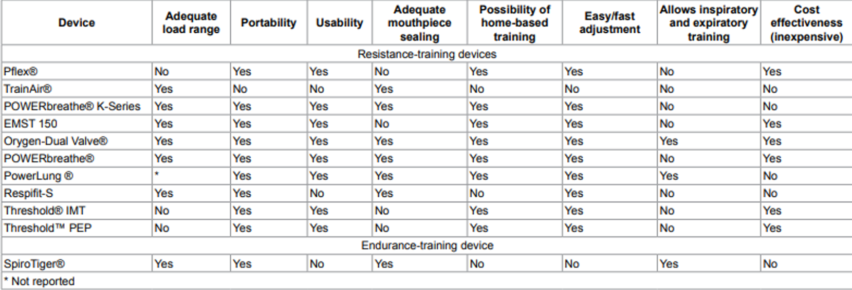
Figure 1. Characteristics of the evaluated respiratory muscle training devices (Menzes et al., 2018, Open Access).
In clinical settings, pressure threshold devices are more commonly used, and the market has seen a proliferation of such devices. This information is based on an article from 2018, and it is worth noting that there may be new devices on the market or ones that are no longer available, like the Phillips threshold device, which is no longer being produced. The Phillips device was widely recognized for being an affordable and straightforward pressure threshold device.
The purpose of this review is to outline the mechanisms and characteristics of various respiratory training devices available at the time, offering valuable insights for those contemplating the integration of IMT into their practice. Examining the table, it is evident that not all devices provide an adequate load range. While the threshold device was accessible in terms of cost, its limited range made it less beneficial for diverse populations. Additionally, some devices lack an ideal mouthpiece, hindering the ability to achieve a proper seal. This aspect is particularly crucial for populations with neuromuscular challenges that may struggle to achieve a secure seal.
Certain devices allow users to create a seal simply by placing them against their mouths. Additionally, there are devices designed for both inspiratory and expiratory training, although previous evidence suggested that simultaneous training for both might not be ideal—favoring a focus on either inspiratory or expiratory training.
It is important to note that some devices may be cost-prohibitive, potentially limiting accessibility for a broader population. Considering these factors is essential when contemplating the implementation of IMT into practice, ensuring that the chosen devices align with the needs and constraints of the intended user base.
Proposed Mechanisms of Benefit
The proposed mechanism behind the benefits of IMT aligns with the principles of strength training applied throughout the body. When a muscle undergoes training, structural changes often occur, and recent advancements, such as real-time ultrasound, allow for the measurement of diaphragm thickness. Increasing thickness and contractility typically signify improved function.
Research, particularly in athletes, reveals that strengthening the diaphragm enhances the respiratory metaboreflex. Some studies demonstrate an increase in oxygen uptake at the lactate threshold or a reduction in lactate buildup. Essentially, this means individuals can exert themselves a bit more strenuously before anaerobic exercise produces a significant buildup of lactate, necessitating a pause. While there is evidence suggesting that IMT can improve lung function, the improvement may stem from the ability to take larger, more forceful breaths and expel air more efficiently.
This differs from addressing changes associated with airway obstruction, as seen in conditions like asthma and COPD. Anecdotal observations and research findings indicate some improvement in pulmonary function, although it is not a cure-all for conditions characterized by airway obstruction.
Metaboreflex
Metaboreflex is a physiological response that comes into play during exercise, particularly in the context of anaerobic exercise. As one engages in physical activity, metabolites such as lactate, potassium, and phosphate accumulate in skeletal muscles. These metabolites are sensed by receptors within the muscles, which then transmit this information to the dorsal horn of the spinal cord through group three and four muscle afferents. From there, the information travels to various areas in the brainstem, including the nucleus tractus solitarius and other cardiovascular control centers, which regulate blood flow and the contraction of skeletal muscles.
This intricate process leads to a series of hemodynamic adjustments. Interestingly, the respiratory muscle metaboreflex is considered even more potent than its counterparts, playing a crucial role in regulating various physiological responses during exercise.
In response to increasing metabolites in the inspiratory muscles during exercise, the metaboreflex is triggered. This reflex prompts a redirection of blood flow and oxygen from the periphery to the respiratory muscles. The rationale behind this response is to prevent fatigue in the respiratory muscles; the body prioritizes ensuring that the respiratory system remains functional. Breathing always takes precedence.
For instance, during high-intensity activities like a time trial on a bike, an individual may experience fatigue in their leg muscles, characterized by a sensation akin to having cement-filled legs due to lactic acid accumulation. However, the body strategically ensures that the respiratory muscles do not reach a point of exhaustion. Preserving the integrity of the respiratory system becomes a priority even when other muscle groups face fatigue.
The metaboreflex, triggered by the accumulation of metabolites, is influenced by IMT. Studies, such as Dempsey et al. (2002), suggest that IMT can effectively delay the onset of this reflex. Using the example of a 40-minute time trial on a bike, if the wall is typically hit at the 30-minute mark, individuals undergoing IMT may experience that challenge closer to the 32 or 33-minute mark. In sports performance, such a shift can have a significant impact.
This observation extends beyond athletics, as research explores the potential impact of the metaboreflex in populations with conditions like COPD. Respiratory muscle weakness and an early onset of the metaboreflex may be contributing factors limiting exercise capacity in these populations. IMT emerges as a potential intervention to address and mitigate these challenges, showcasing its relevance not only in sports performance but also in managing respiratory limitations in various disease states.
Precautions and Contraindications
- A history of spontaneous pneumothorax
- Pulmonary hypertension or large bullae on chest x-ray.
- Marked osteoporosis with a history of rib fractures.
- Desaturation during or following IMT (<94%).
- Asthma patients who have a very low symptom perception and suffer from frequent, severe exacerbations or with an abnormally low perception of dyspnea
- If a patient is suffering from a ruptured eardrum or any other condition of the ear
- Patients with marked elevated left ventricular end-diastolic volume and pressure
- Patients with worsening heart failure signs and symptoms after RMT / IMT
- Active cold, sinusitis, or respiratory tract infection
When considering IMT, it is crucial to be mindful of potential precautions and contraindications, many of which are rooted in the theoretical possibility of generating significant negative pressure swings during training. As individuals draw in a breath, the pressure changes can pose certain risks.
For instance, caution should be exercised if a person has a history of spontaneous pneumothorax or presents with large bullae on a chest x-ray. In such cases, creating intense pressure within the lungs may not be advisable. Similarly, individuals with severe osteoporosis and a history of rib fractures should approach IMT cautiously. The aggressive inhalation involved in the training process may pose risks, especially if individuals experience desaturation during its application. These considerations underscore the importance of tailoring IMT interventions based on an individual's specific medical history and condition.
In the realm of precautions, it is noteworthy to exercise caution with asthma patients who have very low symptom perception and experience frequent severe exacerbations. Additionally, individuals with an active cold, sinus or respiratory infection, or an eardrum issue should approach IMT with care, as the negative pressure generated during training may not be beneficial in these conditions.
When it comes to osteoporosis, it is not necessarily a contraindication but a precaution. In such cases, the approach to IMT may involve adjusting the load and repetitions. Opting for lower loads and longer repetitions could be a more prudent strategy, akin to modifying strength training in any other context. The key takeaway is the importance of tailoring the IMT approach based on individual characteristics and conditions, emphasizing a personalized and cautious prescription. This ensures that the potential benefits of IMT are realized while minimizing any associated risks.
Using IMT in Clinical Populations - What is the Evidence?
When to Consider IMT?
- Symptoms present that might be associated with respiratory muscle weakness, or there is an increased load on the pump:
- Exertional or ‘positional’ dyspnea, impaired cough, ventilatory failure/hypercapnia
- Patients at risk for respiratory muscle weakness:
- COPD/CHF/ICU
- Neurological diseases (MS, Parkinson's, CVA, diaphragm paresis)
- Neuromuscular disease
- Patients with an increased load on the respiratory system
- Reduced chest wall compliance: kyphoscoliosis
- Reduced lung compliance: lung fibrosis
- Patients after prolonged mechanical ventilation and weaning failure
In exploring the application of IMT in clinical populations, we will delve into some of the evidence. It is worth noting that the scope here is not exhaustive, given the intricate nature of IMT and its impact across diverse clinical scenarios. The increasing focus on respiratory muscle weakness and its implications in various clinical settings has spurred a wealth of research and interest in understanding the efficacy of IMT for specific populations. The ongoing research and the expanding interest in this domain emphasize the significance of evaluating its potential applications in different contexts.
The selection of populations for IMT consideration hinges on the underlying rationale for incorporating this training. For those keen on a more in-depth exploration, Rik Gosselink, a physiotherapist, has dedicated considerable time to studying IMT. His extensive research and literature offer insights into the benefits of IMT and guide the process of selecting patient populations suitable for this intervention. This information is drawn from a presentation he gave at the Australia Physiotherapy Association. It serves as an excellent summary of the considerations for inspiratory muscle training. Although the slides are not available for viewing, the details can be accessed online, Research Group for Rehabilitation in Internal Disorders.
Symptoms play a pivotal role in deciding when to incorporate inspiratory muscle training. The presence of symptoms potentially linked to respiratory muscle weakness, such as exertional or positional breathlessness, an impaired cough, or indications of ventilatory failure, suggests a scenario where IMT might be beneficial. Another consideration arises when there is an increased load on the respiratory pump, prompting the thought that reinforcing respiratory muscles could be advantageous.
Patients at risk for respiratory muscle weakness, including those with COPD, heart failure, and those who have undergone ICU treatment, present situations where IMT might be particularly relevant. The subsequent discussion will further delve into the implications of IMT in ICU settings. Neurological diseases are certainly a focus, along with neuromuscular diseases and patients experiencing an elevated respiratory system load. This could manifest as reduced chest wall or lung compliance, and it is especially pertinent in patients who have undergone prolonged mechanical ventilation and experienced weaning failure. The ongoing research in this area is quite compelling. A recent study by Lista-Pax et al. (2023) aimed to define when we should consider inspiratory muscle weakness. It could be a valuable reference to have on hand, even though my colleagues and I playfully jest about the idea of having the reference equation readily available for mental calculations.
Methodology (Lista-Pax et al., 2023)
- Measurements Plmax/PEmax
- SEPAR protocol in agreement with ATS/ERS guidelines
- Flanged mouthpiece
- Pressures sustained 3-5 sec
- Min 6 acceptable manoeuvres, 3 of them with variability <5%
- SEPAR protocol in agreement with ATS/ERS guidelines
Results
- Reference equations
- PImax= 61.48 + 0.66 * age + 1.55 * BMI - 0.01* age2
- PImax= 98.60 + 1.18 * age + 0.76 * BMI - 0.02 * age2
- PEmax= 74.75 + 1.67 * age + 1.75 * BMI - 0.02 * age2
- PEmax= 58.11 + 3.71 * age + 2.64 * BMI - 0.04 * age2
- Cut-offs
- Inspiratory muscle weakness
- Female 62 cmH20
- Male 83 cmH20
- Expiratory muscle weakness
- Female 81 cmH20
- Male 109 cmH20
- Inspiratory muscle weakness
Examining the work of Lista-Pax et al. (2023) adds a layer of complexity to our understanding. The study encompassed a sizable sample of 610 individuals, spanning the wide age range of 18 to 80 years. While the substantial sample size is commendable, the broad age spectrum introduces a challenge, given the potential physiological variations across such a diverse population. The study's key findings provide cutoff values for inspiratory and expiratory muscle weakness. In women, inspiratory muscle weakness is identified below 62 centimeters of water, while in men, the cutoff is 83 centimeters of water. For expiratory muscle weakness, the respective cutoffs are 81 centimeters of water for women and 109 centimeters of water for men. Falling below these thresholds indicates the presence of muscle weakness. It is worth noting that these findings contribute valuable insights, particularly for clinicians working with individuals exhibiting respiratory muscle weaknesses. However, the application of inspiratory muscle training may vary across different healthcare practices. Internationally, physiotherapy colleagues specializing in breathing pattern disorders may adopt unique approaches to inspiratory muscle training, reflecting the diversity in clinical methodologies within the field.
When assessing the impact of diaphragm weakness, our approach differs from setting a rigid cutoff, such as the thresholds mentioned in previous studies. Rather than solely relying on specific numerical values, we consider functional weakness. This means that even if an individual's maximal inspiratory pressure exceeds the defined cutoffs, we still evaluate factors like endurance and performance in real-life situations. For instance, if someone experiences breathlessness during an activity requiring significant core activation and body movement, we may consider IMT even before reaching the defined cutoff levels. Our practice takes into account symptoms and functional limitations alongside objective measurements. If an individual falls below their maximum respiratory pressure and exhibits breathlessness during certain activities, we might initiate IMT earlier, prioritizing the improvement of diaphragmatic function and strength.
Prior to the recent study (Lista-Pax et al., 2023), our reference point was a 2013 study that provided some insight into maximum respiratory pressures. However, the information available in the literature on this topic has been somewhat limited. The 2013 study by Sclauser Pessoa et al., while informative, lacked detailed categorization by age and gender. In contrast, the current study offers a more nuanced breakdown, allowing us to consider age-specific variations. For example, a 30-year-old athlete with a maximum inspiratory pressure of 90 centimeters of water might surpass the general cutoff but would still be identified as someone to work with to enhance diaphragmatic function and strength. This nuanced approach acknowledges the variability in respiratory capabilities across different age groups and genders, enabling a more personalized and effective intervention strategy.
COPD
Let's delve into various population groups, particularly focusing on COPD. This area is intriguing due to the diverse evidence available. The rationale for considering it lies in the fact that hyperinflation can lead to diminished diaphragm function and reduced diaphragm thickness. Notably, a decline in inspiratory muscle strength in COPD was observed as early as 1996 by Gosselink et al.
To illustrate the evolution of studies on this topic, Gosselink et al. (2011) findings indicated that inspiratory muscle training enhances not only inspiratory muscle strength and endurance but also functional exercise capacity, dyspnea, and overall quality of life. However, by 2018 (Schultz et al.), a different perspective emerged, suggesting that three weeks of inspiratory muscle training added to pulmonary rehab enhances inspiratory muscle strengthening. Nevertheless, this addition does not result in improvements in exercise capacity, quality of life, and dyspnea. In summary, the evidence surrounding hyperinflation's impact on COPD and the efficacy of inspiratory muscle training is diverse and has evolved over the years. From initial studies showing improvements in various parameters to recent findings indicating specific limitations, the landscape of knowledge in this area is complex.
I would argue that drawing solid conclusions from a mere three weeks of any intervention, particularly in the realm of exercise, might be overly optimistic—an opinion shared by many. Assessing the efficacy of a practice based on only two measures before and after such a short duration may not offer sufficient insight, potentially explaining the lack of observable functional translation.
However, in 2023, Ichiba et al. completed a study involving a more extended period of inspiratory muscle training—spanning two months at 50% of maximum inspiratory pressure—which yielded more encouraging outcomes. This intervention not only improved maximum inspiratory pressure (MIP), diaphragm thickness, and the six-minute walk test but also led to a reduction in dyspnea and leg fatigue. Once again, the outcomes appear varied, prompting a closer examination of the studies to discern the nuances of how the intervention is implemented.
In 2010, Simms et al. conducted a study at the University of Toronto in Canada, aiming to enhance inspiratory muscle implementation. The research presented a nuanced journey marked by both successes and challenges. Upon inspecting the table's data, a distinct pattern emerges. In 1992, the study concluded that IMT lacked effectiveness in treating COPD. However, a pivotal shift occurred in 2002 when IMT alone exhibited a substantial positive impact on inspiratory muscle strength, endurance, and the mitigation of dyspnea both at rest and during exercise.
The narrative took a downturn in 2003, with a declaration of insufficient evidence to endorse the use of IMT in COPD, prompting a call for additional studies. By 2005, a reversal of fortune is evident, showcasing significant improvements in inspiratory muscle strength and endurance. This positive trajectory is reiterated later in the same year. In 2006, the study recommended considering IMT as a potential therapeutic approach, particularly for patients displaying signs of respiratory muscle weakness.
In 2007, the updated Canadian Thoracic Society Guidelines did not include any mention of IMT. The subsequent year, a systematic review in 2008 revealed enhanced outcomes in exercise capacity and a measure of quality of life, though the clinical significance remained unclear. Concurrently in 2008, there were notable advancements reported.
The trajectory continued with a series of significant improvements, reflecting a fluctuating pattern. Despite these positive findings, the practical implementation of IMT remained elusive. This paper advocated for the use of IMT in the population, lamenting the apparent lack of adoption. Even after two decades, the beneficial aspects highlighted in studies were not universally embraced by the respiratory population. Despite the demonstrated benefits and minimal side effects, IMT continues to face resistance in gaining traction within the respiratory community. The potential for positive outcomes persists, yet widespread acceptance appears to be elusive.
Exploring IMT is a worthwhile endeavor, especially when considering its potential application for patients with COPD. A valuable resource in this context is a practical guide from Hill et al. (2010) that delineates the clinical rationale for its utilization. The guide recommends considering IMT for individuals unable to fully engage in whole-body exercise due to various comorbid conditions. This perspective prompts the consideration of IMT not only as a targeted intervention but also as a potential pre-habilitation strategy. For instance, if a patient finds whole-body exercise challenging or feels too breathless to participate, IMT could serve as an initial step. By focusing on strengthening respiratory muscles, IMT lays the foundation for improved core stability, facilitating progress in overall exercise capacity. This dual approach addresses both respiratory challenges and broader physical conditioning, offering a comprehensive strategy for enhancing patient engagement in exercise programs.
Continuing the exploration of IMT as an adjunct, it is essential to adhere to specific recommendations outlined in the guide. Exclusion criteria involve individuals at risk for spontaneous pneumothorax or rib fractures. Supervision during training is crucial, including the monitoring of respiratory rate and oxygen saturation, aligning with common sense safety precautions. The assessment process, both before and after an IMT program, involves measuring maximum respiratory pressure, dyspnea levels, quality of life, and exercise capacity. This systematic evaluation aims to discern the impact of IMT on individual patient outcomes, contributing to a tailored training program.
Guidelines advocate for the use of pressure threshold training devices, emphasizing their accuracy. Interval-based training programs are recommended, with loads exceeding 30% of the MIP, progressing based on perceived exertion. Implementing IMT is not just about the initial gains in strength; it necessitates a sustained effort through a maintenance program. Strength gains are evident initially, but a consistent approach is required to ensure long-term benefits. For healthcare professionals considering IMT in COPD management, this resource serves as a valuable guide, providing comprehensive insights into implementation principles and exercise strategies.
Asthma
- RCT (IMT vs. sham) of 38 participants found improvements in respiratory muscle strength, exercise capacity, quality of life, daily living activities, reduced perception of dyspnea, and fatigue (Duruturk et al., 2018).
- 2023 systematic review and meta-analysis concluded that IMT causes a significant increase in MIP; more evidence is needed to clarify the effect of IMT on respiratory muscle endurance, rescue medication, exercise capacity, healthcare use, and HRQoL (Lista-Paz et al., 2023).
When considering the asthma population, the evidence surrounding IMT appears somewhat mixed. From a clinical perspective, an increase in maximum inspiratory pressure may be observed, but its translation into functional improvement or enhanced quality of life is uncertain. In practical terms, issues related to breathing pattern disorders in asthma seem more prevalent than actual respiratory muscle weakness, based on clinical experience.
IMT may find utility in addressing breathing pattern disorders by providing a resistance that facilitates biofeedback. It can serve as a tool to enhance awareness of proper muscle engagement during breathing. In the athletic context, IMT might be employed to derive performance benefits or to address exercise-induced laryngeal obstruction or vocal cord dysfunction in athletes with asthma.
However, IMT may not be a routine or go-to option for individuals with asthma in day-to-day clinical practice. The research seems to align with this perspective, suggesting that respiratory muscle weakness is not a predominant feature associated with asthma. Thus, the decision to incorporate IMT into asthma management may be contingent on specific clinical considerations, including the presence of breathing pattern disorders or specific performance goals for athletes with asthma.
Restrictive
- 8 weeks of IMT added to PR improved inspiratory muscle strength, 6MWD, QOL, and dyspnea (Zaki et al., 2022).
- Systematic review of IMT in ILD suggests there is a positive effect of IMT on respiratory muscle function, quality of life, exercise capacity, and dyspnea. However, research is still emerging, and existing study designs are of lower quality (Hoffman et al., 2021).
- Decreased MIP and endurance were related to decreased exercise tolerance in sarcoidosis (Marcellis et al., 2013).
- However, weakness tends to be variable in this population.
There's a growing interest in exploring IMT for individuals with restrictive lung diseases, and it seems to be a promising avenue for potential benefits. In conditions characterized by increased respiratory drive but reduced lung compliance, such as restrictive diseases, the mismatch between demand and the ability to generate required pressures becomes apparent. This creates a scenario where strengthening the respiratory muscles could potentially address the challenges posed by decreased lung compliance.
Approaching IMT on an individual basis rather than a population level is emphasized. Assessing whether a person exhibits signs of respiratory muscle weakness and gauging their potential compliance with a training regimen are crucial considerations. The decision to implement IMT should be tailored to the specific needs and characteristics of the individual.
From a physiotherapist's standpoint, evaluating the rib cage's flexibility and pliability is highlighted. A stiff and non-pliable rib cage may necessitate increased effort from respiratory muscles to achieve expansion and facilitate adequate inspiratory volumes. This mechanical perspective underscores the rationale behind considering IMT for individuals with restrictive lung diseases.
I think this is an area where we could see more increased research focus on inspiratory muscle training, particularly within the context of restrictive diseases. The mechanical reasoning behind its potential benefits makes it a compelling area for further exploration and investigation.
ICU
Allow me to delve into this topic, bearing in mind that I am not an ICU physiotherapist. Back in my student days, I had some exposure to acute care, though not extensively in the ICU. I hope to capture your interest in this matter. If anyone has experience with IMT in the ICU, I would appreciate engaging in a discussion about it. To me, it seems like a straightforward concept.
We are familiar with PICS (Post-Intensive Care Syndrome) or ICU-acquired weakness. It logically follows that the diaphragm weakens after prolonged ventilation. Approximately one-third of adult ICU patients ventilated for seven or more days exhibit impaired inspiratory muscle endurance or diaphragm atrophy (Bissett et al., 2015). In a 2018 meta-analysis by Vorona et al., IMT emerged as feasible and well-tolerated in critically ill patients, linked to shorter ventilation and weaning periods. The recommended protocol for this population involves high intensity, at least 50%, and low repetitions (Bissett et al., 2020).
If you are considering exploring this further, Australian physiotherapist Bernie Bissett has researched and applied IMT in the ICU, providing a practical guide for clinicians. You can find it referenced at the end of the slide handout and available online. There is a notable movement to champion inspiratory muscle training because it aligns with the recognized weakness in patients, which can complicate the weaning process.
As part of that guide, Inspiratory Muscle Training for Intensive Care Patients: A Multidisciplinary Practical Guide (Bissett et al., 2019) outlines considerations for IMT. For patients who are ventilator-dependent, alert, cooperative, and able to trigger spontaneous breaths with a respiratory rate of less than 25, incorporating inspiratory muscle training is advisable. This helps in initiating diaphragm training for those recently weaned from ventilation.
Being alert and cooperative involves the ability to maintain a lip seal around the mouthpiece or follow the guidelines depicted in the diagram. Considerations for strengthening the diaphragm and aiding in weaning or post-ventilation progression are crucial. This aspect forms a substantial area for contemplation in inspiratory muscle training. The application of IMT extends to various populations. I could have easily expanded this discussion with additional slides due to the abundance of information and diverse perspectives on inspiratory muscle training.
Other Populations
Several studies suggest that preoperative IMT may mitigate the risk of postoperative complications, such as pneumonia and atelectasis, particularly in thoracic surgeries (Karanfil & Moeller, 2018). The growing trend is the integration of IMT as a pre-surgical measure. The objective is to bolster muscle strength, focusing on the diaphragm and inspiratory muscles, thereby minimizing the likelihood of post-op complications. This approach has shown positive outcomes in reducing respiratory complications after stroke, as highlighted in a systematic review of five studies (Menezes et al., 2016). However, one challenge lies in achieving a secure mouth seal, with certain devices facilitating this by allowing the mouthpiece to be placed against the lips.
In a pilot study conducted by Human & Morrow (2021), inspiratory muscle training in children and teens with neuromuscular disease was deemed safe, feasible, and acceptable. The intervention correlated with enhanced inspiratory muscle strength, as well as improved upper limb function and coordination, opening up another avenue for exploration. A fascinating tangent in the realm of physiotherapy explores how inspiratory muscle fatigue alters postural strategies. Lotte Janssens, a physiotherapist, delved into this area in her doctoral thesis, available online in PDF format. Her series of studies examined the role of inspiratory muscles in postural control and how fatigue might impact postural control, extending these findings to individuals with chronic obstructive pulmonary disease (COPD).
The work of Janssens et al. (2015) reveals that respiratory muscle fatigue can influence balance issues in COPD, expanding our understanding beyond the traditional respiratory benefits of IMT. The study observed alterations in postural strategies during unexpected movements, typically measured by placing participants on a movable plate. Individuals with inspiratory muscle fatigue exhibited a greater tendency to adopt an ankle strategy instead of the usual back strategy, a pattern commonly seen in those with low back pain. This insight underscores a connection between low back pain, reduced balance, respiratory muscle weakness, and fatigue. The findings suggest a potential avenue for addressing balance and proprioceptive challenges through targeted inspiratory muscle interventions.
Enhancing diaphragmatic strength and function can lead to improvements in postural strategies and balance. This becomes particularly crucial when addressing the observed reduction in balance among individuals with COPD. Consequently, we must explore whether respiratory muscle dysfunction contributes to this imbalance and whether inspiratory muscle training can bring about positive changes beyond respiratory outcomes, potentially influencing balance and proprioceptive strategies and thereby reducing the risk of falls.
Expanding our perspective on inspiratory muscle training, there is a noteworthy study out of Colorado that gained attention for its focus on inspiratory muscle training in sleep apnea. Despite the initial intent, the randomized control, double-blind study involving 36 adults revealed unexpected benefits: inspiratory muscle training not only lowered blood pressure but also improved endothelial function (Craighead et al., 2021). This finding emphasizes the potential broader impacts of inspiratory muscle training, demonstrating its capacity to reduce blood pressure with just five minutes of daily breathing exercises.
Considering the connection between inspiratory muscle training and diaphragmatic breathing, it is worth investigating how these practices compare in terms of changes in blood pressure. Engaging in diaphragmatic breathing, tied to the parasympathetic nervous system and the vagus nerve, typically leads to lowered heart rate and blood pressure. Exploring whether inspiratory muscle training and diaphragmatic breathing yield similar effects on blood pressure could provide valuable insights.
In conclusion, the multifaceted benefits of inspiratory muscle training extend beyond respiratory improvements, encompassing aspects of balance, blood pressure regulation, and its potential impact on obesity-related respiratory challenges. Further research is warranted to delve deeper into these areas and compare the effectiveness of inspiratory muscle training with other therapeutic interventions, such as diaphragmatic breathing.
Obesity
- Obesity can cause impairments to chest wall mechanics, reductions in lung volumes, and increased work of breathing (Severin et al., 2019)
- Respiratory muscle fatigue is higher in obese individuals with poor sleep quality (Severin et al., 2021)
- Patients with obesity demonstrate respiratory muscle fatigue after peak aerobic exercise ((Severin et al., 2022)
- 2023 systemic review determined that IMT improves physical capacity and IM strength (Caicedo-Trujillo et al., 2023)
Turning our attention to obesity, it emerges as an emerging area of interest in the context of inspiratory muscle training. Obesity induces impairments in chest wall mechanics, reductions in lung volumes, and increased work of breathing. Respiratory muscle fatigue is notably higher in obese individuals with poor sleep quality, and those with obesity display respiratory muscle fatigue after peak aerobic exercise.
Recent findings indicate that individuals with obesity may experience potentially abnormal respiratory muscle fatigue compared to those without obesity. A systematic review from 2023 underscores the positive impact of inspiratory muscle training on physical capacity and inspiratory muscle strength. This intervention is not only easy to implement and relatively inexpensive but also holds promise as an accessible option, particularly for individuals for whom exercise may present challenges. It becomes a potentially empowering self-directed approach, offering positive gains in physical well-being. This is particularly relevant for populations facing barriers to traditional exercise methods.
In considering the diverse range of populations that might benefit from inspiratory muscle training, it becomes apparent that this intervention has far-reaching implications. Its easy implementation and potential positive outcomes make it a valuable consideration across various patient populations. The ever-growing list of potential beneficiaries underscores the versatility and broad applicability of inspiratory muscle training in improving both respiratory and overall physical health.
More?
- GERD
- Sport Performance
- Post COVID (CAREFULLY!)
- Heart Failure
Fascinating research has explored the relationship between reflux and diaphragm strength, highlighting that diaphragmatic strength can influence the tone of the lower esophageal sphincter. The lower esophageal sphincter, situated within the diaphragm, is shown to be affected by the strength or tone of the diaphragm, influencing its closure and pressure. IMT has been investigated as a potential means to improve gastroesophageal reflux disease (GERD). Anecdotal evidence, supported by patient reports, suggests that reflux symptoms may improve with IMT, although caution is advised as excessively high loads during training may exacerbate reflux.
In the realm of sports performance, numerous studies have explored IMT as an ergogenic aid. The potential benefits lie in delaying the onset of fatigue, allowing athletes to push their limits further. This avenue is gaining interest, and it would not be surprising if it starts appearing in social media discussions related to inspiratory muscle training for enhanced sports performance.
A noteworthy area of exploration is the use of IMT in the post-COVID context. While some evidence suggests its potential utility, there is a crucial need for caution, particularly in individuals with post-exertional malaise—a common symptom in those with long COVID. Given that IMT essentially involves exercise, its application in this population requires careful consideration of individual circumstances, such as the presence of respiratory muscle weakness without post-exertional malaise.
Heart failure has also been a subject of recent study, with emerging evidence suggesting potential benefits of IMT. Additionally, ongoing research in the area of double lung transplant recovery is anticipated, as some researchers are delving into the application of IMT post-transplant.
Lastly, a new study has delved into the realm of low back pain. This could be particularly intriguing for physiotherapists as it aligns with their focus. The diverse applications of IMT across various conditions underscore its versatility, providing potential benefits in areas ranging from respiratory health to sports performance and beyond. Depending on the patient population and healthcare setting, these applications may offer relevant and valuable interventions.
Training Protocol
- Research based on quantity – 30 breaths, 2x/day, approximately 40% MIP
- Some studies will advocate for higher loads
- Clinical experience
- Quality vs. quantity
- Aim for pattern first
- Strength vs. Speed
When it comes to training protocols for IMT, existing research often supports a regimen of 30 breaths twice a day, with the load set at approximately 40-60% of the maximum inspiratory pressure. However, it is important to note that some studies advocate for higher loads, and the choice depends on the specific population being addressed. For instance, in the case of COPD, there is a recognition that these individuals possess good endurance in their diaphragm, albeit with reduced power. As a result, a higher load may be more beneficial for COPD patients, focusing on generating the necessary force for activities that demand increased respiratory effort.
Clinical considerations also play a role, especially in fields like breathing pattern disorders. Emphasis is often placed on achieving the most efficient breathing pattern, typically favoring diaphragmatic breathing when all components are functioning optimally. In this context, the goal may be to prioritize quality over quantity in training. It becomes crucial to steer away from training individuals to rely on accessory muscles, except in specific patient populations.
Deciding between strength and speed is another aspect to consider. For certain patient populations, particularly those with upper airway dysfunction, the emphasis may be on speed of breath. Athletes, for instance, might benefit from focusing on breath speed to address upper airway issues related to the pace of breathing. This consideration, however, may not be as relevant in all patient populations and may need to be tailored based on individual needs and goals. The key is to customize the training approach to the specific requirements of the population being addressed.
Is How You Cue Important?
The importance of how you cue individuals during inspiratory muscle training depends on the specific goals and characteristics of the population you're working with. In scenarios where the primary goal is ventilation, such as in the ICU or with patients experiencing diaphragm paralysis, the focus may be on engaging any available muscles for inspiration. In these cases, you prioritize ventilation over the specific muscles involved. Additionally, for patients with pectus excavatum (a condition involving chest wall deformities), high-load diaphragmatic inspiratory muscle training might not be suitable, as it could exacerbate negative pressure and draw everything inward. In such cases, alternative approaches, like practicing with hands behind the head and adopting more of a chest pattern, may be more appropriate.
Cueing becomes crucial in these situations, tailored to the individual's needs and circumstances. A study in 2016 by Ramsook et al. compared the effects of different cueing strategies on healthy men. The study found that diaphragmatic-targeted cueing led to a significant increase in electromyography (EMG) activity of the diaphragm, increased transdiaphragmatic pressure, and decreased scalene muscle activity.
- Found that training with tapered flow resistive loading or pressure threshold loading from Functional Residual Capacity improved respiratory muscle function at higher lung volumes
- Pressure threshold loading from residual volume (standard protocol) restricted gains at lower lung volumes (Van Hollebeke et al., 2020)
Another aspect to consider is lung volume during inspiratory muscle training. Research has explored whether it is better to start from the end of expiration and then breathe in or to follow a regular inhale-exhale pattern. A study by Van Hollebeke et al. compared tapered flow resistive (electronic devices) and standard manual pressure threshold devices and found that better muscle function was observed when starting from functional residual capacity (end exhale) rather than almost reserve volume.
Using a Personalized Approach
- Consider training load (Ren-J S et al., 2021)
- Strength vs endurance
- “task failure”
- Concurrent training – training with activity
- Lung volumes
- Expiratory muscle training…
Being specific in how you cue individuals during inspiratory muscle training can have a significant impact on outcomes, including muscle engagement and function. Tailoring your approach based on individual characteristics, goals, and potential limitations is crucial for effective and safe training. Are you prioritizing strength or endurance? Is the focus on enhancing ventilation, particularly in patient populations like the ICU or those with spinal cord injuries? Perhaps the goal is to improve breathing patterns. I specifically emphasize diaphragmatic breathing, using it as a biofeedback tool. For instance, I might introduce a straw, aiming for subtle adjustments, or opt for a more functional approach, incorporating movement while breathing. This is crucial to address potential issues like breath-holding during activity, ensuring an integrated approach to core stability, breathing, and movement.
Creativity plays a key role in tailoring interventions. Introducing devices during training sessions, where individuals move while breathing, can be effective. The concern here is addressing potential breath-holding habits during activity, especially when there's a need to integrate core stability, breathing, and movement seamlessly.
The ellipsis after "expiratory muscle training" alludes to a distinct and evolving area of research. The focus shifts to different muscles with unique benefits. Determining if a patient would benefit from expiratory muscle training is a topic for another day, yet it highlights the importance of a personalized approach in treatment decisions.
In conclusion, a personalized approach is fundamental to all aspects of treatment. Whether considering strength, endurance, ventilation, or breathing patterns, tailoring interventions to individual needs is key. The evolving nature of research, as seen in expiratory muscle training, further underscores the need for adaptability and personalized care. It seems I've managed to navigate this discussion within the allotted time. If you have further questions or need clarification, feel free to ask.
Questions and Answers
Would it be reasonable to expect stronger voluntary cough reflex in COPD patients?
If your concern is coughing, then directing attention to expiratory muscle training may be beneficial. Improved inspiratory effort, resulting from effective inspiratory muscle training, may enhance expiratory effort as well. This improvement is attributed to a larger lung volume, supporting increased pressure generation for exhalation. When individuals seem to be taking in limited air, inspiratory muscle training becomes a potential avenue for addressing this concern. Moreover, it is noteworthy that inspiratory muscle training might contribute to improved mucus clearance facilitated by the adoption of deeper and more substantial breaths.
The active cycle of breathing, coupled with the use of the inspiratory muscle trainer followed by the positive expiratory pressure (PEP) device, can be an effective strategy for mucus clearance. This approach is particularly noteworthy in the case of emphysema, where the individual finds value in incorporating both devices. The combination of inspiratory muscle training and the PEP device is perceived as beneficial, aiding in the movement and clearance of mucus. This underscores the versatility and individualized nature of respiratory therapy interventions.
What is the major difference between IMT and EMT?
If the primary focus is on enhancing cough, then directing attention to expiratory muscle training becomes crucial. This type of training primarily targets abdominal muscles, offering improvements not only in the abdominal region but also extending benefits to some of the throat muscles. If optimizing cough effectiveness is the goal, expiratory muscle training would be the more targeted and relevant approach in this context.
- IMT Focus: Primarily targets the muscles involved in inhalation or inspiration.
- Purpose: Aims to strengthen and improve the endurance of the muscles responsible for expanding the lungs during inhalation.
- Training Devices: Involves exercises and devices that provide resistance during inhalation, such as inspiratory muscle trainers.
- EMT Focus: Primarily targets the muscles involved in exhalation or expiration, including abdominal and throat muscles.
- Purpose: Aims to strengthen and enhance the endurance of muscles responsible for compressing the lungs and facilitating exhalation.
- Training Devices: Involves exercises and devices that provide resistance during exhalation, often focusing on abdominal muscle engagement
What method would you use, possibly without a device?
When considering alternatives to specialized devices for inspiratory muscle training, simple and cost-effective methods can still offer benefits. For instance, using a book on the belly or incorporating a straw may not provide the same specificity in terms of strength gains, but they can serve as effective tools for training diaphragmatic breathing. Adding a thick elastic band, akin to what's commonly known as a THERABAND in physio, around the lower ribs can introduce an element of resistance. While this may offer more of a biofeedback effect than explicit strengthening, it remains a viable option, especially for those with budget constraints.
Considering the affordability factor, a metal straw and a piece of theraband could be a more accessible solution, providing some resistance and yielding noticeable benefits. Although these makeshift methods may not be as specialized or targeted as dedicated devices, they can still contribute to improving inspiratory muscle function, making them a practical alternative for individuals with financial constraints.
References
Select references are listed here (frequently quoted). A complete reference list is provided in the course handout.
Sclauser Pessoa, et al. (2014). Reference values for maximal inspiratory pressure: a systematic review. Canadian Respiratory Journal, 21(1), 43-50.
Gosselink, R., et al. (2011). Impact of inspiratory muscle training in patients with COPD: what is the evidence? European Respiratory Journal, 37(416-425).
Schultz, K., Jelusic, D., Wittmann, M., et al. (2018). Inspiratory muscle training does not improve clinical outcomes in 3-week COPD rehabilitation: results from a randomised controlled trial. European Respiratory Journal, 51(1702000).
Dempsey, J et al. (2002). Respiratory influences on sympathetic vasomotor outflow in humans. Respiratory Physiology & Neurobiology, Volume 130, Issue 1, Pages 3-20, ISSN 1569-9048.
Ichiba, T., Miyagawa, T., Tsuda, T., Kera, T., & Yasuda, O. (2023). Changes in diaphragm thickness and 6-min walking distance improvement after inspiratory muscle training in patients with chronic obstructive pulmonary disease: Clinical trial. Heliyon, 9(9), e20079.
Simms, A. L., Li, L. C., & Reid, W. D. (2011). Development of a theory-based intervention to increase prescription of inspiratory muscle training by health professionals in the management of people with chronic obstructive pulmonary disease. Physiotherapy Canada, 63(3), 315–323.
Hill, K., et al. (2010). Inspiratory Muscle Training for Patients with Chronic Obstructive Pulmonary Disease: A Practical Guide for Clinicians. Archives of Physical Medicine and Rehabilitation, 91(1466-1470).
Duruturk, N., Acar, M., & Doğrul, M. I. (2018). Effect of Inspiratory Muscle Training in the Management of Patients With Asthma: A RANDOMIZED CONTROLLED TRIAL. Journal of Cardiopulmonary Rehabilitation and Prevention, 38(3), 198-203.
Lista-Paz, A., et al. (2023). Effect of respiratory muscle training in asthma: A systematic review and meta-analysis. Annals of Physical and Rehabilitation Medicine, 66(3), 101691.
Hoffman, M. (2021). Inspiratory muscle training in interstitial lung disease: a systematic scoping review. Jornal Brasileiro de Pneumologia, 47(4), e20210089.
Marcellis, R. G., Lenssen, A. F., de Vries, J., & Drent, M. (2013). Reduced muscle strength, exercise intolerance and disabling symptoms in sarcoidosis. Current Opinion in Pulmonary Medicine, 19(5), 524-530.
Bissett, B., Leditschke, I. A., Neeman, T., Boots, R., & Paratz, J. (2015). Weaned but weary: one third of adult intensive care patients mechanically ventilated for 7 days or more have impaired inspiratory muscle endurance after successful weaning. Heart & Lung, 44(1), 15-20.
Zaki, S., et al. (2022). Does inspiratory muscle training provide additional benefits during pulmonary rehabilitation in people with interstitial lung disease? A randomized control trial. *Physiotherapy Theory and Practice.
Bissett, B., et al. (2020). Respiratory Muscle Rehabilitation in Patients with Prolonged Mechanical Ventilation: A Targeted Approach. Critical Care, 24(103).
Bissett, B., et al. (2019). Inspiratory muscle training for intensive care patients: A multidisciplinary practical guide
Select references are listed here (frequently quoted). A complete reference list is provided in the course handout.
Menzes, K. K. P., Nascimento, L. R., Avelino, P. R., Polese, J. C., & Salmela, L. F. T. (2018). A Review on Respiratory Muscle Training Devices. Journal of Pulmonary & Respiratory Medicine, 8(451).
Gosselink, R., Troosters, T., & Decramer, M. (1996). Peripheral muscle weakness contributes to exercise limitation in COPD. American Journal of Respiratory and Critical Care Medicine, 153(976-980).
Vorona, S., et al. (2018). Inspiratory Muscle Rehabilitation in Critically Ill Adults: A Systematic Review and Meta-Analysis. Annals of the American Thoracic Society, 15(6), 735-744.
Karanfil, E. O. T., & Moller, A. M. (2018). Preoperative inspiratory muscle training prevents pulmonary complications after cardiac surgery – a systematic review. Danish Medical Journal, 65(3), A5450.
Menezes, K. K. P., et al. (2016). Respiratory muscle training increases respiratory muscle strength and reduces respiratory complications after stroke: a systematic review. Journal of Physiotherapy, 62(3), 138–144.
Human, A., & Morrow, B. M. (2021). Inspiratory muscle training in children and adolescents living with neuromuscular diseases: A pre-experimental study. South African Journal of Physiotherapy, 77(1), a1577.
Craighead, D., et al. (2021). Time-Efficient Inspiratory Muscle Strength Training Lowers Blood Pressure and Improves Endothelial Function, NO Bioavailability, and Oxidative Stress in Midlife/Older Adults with Above-Normal Blood Pressure. *Journal of the American Heart Association, 10.1161/JAHA.121.020980.
Janssens, L., McConnell, A. K., Pijnenburg, M., Claeys, K., Goossens, N., Lysens, R., Troosters, T., & Brumagne, S. (2015). Inspiratory muscle training affects proprioceptive use and low back pain. Medicine and Science in Sports and Exercise, 47(1), 12–19.
Severin, R., et al. (2019). Obesity and Respiratory Skeletal Muscles. In S. Walrand (Ed.), Nutrition and Skeletal Muscle (pp. 197-215). Academic Press.
Severin, R., Phillips, S. (2021). Respiratory muscle fatigability is higher in obese individuals with poor sleep quality. ERJ Open Research, 7(6). DOI: 10.1183/23120541.sleepandbreathing-2021.6.
Severin, R. S., Alshammari, S., Sherman, S. R., & Phillips, S. A. (2022). Acute Respiratory Muscle Fatigue in Patients with Obesity Following Peak Aerobic Exercise. The FASEB Journal, 36.
Caicedo-Trujillo, S., et al. (2023). Inspiratory muscle training in patients with obesity: a systematic review and meta-analysis. Frontiers in Medicine, 10, 1284689.
Ramsook, A. H., Molgat-Seon, Y., Schaeffer, M. R., Wilkie, S. S., Camp, P. G., Reid, W. D., Romer, L. M., & Guenette, J. A. (2016). Effects of inspiratory muscle training on respiratory muscle electromyography and dyspnea during exercise in healthy men. Journal of Applied Physiology, 122(5), 1267–1275.
Van Hollebeke, M., Gosselink, R., & Langer, D. (2020). Training Specificity of Inspiratory Muscle Training Methods: A Randomized Trial. Frontiers in Physiology, 11, 576595.
Ren, J. S., et al. (2022). Time to move beyond a "one-size fits all" approach to inspiratory muscle training. Frontiers in Physiology, 12.
Citation
DeMars, J. (2024). Inspiratory Muscle Training: A Review of the Evidence. Continued.com - Respiratory Therapy, Article 213. Available at www.continued.com/respiratory-therapy
Dec 12, 2023
1 CE HourNon-invasive Ventilation in the Post-Acute Care Setting
Claire Aloan, MS, RRT-NPS, FAARC
Editor’s note: This text-based course is an edited transcript of the webinar, Non-invasive Ventilation in the Post-Acute Care Setting, presented by Claire Aloan, MS, RRT-NPS, FAARC.
It is recommended that you download the course handout to supplement this text format.
Learning Outcomes
After this course, participants will be able to:
- Describe the patients who may benefit from non-invasive ventilation in the post-acute care setting
- List the available methods of non-invasive ventilation for post-acute care
- Explain the advantages and disadvantages of each method
Terminology
- Non-invasive ventilation (NIV) is a form of mechanical ventilation where positive pressure is provided to the airway and lungs through a mask or cannula, or mouthpiece
- Negative pressure ventilation (NPV) is a form of ventilation that promotes air flow using negative pressure applied to the chest wall
- POV® proportional open ventilation, a technology that augments inspired tidal volume
- TAV®: Tidal assist ventilator. Adds bolus to spontaneous tidal volume
- Post-acute care settings: usually home but may also include long-term care facilities and rehab settings
During this presentation, I will be using certain terminology. Hopefully, most of it is familiar, although some might be new to you. We will be referring to non-invasive ventilation as NIV, which involves mechanical ventilation providing positive pressure to the airway lungs through a mask, cannula, or mouthpiece. Sometimes, you will see this abbreviated as NPPV, denoting non-invasive positive pressure ventilation.
There are a couple of other abbreviations to keep in mind. However, when discussing non-invasive ventilation, our main focus will be on positive pressure. This sets the tone for our discussion. We will also touch upon NPV, which stands for negative pressure ventilation—a method where airflow is encouraged by applying negative pressure to the chest wall.
We will delve into the concept of proportional open ventilation, a technology that boosts inspired tidal volume. We will discuss the tidal assist ventilator (TAV), which essentially supplements spontaneous tidal volume with a bolus.
To clarify, post-acute care primarily refers to the home setting. Usually, when we talk about non-invasive ventilation beyond acute care, whether short-term or long-term, we are considering patients transitioning to home care. However, these interventions may also be relevant in long-term care facilities and rehabilitation settings, catering to the diverse needs of our patients.
Why NIV in Post-Acute Care?
Why do we want to even think about non-invasive ventilation in post-acute care? Well, when we use this, especially in the appropriately selected patients, we can do a number of things. One of the most important things we can do is to improve the quality of life of our patients. Many of our patients with disorders that affect the respiratory system are very limited in what they can do their quality of life tends to go downhill fairly rapidly.
Being able to reverse that and improve their quality of life is such a huge benefit of non-invasive ventilation. If you are involved in this at all with these patients, it is really a very feel-good place to be as a respiratory therapist. We also like the idea of reducing hospital admissions. That is a good thing for a number of reasons. Obviously, the expense of being in the hospital also affects your quality of life. Patients really do not want to be in the hospital, and it is not a great place for them to be. Reducing mortality is something that we work on patients can live longer, better lives with non-invasive ventilation. Oftentimes, we are looking at reducing dyspnea. We will talk specifically about how with some devices improving patients' exercise capacity, non-invasive ventilation has been shown to be helpful in all of those things.
Patients Who May Need NIV
- COPD
- Thoracic restrictive disorders
- Obesity hypoventilation syndrome
- ? Interstitial lung disease
When considering patients who might benefit from non-invasive ventilation beyond the acute care setting, we often think of individuals with a variety of respiratory conditions. While non-invasive ventilation is commonly utilized in acute care for COPD patients to avoid invasive intubation and its associated complications, its potential extends well beyond this setting.
As patients transition from hospitalization to home, rehabilitation facilities, or long-term care, the need for non-invasive ventilation may persist. This includes COPD patients who require ongoing respiratory support beyond the acute phase. However, it is essential to broaden our perspective to encompass other respiratory conditions such as thoracic restrictive disorders, obesity hypoventilation syndrome, and possibly interstitial lung disease.
Thoracic restrictive disorders encompass a range of conditions characterized by reduced lung expansion, potentially leading to hypercapnic respiratory failure. Similarly, obesity hypoventilation syndrome, defined by obesity, daytime hypercapnia, and coexisting obstructive sleep apnea, may necessitate non-invasive ventilation to address respiratory insufficiency.
While the role of non-invasive ventilation in interstitial lung disease remains somewhat uncertain, it is an area that warrants further exploration. By considering these diverse patient populations, we can better recognize the potential benefits of non-invasive ventilation and ensure its appropriate utilization beyond the acute care setting.
COPD
In discussions regarding COPD, there has been considerable controversy in recent years regarding its efficacy. However, the most recent study by William et al. (2021), which serves as the cornerstone publication summarizing various aspects of non-invasive ventilation by Williams and other pulmonologists, presents compelling evidence.
The prevailing belief now is that non-invasive ventilation indeed enhances survival and reduces healthcare utilization. This focus is increasingly favored. Often, in intensive care or step-down units, patients initially on invasive ventilation can be transitioned to non-invasive ventilation as a bridge to home respiratory support. This approach evolves alongside advancements in non-invasive ventilation technology. We can consider weaning non-invasive ventilation patients to settings conducive to home use.
Frequently, we administer non-invasive ventilation at relatively high settings during exacerbations, gradually tapering down but not necessarily discontinuing it entirely. Instead, we adjust the settings to accommodate home use. The timing of initiating non-invasive ventilation is crucial for COPD patients during exacerbations, particularly for those persistently hypercapnic, as they demonstrate the most significant benefits. It is important to note that non-invasive ventilation is not universally applicable to all COPD patients. It is most beneficial for those persistently hypercapnic.
There is evidence suggesting the need for more aggressive settings. Research indicates that patients receiving high-intensity pressure support fare better than those receiving low-intensity pressure support. When prescribing settings for home use, it may be advantageous to opt for slightly higher pressure differentials between inhalation and exhalation, defining pressure support. This calls for a reconsideration of our approach, perhaps leaning towards a more proactive stance than previously adopted.
Hypercapnic COPD patients constitute a distinct phenotype driven by specific physiological factors, such as air trapping, which imposes a significant mechanical disadvantage. Many of these individuals experience nocturnal hyperventilation and are prone to sleep apnea, placing them at heightened risk for adverse health outcomes. They represent a priority target for intervention, as highlighted in the latest GOLD guidelines, which offer a comprehensive discussion of COPD phenotypes worth exploring.
Home non-invasive ventilation holds promise in addressing the underlying physiological issues contributing to hypercapnia. While older literature yielded mixed results regarding its efficacy in various populations, recent clinical trials have demonstrated significant benefits, including improvements in quality of life, reduced hospitalizations, and mortality rates. This emerging body of literature supports a more assertive approach towards its utilization. Additionally, advancements in non-invasive ventilation technology, particularly in optimizing inspiratory pressure, may account for the observed success in home settings.
Effective implementation necessitates a thorough understanding of patient selection criteria and familiarity with available device modes and titration strategies. While literature offers insights into these aspects, practical constraints often limit our options in device selection and mode availability, both in hospital and home settings. Providers typically operate within the confines of specific devices offered by their institution or supplier, thereby influencing treatment decisions. Nevertheless, advocating for access to appropriate devices and modes remains crucial in optimizing patient outcomes.
We are excited about the emergence of telemonitoring, as it addresses a key challenge in the management of patients on non-invasive ventilation transitioning from hospital to home care. In the hospital setting, we have the luxury of being present, continuously titrating the device, and monitoring patient response. However, once patients return home, this level of oversight diminishes significantly. While in-home visits are feasible, they cannot provide round-the-clock surveillance, making telemonitoring an invaluable tool.
Telemonitoring enables us to remotely adjust device settings, enhance patient adherence, troubleshoot issues, and even anticipate exacerbations. Its evolving capabilities hold promise in optimizing non-invasive ventilation outcomes in home settings. Studies have demonstrated the efficacy of non-invasive ventilation in prolonging exercise endurance in COPD patients. However, implementing this during routine activities outside clinical settings poses logistical challenges, as most devices lack portability and usability during normal activities.
Nevertheless, there are exceptions to this limitation, which we will delve into further. Overall, telemonitoring represents a significant advancement in the management of COPD patients on non-invasive ventilation, offering enhanced support and optimization beyond the confines of traditional care settings.
Thoracic Restrictive Disorders
Another group of conditions we frequently consider is what we refer to as thoracic restrictive disorders. This encompasses a broad spectrum of conditions characterized by hypercapnic respiratory failure, where ventilatory defects impede adequate air movement in and out of the chest, resulting in elevated CO2 levels exceeding 45 millimeters of mercury.
A prominent category within this group comprises neuromuscular disorders, including conditions such as ALS, spinal cord injuries, muscular dystrophy, diaphragm paralysis, and spina bifida. Additionally, certain congenital central hypoventilation syndromes may hinder sufficient air exchange. Chest wall deformities, notably kyphoscoliosis, also fall under this classification. Furthermore, ventilatory drive defects, where patients lack the innate urge to breathe adequately, are pertinent considerations.
It is crucial to note that interstitial lung diseases are not typically classified under thoracic restrictive disorders despite causing restrictive pulmonary dysfunction. Consequently, thorough assessment is imperative to rule out obstructive sleep apnea (OSA), which, although less common in these conditions compared to COPD, may still occur. Monitoring CO2 and oxygen levels during wakefulness and sleep is essential post-therapy initiation, with sleep testing reserved for cases suggestive of OSA. Non-invasive ventilation remains a viable therapeutic option for these patients.
Obesity Hypoventilation Syndrome (OHS)
Another disorder to consider is obesity hypoventilation syndrome (OHS), characterized by the combination of obesity (with a BMI greater than 30), daytime hypercapnia exceeding 45 millimeters of mercury for CO2, and a diagnosis of obstructive sleep apnea (OSA). It is crucial to exclude other potential causes of alveolar hypoventilation before confirming this diagnosis.
Given the co-existence of OSA in these patients, continuous positive airway pressure (CPAP) is typically the initial treatment choice. However, CPAP may prove ineffective or poorly tolerated in correcting the ventilation disorder, especially when patients are hypoventilating alongside their OSA. In such cases, non-invasive ventilation becomes necessary to address their respiratory needs effectively.
Careful titration is essential for these patients, particularly regarding pressure selection, to reduce the apnea-hypopnea index to normal levels (less than five). Additionally, treatment must aim to alleviate both hypoventilation and hypoxia. Patients with OHS require comprehensive monitoring and follow-up, including sleep lab titration and clinic visits to assess blood gases and device adherence. Typically, follow-up appointments are scheduled four to six weeks post-initiation of non-invasive ventilation, allowing patients sufficient time to adjust to and utilize the device properly.
Interstitial Lung Disease (ILD)
Interstitial lung disease (ILD) encompasses a diverse array of progressive and debilitating lung conditions characterized by similarities in lung pathology and radiographic findings. However, with hundreds of distinct ILDs identified, the patient population exhibits considerable heterogeneity.
Management of ILD often involves oxygen therapy to maintain adequate oxygen saturation levels, yet achieving this can be challenging due to various factors contributing to limited exercise tolerance in these patients. In instances where oxygen therapy alone proves insufficient, considering adjunctive non-invasive ventilation may be beneficial. Non-invasive ventilation can help improve the ventilation-perfusion (VQ) ratio, reduce respiratory effort, and enhance exercise tolerance, particularly during episodes of exercise-induced desaturation.
While evidence supporting the use of non-invasive ventilation in ILD remains limited compared to other respiratory conditions, preliminary studies have shown promising results. Some studies have demonstrated improved oxygen saturation levels during exercise with the use of non-invasive ventilation, while others have observed enhancements in physical functioning and quality of life following non-invasive ventilation therapy.
Though the evidence base for non-invasive ventilation in ILD is still evolving, it remains a viable option to consider, especially for patients experiencing significant functional impairment. While more research is needed to establish its efficacy definitively in this patient population, non-invasive ventilation stands as a potential intervention to enhance the management and quality of life for individuals with ILD. Now that we have identified the patient groups that may benefit from non-invasive ventilation. Let's discuss the practical aspects of its implementation.
Methods for Non-Invasive Ventilation
- Traditional positive pressure ventilation with various interfaces (NIV)
- Proportional Open Ventilation (POV®)
- Negative pressure ventilation (NPV) or Biphasic Cuirass Ventilation (BCV)
- Tidal Assist Ventilation (TAV®)
Our discussion briefly touches on traditional positive pressure ventilation, which is a commonly utilized method known as non-invasive ventilation. This approach involves delivering positive pressure to the airway through various interfaces, a technique frequently employed in hospitalized patients and increasingly utilized in home care settings.
Next, explore proportional open ventilation, a distinct technique offering notable advantages. Following that, we will review negative pressure ventilation, an older yet still relevant method historically associated with the iron lung, originally developed during the polio epidemics. Negative pressure ventilation, often referred to as Biphasic Cuirass Ventilation (BCV) by the sole provider of this technology, presents unique benefits worth considering. Finally, we will touch briefly on Tidal Assist Ventilation, a newer approach that may gain prominence in the future.
Non-Invasive Ventilation
Traditional NIV
Traditional non-invasive ventilation (NIV), is a widely employed method utilizing interfaces to deliver either volume or pressure ventilation to patients across all age groups, from neonates to adults. These interfaces are meticulously designed to provide a snug fit while allowing for minor leaks to prevent potential pressure ulcers. With hundreds of interface options available, the selection process is critical, considering factors such as patient comfort and tolerance.
One noteworthy interface worth mentioning is mouthpiece ventilation, which is often overlooked in acute care settings but offers substantial benefits to many patients. While some devices feature preset values, others, like AVAPs, offer auto-adjustment capabilities, ensuring consistent tidal volume delivery with each breath.
A primary advantage of NIV is its avoidance of invasive procedures like endotracheal intubation, which come with a host of potential complications. Patients previously tracheostomized due to prolonged invasive mechanical ventilation can potentially transition to NIV, a more tolerable option for many.
However, challenges persist, primarily centered around patient intolerance or discomfort with the interface. Addressing these issues often involves exploring various interface options and rotating between them to alleviate pressure points and irritation. Ensuring access to a diverse range of interfaces is essential, particularly in home care settings, where patient comfort and adherence are paramount.
While traditional NIV offers a plethora of interface options—from full face masks to nasal prongs and minimalist designs—it is crucial to recognize that access to every interface may be limited depending on the clinical setting. Nonetheless, prioritizing interface comfort and patient preference can significantly enhance the effectiveness and acceptance of NIV therapy, mirroring the challenges faced with CPAP therapy. Thus, a comprehensive approach that incorporates patient-centered care and interface variety is essential to optimize outcomes in NIV implementation.
Mouthpiece Ventilation (MPV)
Mouthpiece ventilation (MPV), often overlooked in acute care settings, serves as a vital daytime non-invasive respiratory support method, particularly for patients with neuromuscular disorders. With MPV, patients utilize a mouthpiece or straw to facilitate ventilation. During MPV, patients intentionally initiate a series of disconnections, whether partial or complete, from the ventilator, followed by reconnections. Essentially, with each inhalation, patients have the autonomy to determine the volume of air they wish to intake. By adjusting the seal with their lips on the mouthpiece, patients can modulate their breaths.
A tight, complete seal ensures nearly full tidal volume (approximately 100%), as dictated by the settings of the volume-cycled ventilator. Conversely, a more relaxed or incomplete seal results in either partial tidal volume or, in some instances, no tidal volume intake at all, ranging from 0 to 99%. This flexibility allows patients to tailor their ventilation to their comfort and respiratory needs, promoting a more personalized and effective approach to non-invasive respiratory support.
Devices
A Breas ventilator is equipped with a mouthpiece that simplifies mouthpiece ventilation. It is typically mounted on an adjustable arm for easy positioning directly in front of the patient's mouth. Theoretically, leaks during mouthpiece ventilation will not cause discomfort because they are determined and managed by the patient. However, this holds true specifically for volume-cycled modes of ventilation. In volume-cycled modes, patients have control over the amount of air they inhale, allowing them to adjust the seal with their lips on the mouthpiece to accommodate intentional leaks without discomfort.
Conversely, in pressure-cycled modes of ventilation, leaks can indeed result in discomfort. This discomfort stems from the ventilator's automatic compensation for leaks by delivering additional flow, which can be excessive and difficult for the patient to manage. As a result, patients may experience challenges in speaking and swallowing during MPV, highlighting the importance of utilizing volume-cycled modes to optimize patient comfort and adherence. Mouthpiece ventilation provides a range of devices, and while the following list is not exhaustive, it highlights the importance of identifying the most appropriate option for each patient.
- Vyaire LTV
- ResMed Astral
- Breas Vivo65
- Breas Vivo45
- PhillipsTrilogy EV300
- Ventec VOCSN
- Movair Luisa
- Phillips Trilogy 100
Common options include the Vyaire LTV, ResMed Astral, and various models from Breas, with notable features such as integrated end-tidal CO2 monitoring in Breas devices, enhancing patient care. The Phillips Trilogy and Ventec VOCSN are also prominent choices. The VOCSN, notable for its multifunctionality, integrates ventilation, oxygen concentrator, suction, and nebulizer functions into a single device, streamlining mobility for active patients.
For instance, without the Ventec VOCSN, patients relying on traditional devices like the Phillips Trilogy would need additional equipment for oxygen supply, suctioning, and nebulization, complicating mobility. The Movair Lusia is a newer addition generating considerable excitement, alongside established options like the Phillips Trilogy 100, commonly employed for home use.
While these examples offer insight, collaborating with durable medical suppliers remains crucial to explore available options tailored to individual needs in standard non-invasive ventilation. In addition to the devices mentioned, it is worth noting that advancements in non-invasive ventilation technology continue to evolve, offering enhanced features and capabilities. For instance, some newer models may incorporate innovative algorithms for more precise patient monitoring and therapy delivery. Features such as remote monitoring capabilities, wireless connectivity, and user-friendly interfaces are becoming increasingly common, facilitating seamless integration into patients' daily lives.
Furthermore, considerations such as portability, battery life, and ease of maintenance play pivotal roles in device selection, particularly for patients who lead active lifestyles or require frequent travel. As technology advances, the aim is to provide patients with greater autonomy, comfort, and flexibility in managing their respiratory conditions.
It is important for healthcare providers to stay abreast of the latest developments in non-invasive ventilation technology and collaborate closely with patients and durable medical equipment suppliers to ensure optimal device selection and patient outcomes. By leveraging the benefits of cutting-edge technology, healthcare professionals can enhance the quality of care and improve the overall experience for patients relying on non-invasive ventilation.
Novel Methods of Non-invasive Ventilation
Proportional Open Ventilation (POV)
Let's delve into one of the novel methods of non-invasive ventilation, known as proportional open ventilation (POV). Originally developed by Brief Technologies under the name Non-Invasive Open Ventilation (NIOV), this innovative approach has evolved and is now trademarked by Hillrom as proportional open ventilation or POV. While NILV may still be referenced in the literature as the original terminology, it is essential to recognize the transition to POV.
Proportional open ventilation synchronously delivers positive inspiratory pressure and supplemental oxygen, typically via a nasal pillow interface. It operates by augmenting tidal volume during spontaneous breathing, making it particularly suitable for individuals with limited tolerance for physical activity due to breathlessness. The device offers a myriad of programmable settings tailored to meet the unique needs of each patient. Notably, patients experiencing significant dyspnea during exertion may find proportional open ventilation beneficial.
Baxter Hillrom Life 2000 Ventilator
The interface of this ventilation method incorporates openings that leverage the Venturi principle to entrain room air, which combines with the preset volume from the device and the patient's own effort. The total delivered tidal volume results from the interplay of these factors. Currently available from Baxter Hillrom under the name Life 2000 Ventilator, this technology represents a promising advancement in non-invasive ventilation, offering enhanced support for patients with respiratory challenges.
The Baxter Hillrom Life 2000 Ventilator device has three patient-selectable prescription settings, denoted by pictograms on the right-hand side of the interface. The top pictogram illustrates someone walking, representing the highest level of support suitable for mobility. The middle pictogram depicts someone sitting, which is ideal for home activities and daily routines. The bottom pictogram is designated for restful states.
These three distinct settings offer patients considerable flexibility in tailoring their support levels to match their varying activities and ventilatory needs. Whether they require continuous support or wish to adjust settings periodically, the interface simplifies the process with the push of a button, adapting seamlessly to their breathing patterns.
This adaptability proves invaluable in both clinical and home settings. In the hospital, clinicians can readily titrate ventilation settings to accommodate patients' evolving needs. Conversely, in-home care settings, periodic adjustments by healthcare providers ensure optimal support, as patients may not have the expertise to modify settings independently.
Approved for both invasive and non-invasive use, this device holds promise not only for post-acute care but also for promoting mobility in acute care settings. Streamlining equipment and reducing bulk facilitates patient mobility with ease. Powered by a rechargeable lithium-ion battery, it weighs only about a pound, making it remarkably lightweight and portable—a stark contrast to traditional ventilator setups. Its compact size, akin to a large home telephone, further enhances its practicality and accessibility for patients.
- Clinician adjustable settings:
- PEEP: 0 to 10 cmH2O
- Breath Rate: 1 to 40 BPM
- Breath Timeout Period: 20 or 60 seconds
- Breath Timeout Action: 12 BPM or 3 LPM
- Delivered Volume: 50 mL to 750 mL
- Gas source: Oxygen or Air
- I-Time: 0.15 to 3.00 seconds
- Trigger Sensitivity
The clinician has the flexibility to fine-tune settings within each activity level—rest, sitting, and walking. These adjustable parameters include PEEP (positive end-expiratory pressure), breath rate, and breath timeout period—the duration the machine waits before detecting a cessation in breathing. In the event of breath absence, the device can be programmed to maintain a continuous flow of oxygen or revert to a predefined breath rate, typically 12 breaths per minute.
Furthermore, the clinician can customize settings such as delivered volume, gas source (air compressor or oxygen tank), and inspiratory time—a critical consideration, especially for patients with COPD. Ensuring the device accurately detects patient breaths is paramount, and sensitivity adjustments allow for precise responsiveness.
A comprehensive array of clinician-adjustable alarms is available, covering low respiratory limits, low and high-pressure thresholds, and the breath timeout period. These alarms serve as essential safeguards, alerting clinicians to deviations from desired parameters, including excessively high or low breath rates. Adjustments can then be made promptly to optimize patient comfort and safety—a standard feature across most ventilators.
- Clinician adjustable alarms:
- Low Respiratory Limit: 0 to 119 BPM
- High Respiratory Limit: 5 to 120 BPM
- Low PIP Limit: 0 to 15 cmH2O
- High PIP Limit: 5 to 40 cmH2O
- Breath Timeout Period: 20 or 60 seconds
- High Breath Rate: 5 to 120 BPM
- Low Breath Rate: 0 to 119 BPM
Then there are also some fixed alarms:
- Low/High Source Pressure
- Low Battery
- Low/High Delivery Pressure
- High PEEP Pressure
- System fault
The Life 2000 ventilator incorporates an extensive alarm system designed to alert clinicians to various potential issues. These alarms encompass discrepancies in source pressure, battery status, and delivery pressure, as well as high PEEP (positive end-expiratory pressure) levels, among others. This comprehensive alarm functionality distinguishes the Life 2000 as a full-fledged ventilator, ensuring thorough monitoring and prompt identification of critical situations. The interface commonly paired with the Life 2000 ventilator optimizes its functionality and usability, facilitating seamless integration into patient care protocols.
Breathe Pillows Entrainment Interface
The interface used with the Life 2000 ventilator, known as Breathe Pillows, embodies the principles of POV NIOV (proportional open ventilation, non-invasive open ventilation). It comprises cannulas equipped with tubes for delivering both oxygen and positive pressure alongside small air entrainment ports designed to remain open to allow the machine to draw in air as needed to meet the patient's ventilation requirements. Additionally, the cannulas feature sensing ports to detect the patient's breathing patterns. Despite its advanced functionality, the Breathe Pillows interface resembles a standard nasal cannula, albeit with slightly larger ports entering the nose, available in various sizes ranging from extra small to extra large. Moreover, users have the flexibility to connect this ventilator to alternative interfaces if desired.
Breathe Universal Circuit Connector
The Life 2000 ventilator features a universal circuit connector that offers compatibility with various types of interfaces. Whether the patient prefers using a mask typically utilized for CPAP or BiPAP therapy at night or requires a full face mask or tracheostomy tube during the day, this ventilator can be seamlessly connected to accommodate their needs. This versatility extends to patients with tracheostomies who may benefit from additional respiratory support during periods of activity. The ease of switching between different interfaces makes it convenient for healthcare providers to transition patients from the hospital or acute care setting to the home environment. This universal circuit connector enhances flexibility and adaptability in providing non-invasive ventilation tailored to each patient's requirements.
Versatile Configurations
The Life2000 Ventilation System offers three configurations to maximize versatility. The first is the stationary setup, where the patient remains at home, seated in their chair, connected to a compressor. Additionally, up to 50 feet of tubing can be attached to the compressor, allowing the patient to move around the home while still receiving respiratory support.
For patients requiring support during daily activities such as using the bathroom or preparing meals, the system can be worn while being active within the home environment. It can also be worn outside the home by connecting it to an alternate pressure source, typically an oxygen cylinder. The device is compact and attaches to the patient's waist, with a belt clip for easy carrying. A carrying case is also provided for added convenience.
Research indicates that the Life2000 system can enhance activities of daily living (ADL) performance at home by significantly increasing endurance time, improving oxygenation, and reducing dyspnea, fatigue, and discomfort. This practical solution proves beneficial for oxygen-dependent COPD patients seeking to improve their activity and exercise tolerance.
NIV has demonstrated effectiveness in improving exercise endurance in COPD patients. However, traditional NIV devices are often not designed for portability, making routine use outside of clinical settings challenging. With lightweight devices like POV, providing non-invasive ventilation during exercise becomes much more feasible and convenient for patients, thereby potentially enhancing their exercise tolerance and quality of life.
Novel Methods of Non-Invasive Ventilation
Negative Pressure Ventilation (NPV)
The next type of ventilation we will discuss is NPV or non-invasive positive pressure ventilation. Mechanical ventilation originally began with negative pressure ventilation in the early 1930s, particularly during the polio epidemics of the mid-20th century when many patients developed respiratory failure. Devices such as iron lungs were used, but they required patients to be enclosed, making care challenging. Despite this, negative pressure ventilation saw considerable success. Nowadays, more comfortable devices have been developed, leading to increased interest in using non-invasive negative pressure ventilation due to its improved convenience and comfort compared to earlier iterations.
Hayek Biphasic Cuirass ventilator (BCV)
The Hayek Biphasic Cuirass ventilator (BCV) is a non-invasive ventilator that has gained popularity for its efficacy and versatility. Unlike traditional positive pressure ventilation devices, the Hayek ventilator does not require a mask, eliminating issues related to mask discomfort or interface complications. It utilizes negative pressure ventilation, which helps with lung recruitment, volume expansion, and gas exchange improvement.
One notable feature of the Hayek ventilator is its ability to deliver both negative and positive pressure to the chest wall. This biphasic approach aids in promoting inspiration and expiration, making it particularly beneficial for patients struggling with elevated CO2 levels. With multifunction capabilities, including controlled ventilation, the Hayek ventilator offers clinicians flexibility in tailoring ventilation support to individual patient needs.
The Hayek Biphasic Cuirass ventilator offers several advanced features to enhance patient comfort and respiratory support. One notable feature is its ability to synchronize ventilation with the patient's spontaneous breathing, improving comfort and synchronicity. Additionally, the ventilator includes a built-in high-frequency chest wall oscillation function, which can aid in mobilizing secretions for patients with secretion clearance issues. It also incorporates a cough assist feature, providing additional support for patients with excessive secretions.
The device consists of a shell that is placed over the patient's chest, with a foam backing for comfort. It comes in various sizes to ensure proper fit and comfort for different patients. The ventilator itself is connected to the shell via a simple hose, through which settings such as negative and positive pressure, as well as respiratory rate, can be adjusted. Despite its advanced capabilities, the device is relatively straightforward to use, making it suitable for a wide range of patients, including pediatrics and even infants. Its ability to mimic natural breathing patterns with negative pressure on inspiration makes it particularly advantageous for patients with various respiratory conditions, including those with congenital heart defects who may require extensive surgeries.
The Hayek BCV offers significant advantages in promoting cardiac output and blood return to the chest, thereby supporting better cardiac function. Its multifunctional capabilities, combined with a single interface, simplify respiratory support for patients, eliminating the need to switch between different devices for chest wall oscillation or ventilation. This integrated approach reduces patient discomfort and the need for sedation, particularly in the acute care setting where sedation may be required due to endotracheal tube discomfort.
The synchronization of the ventilator with the patient's spontaneous breathing reduces the risk of hospital-acquired infections associated with intubated patients. Overall, the use of the Cuirass ventilator has been associated with shorter hospital stays, making it a valuable option to consider for patients who could benefit from non-invasive ventilation in both acute and post-acute care settings.
The use of negative pressure ventilation, such as with the Hayek Biphasic Cuirass ventilator, offers several advantages over positive pressure ventilation. Negative pressure ventilation provides more homogeneous lung inflation, ensuring that all parts of the lung receive adequate ventilation, unlike positive pressure ventilation, which may inflate only the already inflated areas. This approach is safer and more effective, reducing the need for lung recruitment maneuvers and potentially harmful interventions.
Negative pressure ventilation mimics the natural intrathoracic pressure wave of spontaneous respiration, promoting cardiac function by facilitating the return of blood to the chest cavity. In contrast, positive pressure ventilation, particularly with PEEP, may decrease cardiac output by impeding blood return to the chest. The biphasic thoracic ventilation provided by the Cuirass ventilator supports cardiac function similarly to spontaneous breathing, offering a significant advantage in patient care. Additionally, the cost-effectiveness and versatility of the device make it suitable for use in both hospital and home settings.
Nocturnal negative pressure ventilation is a highly effective method for preventing sleep-induced reductions in alveolar ventilation, particularly in patients experiencing elevated CO2 levels during sleep. This approach serves as a practical long-term management strategy for individuals with non-obstructive chronic respiratory failure. Unlike positive pressure techniques, negative pressure ventilation supports both inspiratory and expiratory work of breathing in a natural manner, offering patients the ability to eat, speak, and drink without interference.
Moreover, negative pressure ventilation minimizes the risks and complications associated with other ventilation methods, making it a preferred choice for many patients. It has demonstrated effectiveness across various patient groups, including those with neuromuscular disorders, COPD, cystic fibrosis, and even acute respiratory distress syndrome (ARDS). The versatility and favorable patient outcomes associated with negative pressure ventilation highlights its significance in respiratory care.
Inogen TAV
The Inogen Tidal Assist Ventilator (TAV) offers a compact and non-obtrusive solution for patients requiring respiratory support. The device is equipped with three settings: Tidal Assist, Pulse, and Constant. In the Tidal Assist mode, patients can select from five settings ranging from one to five, with each setting delivering varying volumes of air assistance. This mode is particularly beneficial for patients who require additional respiratory support during activities or periods of exertion.
The Pulse mode functions similarly to traditional pulse dose oxygen concentrators, delivering oxygen in short bursts based on the patient's inhalation. The Constant mode provides a continuous flow of oxygen as needed. Patients can adjust the settings based on their oxygen requirements and activity levels.
The device comes with a regulator and specific adapter to ensure compatibility and proper functioning. It offers flexibility and convenience for patients needing respiratory assistance in various situations, whether at home or on the go. The TAV is specifically designed for patients with COPD, providing assisted non-invasive ventilation in a portable and lightweight unit. Similar to the Life 2000 ventilator, it offers simplicity and ease of use. The device features settings ranging from one to five, where each setting delivers a different volume boost to the patient's breathing, ranging from 50 to 250 milliliters.
The technology is integrated into the nasal interface, utilizing two tiny discs surrounded by pillows to provide higher oxygen flow pressure, up to five times the volume per breath compared to oxygen alone. The device operates on a single AA battery, making it convenient for patients to carry with them, weighing only 3.8 ounces with the battery installed.
Although there is limited published literature and reimbursement challenges, studies have shown promising results. A study conducted at a pulmonary rehab center in California demonstrated that patients using oxygen therapy with the TAV were able to increase their exercise tolerance compared to those using oxygen therapy alone. This indicates the potential for the TAV to improve patient outcomes and increase exercise capacity in COPD and other respiratory conditions.
NIV Comparisons
Indeed, traditional non-invasive positive pressure ventilation (NIPPV) with a mask or other interface has several advantages:- Well-established: NIPPV has been extensively used and studied, making it a familiar and reliable option for healthcare providers.
- Hospital to home transition: Hospitals are proficient at setting up NIPPV for patients, facilitating a smooth transition to home care.
- Variety of interfaces and devices: There are numerous interface options available, allowing for customization to meet individual patient needs and preferences.
- Useful across multiple disorders: NIPPV has shown efficacy in various respiratory disorders, including COPD and thoracic restrictive disorders, based on extensive clinical experience and research.
On the other hand, proportional open ventilation (POV) offers unique advantages, particularly for activities of daily living (ADLs):
- Portability: POV devices are designed to be portable, allowing patients to engage in ADLs and other activities while receiving ventilation support.
- Adaptability: POV devices have multiple settings that can accommodate changes in activity level, providing flexibility for patients as they move about.
- Comfortable interface: The nasal pillows interface used with POV devices is comfortable and minimally obtrusive, enhancing patient comfort and compliance.
Overall, while traditional NIPPV is well-established and effective, POV offers additional advantages in terms of portability, adaptability, and comfort, particularly for patients engaging in daily activities. The choice between these options should be based on individual patient needs and preferences, as well as clinical considerations. Certainly, while non-invasive ventilation (NIV) offers many benefits, it also comes with some disadvantages:
- Cumbersome with activity: Standard NIV devices can be challenging to use during physical activity. Patients may require assistance, and the equipment can be cumbersome, hindering mobility.
- Difficulty in adjusting to the interface: Some patients find it challenging to adjust to the interface, such as masks or nasal cannulas used in traditional NIV. Discomfort or dislike of the interface may lead to non-compliance with treatment.
- Risk of pressure sores: Proper application of NIV devices is crucial to prevent leaks, which can lead to pressure sores or skin irritation. Achieving a tight seal without causing discomfort can be challenging.
- Minimal leak requirement: NIV devices typically require a minimal leak to maintain effective ventilation. Balancing the need for a seal with the risk of pressure sores can be challenging for healthcare providers.
In contrast, POV devices, such as the Breathe Pillows, offer a comfortable interface and may mitigate some of these disadvantages. The nasal pillow interface used with POV devices is designed for comfort and may be more tolerable for patients. Additionally, the ability to adjust settings and accommodate changing conditions provides flexibility and may improve patient compliance. Overall, while NIV has its challenges, advancements in technology, such as POV devices, aim to address some of these issues and improve patient comfort and compliance with non-invasive ventilation therapy.
We have encountered some pressure sore issues with NPV, where the pressure may build up on the chest due to the cuirass shell's contact with the skin. However, we have implemented effective strategies to mitigate this problem, so it is typically not a major concern. Negative pressure ventilation, or NPV, is not as widely embraced as other methods. Many healthcare professionals have moved away from NPV, as it may not be as comfortable or familiar as positive pressure ventilation. This highlights a disadvantage that warrants further consideration and exploration. NPV also necessitates good upper airway function, as patients must manage secretions and cough effectively. If a patient struggles with these aspects, NPV may not be suitable for them. Additionally, NPV lacks mobility, meaning it cannot be easily used while performing daily activities or moving around. In summary, I hope this overview has provided valuable insights into the various available non-invasive ventilation methods, along with important considerations for patient care.
Questions and Answers
How are hospitals adapting to the use of newer non-invasive ventilation devices in the post-acute care setting?
Hospitals face limitations in accessing a wide range of positive pressure devices due to budgetary constraints. While some hospitals may have a variety of options, many are restricted to one or two devices. This limitation necessitates careful consideration of available options and familiarity with patient needs. Additionally, hospitals may have access to programs like the hospital-to-home program, which assists in setting up patients for home use of specific devices.
What advice is offered for selecting the appropriate non-invasive ventilation device?
It is crucial to explore all available options and consider patient-specific needs when selecting a device. Understanding the patient population and their requirements is essential. However, certain devices, such as the Life 2000 POV, may require involvement from external companies like Hillrom for setup and support.
Are there specific considerations for non-invasive negative pressure ventilation?
Yes, non-invasive negative pressure ventilation requires specialized equipment and expertise. Companies like Hayek provide assistance in selecting and implementing these devices, offering training and ongoing support. This option presents a unique alternative for patients with specific respiratory needs.
How important is it to recognize that there is no one-size-fits-all approach in selecting non-invasive ventilation devices?
Recognizing the diversity of patient needs is paramount. Each patient may require a tailored approach, and what works for one individual may not be suitable for another. Therefore, understanding the available options and patient-specific requirements is crucial for effective care delivery.
References
Select references are listed here. A complete reference list is provided in the course handout.
Annunziata, A., Calabrese, C., Simioli, F., Coppola, A., Flora, M., Marotta, A., Di Spirito, V., Didonna, F., Cicalese, M., & Fiorentino, G. (2022). Negative-pressure ventilation in neuromuscular diseases in the acute setting. Journal of Clinical Medicine, 11(9), 2589.
Brochard, L. J. (2023). Mechanical ventilation: Negative to positive and back again. Critical Care Clinics, 39(3), 437–449.
Cooksey, J. A., & Sergew, A. (2020). Noninvasive ventilation in amyotrophic lateral sclerosis. Sleep Medicine Clinics, 15(4), 527–538.
Cuerpo, S., Palomo, M., Hernández-González, F., Francesqui, J., Albacar, N., Hernández, C., Blanco, I., Embid, C., & Sellares, J. (2020). Improving home oxygen therapy in patients with interstitial lung diseases: Application of a noninvasive ventilation device. Therapeutic Advances in Respiratory Disease, 14, 1753466620963027.
Dong, S., Lu, W., & Wang, Y. (2023). Positive- and negative-pressure ventilation characterized by local and global pulmonary mechanics. American Journal of Respiratory and Critical Care Medicine, 207(6), 800.
Dretzke, J., Wang, J., Yao, M., Guan, N., Ling, M., Zhang, E., Mukherjee, D., Hall, J., Jowett, S., Mukherjee, R., Moore, D. J., & Turner, A. M. (2022). Home non-invasive ventilation in COPD: A global systematic review. Chronic Obstructive Pulmonary Diseases, 9(2), 237–251.
Frazier, W., van Eijndhoven, E., Murphy, R., & Jena, A. B. (2021). Noninvasive ventilation at home reduces mortality in COPD with CRF. American Journal of Managed Care, 27(9), e308–e315.
Gong Y, Sankari A. Noninvasive Ventilation. [Updated 2022 Dec 11]. In: StatPearls [Internet]. Treasure Island (FL): StatPearls Publishing; 2024 Jan-. Available from: https://www.ncbi.nlm.nih.gov/books/NBK578188/
Citation
Aloan, Claire (2024). Non-invasive Ventilation in the Post-Acute Care Setting. Continued.com - Respiratory Therapy, Article 218. Available at www.continued.com/respiratory-therapyAug 28, 2023
1 CE HourRespiratory Management of Patients With Neuromuscular Weakness Series: Review Recommendations and Best Practice
Duane Reed, RRT, RCP, EdD
Editor’s note: This text-based course is an edited transcript of the webinar, Respiratory Management of Patients With Neuromuscular Weakness Series: Review Recommendations Best Practice, presented by Dr. Duane Reed, EdD RRT, RCP. Any errors in transcription or editing are the responsibility of Continued.com and not the course presenter. It is recommended to download the course handout to supplement this text format.
Learning Outcomes
After this course, participants will be able to:
- Identify neuromuscular diseases that affect the respiratory system.
- Identify evidence-based recommendations for patients with neuromuscular diseases.
- Discuss best practice management for patients with neuromuscular diseases.
Overview of Neuromuscular Diseases (NMD)
Many forms of neuromuscular disease exist:
- Amyotrophic Lateral Sclerosis (ALS)
- Charcot Marie Tooth Disease (CMT)
- Multiple Sclerosis (MS)+
- Muscular Dystrophy
- Myasthenia Gravis
- Myopathy
- Myositis
- Guillain Barre Syndrome (not a disease, but when the immune system attacks the neuro system)
The foundation for our discussion comes from a panel discussion centered around 128 studies presented in the Journal of Chest. This panel has yielded 15 graded recommendations and a good practice and consensus-based statement. Our primary aim is to uncover evidence-based best practices for effectively managing the respiratory aspects of neuromuscular diseases.
Neuromuscular diseases manifest when the nerves governing movement are negatively impacted. This brings our attention to the nervous system. The resulting muscle weakness, a notable complication stemming from neuromuscular diseases, significantly affects the respiratory system. This connection becomes evident when considering the role of the diaphragm, a pivotal component of the respiratory system. The diaphragm relies on nerve impulses for stimulation. Thus, any damage to the nerves jeopardizes the function of the respiratory system. This is where the link between nerve damage and respiratory system vulnerability emerges. Respiratory failure is a prevalent complication closely linked to neuromuscular diseases.
This serves as a pivotal reason for us to delve into a discussion about various neuromuscular diseases. When nerve functionality is compromised, complexities can emerge, especially concerning respiratory function. This can lead to disruptions in the processes necessary for breathing and lung function. While you might be familiar with some of these conditions, others may be less known, yet all are intrinsically tied to the risk of respiratory failure. Understanding neuromuscular diseases' influence on the respiratory system is crucial because they often lead to respiratory complications due to their impact on nerves and muscles involved in breathing.
Figure 1. Diagram illustrating motor neuron of ALS (Continued Getty Images).
Many forms of neuromuscular diseases exist, with some being more familiar to us than others. One well-known example is ALS or amyotrophic lateral sclerosis in Figure 1. This disease primarily affects the nervous system, leading to a gradual weakening of the muscles. You may recognize it by its former name, Lou Gehrig's disease. It leads to muscle cell degeneration, resulting in a decline in muscle functionality and overall mobility.
Figure 2. The foot of a person with Charcot-Marie-Tooth. (Benefros at English Wikipedia, CC BY-SA 3.0 via Wikimedia Commons).
Another example is Charcot-Marie-Tooth disease, commonly referred to as CMT. This condition is essentially a hereditary disorder that primarily induces nerve damage in the arms and legs seen in Figure 2. CMT disease leads to reduced muscle size and strength, accompanied by potential sensory loss, muscle contractions, and challenges in walking. Commonly observed are foot deformities like hammertoes and high arches. While symptoms often initiate in the lower extremities, progression to the hands and arms can occur over time. The manifestations of CMT disease generally emerge during adolescence or early adulthood, although they can also manifest in midlife.
Figure 3. Damage myeline sheath of Multiple Sclerosis. (Continued Getty Images).
Multiple sclerosis in Figure 3, often referred to as MS, is a widely recognized condition. It is a potentially disabling disease affecting the brain, spinal cord, and central nervous system (CNS). In this disease, the immune system erroneously targets the protective sheath, known as myelin, covering nerve fibers. This leads to various issues, as the degradation of myelin disrupts the proper functioning of nerves, resulting in communication problems within the body. This condition can cause various symptoms and challenges, and we will provide an illustrative image to convey its effects better.
Muscular dystrophy is a widely recognized condition known for inducing muscle-related issues. This disorder also intricately affects the surrounding protective sheath of nerves. Over time, these issues inevitably extend their reach to the respiratory system, raising a pivotal concern. Delving into the context of ALS, a compelling image illustrates the contrast between a healthy motor neuron and one that is emblematic of ALS. These distinctions become apparent in the cell bodies and axons, both crucial for maintaining optimal nerve function. These transformations culminate in muscle atrophy, setting apart ALS-affected motor neurons from their healthy counterparts within the spinal cord. ALS is unquestionably a profoundly debilitating condition.
Moreover, we have previously explored Charcot-Marie-Tooth disease. A comparison between unaffected and affected nerves visually portrays the scarring of the myelin sheath, a distinctive feature of this hereditary disorder. The damage predominantly manifests in the arms and legs, impairing nerve and myelin sheath. This disease can significantly impair affected individuals, and its impact may even extend to lung function. It is important to note that while these diseases, such as multiple sclerosis, exhibit distinct origins, they share commonalities in terms of their repercussions.
Regarding multiple sclerosis, a parallel arises concerning the damage inflicted upon the myelin sheath. A visualization on the right effectively underscores the compromised state of the myelin, underscoring its pivotal role in safeguarding nerves. This assumes particular significance when considering nerves stemming from the cervical spine, which extend to the diaphragm. Any compromise in these nerves disrupts the necessary transmission for stimulating impulses, posing a considerable challenge to respiratory function. This emphasizes the significance of delving into neuromuscular diseases, given their substantial influence on the respiratory system, particularly within the realm of inherited disorders.
Among the array of disorders highlighted, myasthenia gravis emerges as a notable case. This condition typically initiates in the brain and progresses downward, impacting muscles and culminating in rapid fatigue. This importance cannot be understated, as it targets voluntary muscle groups, including those implicated in respiration. Conversely, while Guillain-Barre syndrome is classified as a syndrome rather than a disease, its effects on muscle function are noteworthy. Additionally, it is essential to distinguish myositis, characterized by inflammation of muscles, as a rare disorder. Both Guillain-Barre syndrome and myositis present substantial challenges.
All these diseases collectively manipulate nerve function. Once nerve integrity is compromised, the essential nerve impulses the diaphragm relies upon to generate pressure changes are disrupted. This underscores the potential complexities that can arise when nerve functionality is compromised, particularly concerning respiratory function.
Advancing Research on Respiratory Management of Patients with Neuromuscular Weakness
An expert panel conducted a systematic review focusing on the respiratory management of neuromuscular diseases (NMD). They employed the Grading of Recommendations, Assessment, Development, and Evaluations approach to assess the certainty of evidence, formulate recommendations, and assign grades. The consensus was reached through a modified Delphi technique.
Drawing insights from 128 studies, the panel established 15 graded recommendations, one good practice statement, and one consensus-based statement. It is important to note that the evidence supporting best practices for NMD respiratory management is limited, primarily relying on observational data from amyotrophic lateral sclerosis.
The guidelines offered an understanding of areas such as mouthpiece ventilation, the transition to home mechanical ventilation, salivary secretion management, and airway clearance therapies. It's emphasized that the pathologic features of NMD encompass a wide spectrum of disorders, each with varying rates of lung function decline. In clinical practice, the role of clinicians extends beyond medical evaluation to shared decision-making with patients and families. This involves respecting patient preferences and treatment goals, considering the quality of life, and judiciously utilizing available resources in decision-making processes.
Panel Recommendation 1: Use and Timing of Pulmonary Function Test (PFT)
In the context of patients with neuromuscular diseases at risk of respiratory complications, experts advise incorporating the timing of Pulmonary Function Testing (PFT) as a valuable tool for management. The panel strongly recommends the integration of PFT into the management process. These recommendations aim to serve as a basis for developing protocols and guidelines.
Panel Suggestions:
The panel proposes PFT as a cost-effective intervention. They recommend spirometry, focusing on FVC (forced vital capacity) and SVC (slow vital capacity), along with MIP (maximum inspiratory pressure), MEP (maximum expiratory pressure), and SNIP (nasal inspiratory pressure) measurements for patients with neuromuscular conditions. Additionally, peak cough flow should be considered. FVC and SVC techniques should be employed to assess respiratory health comprehensively. A slower version like SVC is crucial for patients with COPD who may experience prolonged air trapping. Evaluating respiratory reserve and muscle strength involves measuring MIP and SNIP. These parameters are indispensable when managing neuromuscular patients, expanding our toolkit. Panel recommendation two emphasizes utilizing PFTs and understanding their optimal timing.
Panel Recommendation 2: Use and Timing of PFT for Patients with NMD at Risk of Respiratory Failure
For patients with neuromuscular disease prone to respiratory complications. The panel advises conducting PFTs at least every six months, as deemed suitable. This recurring six-month interval aligns with the recommended frequency.
Panel Suggestions:
Employing a consensus-based approach, the panel arrived at a six-month testing interval. While recognizing that the progression rates vary among NMDs, particularly evident in cases like ALS, where significant respiratory parameter changes could manifest in 3 to 6 months, the panel acknowledged that for more stable or slowly progressing conditions like Duchenne muscular dystrophy, PFTs could be conducted at less frequent intervals, such as every 12 months. The panel proposes considering one or more of the following assessments within this timeframe. While not mandatory to perform all, options include measuring vital capacity, slow vital capacity, MIP, MEP, SNIP, and cough.
Panel Recommendation 3: Screening for Respiratory Failure and Sleep-Related Breathing Disorders
Let's delve into recommendation three, which revolves around sleep-related disorders in the context of neuromuscular diseases. When working with patients with symptomatic patients with NMD who have normal PFT, the panel proposes employing overnight oximetry, often referred to as ONO. Additionally, the suggestion involves utilizing polysomnography to evaluate the necessity for non-invasive ventilation. This step relates to the consideration of sleep studies as well.
Panel Suggestions:
The panel highlights polysomnography as a valuable tool for determining non-invasive ventilation's appropriateness. Clinicians, consider polysomnography to assess whether noninvasive ventilation (NIV) is clinically indicated. This particular ventilation approach includes methods like BiPap.
Panel Recommendation 4: Noninvasive Ventilation (NIV)
Moving on to recommendation four primarily centers around patients with neuromuscular diseases experiencing chronic respiratory failure. This scenario necessitates a closer look at non-invasive ventilation strategies. When discussing chronic respiratory failure, it is important to note that this often pertains to COPD patients.
Panel Suggestions:
The panel's recommendation of employing non-invasive ventilation aligns logically with this context. However, it is worth highlighting that while clinical indications for non-invasive ventilation are present, they can vary based on factors such as the specific neuromuscular disease, patient age, and the pace of disease progression. In essence, these multifaceted variables influence the suitability of non-invasive ventilation.
The evidence was primarily derived from adults with ALS, given its prevalence as the most common NMD. However, its application should be approached thoughtfully when considering younger children, adolescents, and adults with non-ALS diagnoses. The panel further suggests that its applicability remains subject to these diverse factors even with established indications for non-invasive ventilation. The panel highlights key indicators, such as a decrease in forced vital capacity (FVC) to less than 80% of predicted accompanied by symptoms or an FVC below 50% of predicted symptoms. Similarly, a SNIP or MIP measurement indicative of a reserve of -40 centimeters of water can signify the need for non-invasive ventilation.
The guidelines have transitioned from recommendations and marked a significant development in 2023. Specifically, if a patient's FVC falls below 80% with symptoms or below 50% without symptoms, the situation warrants the implementation of non-invasive ventilation.
Panel Recommendation 5: Sleep-related Breathing Disorders
Panel recommendation number five pertains to the intersection of neuromuscular diseases and sleep-related breathing disorders. The panel highlights the utilization of non-invasive ventilation as a treatment approach for individuals with neuromuscular diseases facing sleep-related breathing disorders.
Panel Suggestions:
Based on limited available data, transcutaneous and end-tidal CO2 measurements exhibited potential in identifying hypoventilation and initiating as well as managing NIV. Home-based overnight capnography emerged as a practical option and criterion for initiating respiratory support. Furthermore, the panel advises referencing the criteria outlined by the American Academy of Sleep Medicine (AASM). Familiarity with these criteria is valuable, and considering a sleep course might be beneficial if you are not already acquainted. The AASM criteria pertain to sleep disorder-related hypoventilation in adults. It is also important to note that the European Respiratory Society (ERS) offers its own guidelines and recommendations tailored specifically for pediatric patients. While the panel endorses the use of non-invasive ventilation, they emphasize the significance of aligning with the appropriate guidelines for comprehensive and effective treatment.
Panel Recommendation 6: Respiratory Parameters for NIV
When it comes to determining the parameters for non-invasive ventilation in patients with neuromuscular diseases, the panel suggests employing diagnostic tests such as FVC, MIP/MEP, ONO, or evidence of sleep-disordered breathing or hypoventilation on polysomnography to predict the timing of NIV initiation. Additionally, the panel proposes using parameters observed on the polygraph to predict the appropriate timing for initiating non-invasive ventilation in sleep-related breathing disorders or hyperventilation cases. Polysomnography is not deemed obligatory for adult NMD patients under NIV management if PFT or ONO criteria validate its use. In situations where concerns linger regarding whether PFT and clinical assessments adequately address potential complications like hypoventilation, sleep testing can prove beneficial.
Panel Suggestions:
The panel emphasizes that initiating non-invasive ventilation does not necessarily require polysomnography for adults. Instead, relying on the criteria established through pulmonary function tests (PFT) alone can be sufficient for guiding the decision. Notably, ONO presents distinct advantages of being cost-effective and easy to repeat. ONO serves dual roles: evaluating nocturnal desaturation to levels below 88% for a 5-minute interval, potentially qualifying NMD patients for NIV devoid of OSA indications, and monitoring NIV effectiveness by ensuring oxygen saturations exceed 90% during over 90% of the NIV recording. This shift in focus minimizes the necessity for polysomnography in the initiation process.
Panel Recommendation 7: Individualizing NIV Treatment
Recommendation number seven addresses the treatment approach for patients with neuromuscular disease necessitating non-invasive ventilation. The panel highlights the importance of tailoring the non-invasive ventilation treatment to align with individual ventilation goals, as It is not a one-size-fits-all solution.
Panel Suggestions:
The panel recommends adjusting variables like mode selection, inspiratory time, and inspiratory/expiratory pressures. While no specific mode is favored over others, It is crucial to select a mode and then fine-tune parameters like inspiratory time and pressures to meet the patient's unique ventilation objectives. This adaptation process varies on an individual basis, taking into account the broader hospital context, yet each patient's distinct needs must be prioritized when making these determinations.
The panel also suggests that implementing a backup respiratory rate could enhance patient-ventilator synchrony and improve gas exchange. It is important to note that patients with bulbar impairment might face challenges in tolerating non-invasive ventilation. Furthermore, the panel advocates for ongoing assessment encompassing factors such as sleep quality, digital data analysis, leak management, oximetry, and capnography. This comprehensive approach ensures an effective treatment strategy and encompasses the multifaceted aspects of patient care.
Figure 4. Diagram of bulbar neuron impairment (Rcchang16, CC BY-SA 4.0 via Wikimedia Commons).
The topic of bulbar impairment is a crucial consideration in non-invasive ventilation. Bulbar impairment in Figure 4, often referred to as bulbar palsy, results from conditions affecting the lower cranial nerves, typically the seventh and eighth nerves. This condition can lead to speech deficits due to paralysis affecting these nerves.
In the accompanying visual in Figure 4, you can observe the damage to the lower motor neuron, which stems from the primary motor cortex of the brain. This damage can significantly impact speech functions. It is important to understand that non-invasive ventilation might not be a suitable solution when we refer to bulbar impairment due to the nature of the impairment itself. Certain populations, particularly individuals with bulbar dysfunction, may face challenges in performing PFTs. Additionally, daytime PFTs can serve as a valuable indicator of sleep-related hypoventilation, exhibiting a correlation with durations of oxygen saturation ≤ 90% and arterial blood gas Pco2 levels ≥ 45 mm Hg.
Diagnosing bulbar impairment is the realm of medical professionals, but It is essential for us to comprehend that non-invasive ventilation would not effectively address this condition. This understanding is pivotal for making informed decisions about the appropriate course of action in such cases.
Panel Recommendation 8: Bulbar Function
Recommendation number eight addresses the use of non-invasive ventilation in patients with preserved bulbar function who are affected by neuromuscular diseases. In such cases, the panel advises considering the utilization of mouthpiece ventilation (MPV) during daytime hours, serving as an adjunct to the traditional nocturnal mask ventilation.
Panel Suggestions:
While mouthpiece ventilation has been employed to delay the need for mechanical ventilation in neuromuscular diseases, it is important to acknowledge that its effectiveness could be limited by bulbar symptoms in some cases. If non-invasive ventilation is necessary, opting for mouthpiece ventilation is recommended. Various designs exist for mouthpiece ventilation, which is distinct from conventional ventilation due to the absence of intubation. Patients are fitted with a mouthpiece instead. This method is endorsed as a viable approach in cases where preserved bulbar function exists.
Panel Recommendation 9: Mechanical Ventilation
In cases where patients with neuromuscular diseases experience non-responsive non-invasive ventilation and their bulbar function deteriorates, leading to issues like aspiration, inadequate cough, and declining lung function, adopting home mechanical ventilation via tracheostomy becomes a viable alternative. The panel strongly recommends initiating mechanical ventilation as early as feasible, alongside careful consideration of care objectives. This move is particularly pertinent if non-invasive ventilation proves ineffective.
Panel Suggestions:
The positive impacts of MPV remain uncertain, yet they might encompass postponing or circumventing tracheostomy, enhancing speech, augmenting cough efficacy, and refining the synchronization of breathing and swallowing. Given the limited evidence, the equilibrium between risks and advantages likely tips in favor of the intervention, although the certainty of evidence remains very low. Even when confronted with worsening bulbar function, aspiration, ineffective cough, and declining lung function, the panel suggests transitioning to regular mechanical ventilation. The key is to ensure appropriate ventilation and care despite these challenges.
Furthermore, the panel advocates for optimizing secretion management and airway clearance, tailoring these approaches according to patient preferences, treatment objectives, quality of life factors, and available resources. This comprehensive strategy assists in making informed decisions regarding patient care. This emphasis on personalized care accounts for various facets and aligns with the overall care approach.
Panel Recommendation 10: Sialorrhea Management
Moving on to panel recommendation number ten, which addresses sialorrhea in patients with neuromuscular diseases. In such cases, the panel suggests commencing with a therapeutic trial of an anticholinergic medication as the initial treatment option. This approach serves as the primary line of therapy for managing sialorrhea in these patients. Sialorrhea is a prevalent issue in NMD, especially notable in ALS, and can elicit significant distress, leading to a diminished quality of life. It also amplifies the risk of aspiration and pneumonia due to challenges in swallowing, safeguarding the airway, and fostering effective coughing. Consequently, the panel strongly recommends initiating treatment with a trial of anticholinergic agents, which offer affordability and accessibility.
Panel Suggestion:
The panel recommends starting with a trial of oral anticholinergic medication. We are familiar with anticholinergic medicines like DuoNeb and Atrovent, which block cholinergic responses. This action not only addresses their intended effects but also has a secondary effect of reducing secretions. This dual function is important to grasp when considering their use for conditions like sialorrhea.
Sialorrhea
Figure 5. Diagram of salivary glands. (Blausen Medical WikiJournal of Medicine, CC BY 3.0).
Sialorrhea, also known as drooling, is characterized by the presence of excessive saliva beyond the lip margin seen in Figure 5. This condition can manifest due to various neurologic disorders, including ALS, cerebral palsy, Parkinson's disease, and medication side effects. In children, cerebral palsy is the most frequent cause of sialorrhea. Anticholinergic medications can be beneficial for managing excessive drooling in children as they offer a drying effect. Beyond its role in this context, anticholinergics are known to block acetylcholine, which can affect bodily functions, including heart rate regulation.
In the accompanying image (Figure 5), you can observe the various affected areas when someone experiences sialorrhea. The buildup of excessive secretions can pose significant challenges. While anticholinergic drugs are a valuable option, they aren't the sole approach available for addressing this issue.
Panel Recommendation 11: Botulinum toxin (BT) Therapy for Salivary Glands
For patients with NMB and sialorrhea who have an inadequate response or are intolerant of the side effects of anticholinergic therapy, the panel suggests using Botulinum toxin (BT) or Botox.
Panel Suggestion:
A potential alternative for individuals facing challenges with anticholinergic therapy due to side effects is Botulinum Toxin (BT) treatment targeted at the salivary glands. Some individuals find it difficult to tolerate anticholinergic agents because they can impact heart rate regulation, accelerating the heart rate by blocking the slower processes. In cases where anticholinergic therapy is not suitable, the panel also suggests considering the use of Botox injections for the salivary glands. This option stands as a valid panel recommendation for addressing sialorrhea.
Panel Recommendation 12: Salivary Gland Radiation
Panel recommendation number 12 addresses the same concern with sialorrhea. It pertains to patients with neuromuscular disease and sialorrhea who face an inadequate response to or cannot tolerate the side effects of anticholinergic therapy. Alongside the option of using Botox injections, the panel recommends considering salivary gland radiation as an alternative. This treatment approach is more comprehensive and specific to the disease context.
Neuromuscular diseases can present significant challenges, particularly when accompanied by uncontrollable drooling due to sialorrhea. In such cases, exploring advanced options like salivary gland radiation underscores the gravity of the issue and the need for tailored interventions to enhance the patient's quality of life.
Panel Suggestion:
The panel suggests using salivary gland radiation as an option when anticholinergic therapy or Botox injections do not yield the desired results. This approach serves as an additional avenue for managing sialorrhea in patients with neuromuscular disease, particularly when other interventions prove ineffective. The aim is to provide a comprehensive range of options to address this challenging condition and improve patients' well-being.
Panel Recommendation 13: Glossopharyngeal Breathing for Lung Volume Recruitment (LVR) and Airway Clearance
For patients with NMB and hypoventilation, the panel recommends Glossopharyngeal Breathing (GPB) for lung volume recruitment (LVR). This technique is preferred due to its low cost and the patient's ability to perform it independently. The technique requires minimal training or assistance.
Panel Suggestion:
For patients experiencing hypoventilation, an approach to consider is glossopharyngeal breathing (GPB) for lung volume recruitment. This technique aims to counter the development of atelectasis by recruiting alveoli. In essence, GPB is a positive pressure breathing technique suggested by the panel to aid failing respiratory muscles, keeping in mind the context of muscle weakness.
The glottis plays a key role in this method, trapping air in the lungs while a gulp of air is drawn in. While numerous breathing techniques exist, GPB emerges as a valuable option highlighted by the panel for its potential to enhance lung function and counter the challenges associated with muscle weakness and hypoventilation.
Glossopharyngeal Breathing (GPB)
Glossopharyngeal breathing offers a distinct approach to enhance voice and cough effectiveness. This technique focuses on trapping air within the lungs through a stepwise process. Beginning in a relaxed position, the patient sequentially employs the glottis to retain air while taking a gulp of air, followed by a passive exhalation. This method effectively maintains lung expansion and function by harnessing the glottis to hold air, ensuring optimal lung capacity and assisting in voice and cough improvement. This recommendation presents an alternative breathing technique to optimize lung function and address issues like muscle weakness.
Panel Recommendation 14: Reduced Cough
Panel recommendation number 14 addresses patients with neuromuscular diseases who experience diminished cough. Recognizing the critical significance of effective coughing, the panel suggests the adoption of manually assisted cough techniques independently or added to other modalities such as LVR. These techniques encompass various approaches to aid patients in achieving a productive cough.
Panel Suggestion:
The panel recommends employing these cough techniques independently or integrating them with other modalities, like lung volume recruitment (LVR). Essentially, any effective cough technique that can be utilized is encouraged. The panel emphasizes the value of attempting various assisted cough techniques, including those previously employed, to enhance the patient's ability to clear their airways effectively.
Panel Recommendation 15: Reduced LVR
Panel recommendation number 15 pertains to patients with neuromuscular disease experiencing diminished lung volume recruitment (LVR) and cough capabilities. In such cases, the panel advises the utilization of standard LVR techniques employing a handheld resuscitation bag or a mouthpiece.
Panel Suggestion:
The technique itself is cost-effective, yet it does require training and assistance. Notably, manual-assisted coughing proves more effective when combined with volume recruitment or expiratory cough assist. The essence of this technique lies in utilizing a handheld resuscitation bag or a mouthpiece to facilitate coughing. However, it's essential to provide patient training for proficient execution. This recommendation emphasizes a comprehensive approach to enhance lung function and cough effectiveness.
Panel Recommendation 16: Reduced Cough Effectiveness
For patients with neuromuscular disease experiencing compromised cough effectiveness that is not responsive to alternative techniques, the panel recommends integrating a regular mechanical insufflation-exsufflation (MI-E) cough assist device. You might be familiar with these devices, which aid in clearing secretions.
Panel Suggestion:
The panel's suggestion to employ MI-E devices underscores their importance in addressing excessive secretions and the associated risks posed by muscle weakness. The accumulation of excess secretions can lead to serious issues, including atelectasis. Consequently, the recommendation urges the use of cough assist devices like MI-E to mitigate these concerns. While various types of devices are available, the one shown here represents the approach. Assisting individuals with coughing is vital for maintaining respiratory health, whether they have neuromuscular diseases or not. However, providing assistance becomes even more critical for those with neuromuscular conditions due to the challenges they face. Handheld devices play a significant role in aiding individuals in effectively clearing their airways and managing secretions. The goal is to ensure optimal lung function and minimize the potential complications associated with muscle weakness and compromised cough effectiveness.
Panel Recommendation 17: Secretions
Panel recommendation number 17 focuses on secretion management, introducing various devices. For patients experiencing challenges in clearing secretions due to neuromuscular diseases, the panel suggests the utilization of High-Frequency Chest Wall Oscillation (HFCWO), commonly known as vest therapy, for secretion mobilization. In addition, HFCWO is suggested to be combined with airway clearance therapies such as cough assistance or LVR.
Panel Suggestion:
This approach is recommended and can be combined with other airway clearance devices like the cough assist or lung volume recruitment (LVR). These familiar devices, commonly used in our practice, gain enhanced significance in the context of neuromuscular diseases. Notably, the panel recommends using the vest as part of a comprehensive strategy to ensure effective coughing and secretion management. The primary objective is to prevent potential complications that may arise when patients struggle to clear their secretions effectively.
The panel has devised a flowchart (click on the link to access or refer to the course handout) for initiating non-invasive ventilation (NIV) in patients with NMD who exhibit respiratory failure symptoms. Beginning at the top left with the neuromuscular disease, the flowchart outlines a structured process. This includes recommendations for performing PFTs every six months. If criteria are met, progression to the next phase occurs. In instances where criteria are not met, scheduling a follow-up PFT in six months is suggested. Monitoring for current symptoms is essential. If the criteria are met, NIV initiation can be considered. Additionally, ABG and sleep study results are considered, demonstrated in the flowchart.
This flowchart effectively breaks down the discussed criteria, including forced vital capacity thresholds and symptoms, providing a visual representation that aids in understanding and retention. Consider utilizing this informative slide as a resource to reinforce the concepts discussed in our conversation. While not mandatory, this new research offers valuable insights into managing patients with neuromuscular diseases.
Clinical Scenarios
Question 1:
When caring for a patient with an NMD, the panel suggested that a PFT be done at least how often?
A. Once a year
B. Every 3 months
C. Every 6 months
D. PFTs are not recommended for NMD patientsRationale:
For patients with neuromuscular diseases prone to respiratory failure, the recommendation is to schedule evaluations at least every six months, aligned with the progression of the particular neuromuscular condition. When discussing Pulmonary Function Tests (PFT), it is crucial to focus on Forced Vital Capacity, Slow Vital Capacity, as well as assessing respiratory muscle strength through Maximum Inspiratory Pressure (MIP) and Maximum Expiratory Pressure (MEP). Clear comprehension of these elements is essential as they form an integral aspect of the treatment protocol for patients afflicted by neuromuscular disorders.
Question 2:
For patients with systemic NMD who have a normal PFT and overnight oximetry (ONO), what is suggested by the panel to assess whether noninvasive ventilation (NIV) is needed?
A. Polysomnography
B. Chest X-Ray
C. ECG
D. EEGRationale:
In the case of patients affected by systemic neuromuscular diseases who exhibit normal overnight oximetry results but require assessment for the necessity of non-invasive ventilation, the recommended approach by the panel involves polysomnography. Polysomnography serves as a diagnostic tool to determine the suitability of non-invasive ventilation for systemic patients with normal Pulmonary Function Test (PFT) results. Therefore, when patients present with normal overnight oximetry data, the appropriate course of action involves conducting a polysomnography, commonly referred to as a sleep study, to ascertain the potential requirement for non-invasive ventilation.
Question 3:
For the treatment of patients with NMD and sleep-related breathing disorders, the panel suggests which of the following?
- Tracheostomy
- Bronchodilator therapy
- PFT
- Noninvasive Ventilation (NIV)
Rationale:
When addressing the treatment approach for patients with neuromuscular diseases who exhibit sleep-related disorders, and considering options such as tracheostomy, bronchodilator therapy, Pulmonary Function Tests (PFT), or non-invasive ventilation, the recommended course of action is to opt for non-invasive ventilation.
This decision aligns with the criteria established by the American Academy of Sleep Medicine (AASM) for adults and the European Respiratory Society's guidelines for pediatric patients experiencing sleep-related breathing hypoventilation. As per these guidelines, once patients meet the specified criteria during evaluations, it becomes appropriate to initiate non-invasive ventilation. It is noteworthy that the decision to employ non-invasive ventilation applies to both adult and pediatric patients, depending on which set of guidelines is applicable to the specific case.
Question 4:
For patients with NMD and sialorrhea, the panel suggests the use of which treatment?
- Sympathomimetic bronchodilator medication
- Anticholinergic medications
- Steroidal medications
- Nonsteroidal anti-inflammatory drug
Rationale:
In the scenario where a patient with a neuromuscular disorder is experiencing issues with excessive drooling and impaired fluid control, the recommended solution is the utilization of anticholinergic medication. The panel suggests an initial trial of a cost-effective oral anticholinergic medication as a suitable intervention.
When considering anticholinergic drugs, examples such as Atrovent and Spiriva are relevant. These medications work to address the symptoms associated with excessive drooling and inadequate fluid control caused by neuromuscular disease.
Question 5:
After evaluating patients with NMD and difficulties with secretion clearance, the panel recommends using which for secretion mobilization?
- Suctioning with a large catheter
- High-Frequency Chest Wall Oscillation (HFCWO)
- Incentive spirometer
- Tracheostomy
Rationale:
To facilitate the movement and clearance of secretions, a strategy often employed is secretion mobilization. The term "mobilization" signifies movement in this context. An effective solution, as recommended by the panel, is the utilization of high-frequency chest wall oscillation, commonly known as the vest. This approach has been suggested by the panel for enhancing secretion mobilization.
Furthermore, it is worth noting that incorporating vests, like the high-frequency chest wall oscillation device, can be enhanced by combining them with other airway clearance devices. This combined approach aligns with the panel's suggestions for optimizing the effectiveness of secretion mobilization techniques.
Summary
Our discussion has revolved around neuromuscular diseases and their respiratory management. The panel's recommendations have been formed based on an amalgamation of 128 studies. These recommendations are categorized as graded and consensus-based statements.
These guidelines underscore the challenges posed by limited research, largely reliant on observational data from ALS patients. Our primary focus has been on pulmonary function assessment. Low-cost interventions have been highlighted, particularly forced vital capacity (FVC), slow vital capacity, maximum inspiratory and expiratory pressures, including SNIP (nasal inspiratory pressure), and peak cough flow. These parameters should be considered in patients with neuromuscular diseases, bearing in mind clinical practice variances.
Furthermore, the panel endorsed the utilization of pulmonary function tests (PFTs) at least every six months, based on the specific neuromuscular disease progression. The essentials include FVC, MIP, and SNIP. For those with normal PFTs but sleep-related disorders, polysomnography is recommended to assess the need for non-invasive ventilation.
In the context of chronic respiratory failure in neuromuscular disease patients, non-invasive ventilation is advocated. The criteria include FVC dropping below 80% of predicted with symptoms or below 50% without symptoms, as well as SNIP/MIP falling below 40 cmH2O or the presence of hypercapnia.
Polysomnography aids in predicting the timing of non-invasive ventilation. In specific cases, PFT criteria alone suffice. Non-invasive treatment aligns with hospital goals, emphasizing an individualized approach. For patients with bulbar impairment, toleration of non-invasive ventilation can be challenging. Mouthpiece ventilation is considered, along with nocturnal mechanical ventilation for those with preserved bulbar function. Management includes anticholinergic medication or Botox for sialorrhea. In severe cases, salivary gland radiation might be necessary.
For lung recruitment, glossopharyngeal breathing can be explored. Reduced cough capacity necessitates assisted cough techniques, potentially utilizing insufflation-exsufflation devices. Secretion mobilization is vital, with options including vests and PEP devices to aid in preventing complications from impaired cough reflexes. This comprehensive approach, from PFTs to non-invasive ventilation, underscores the commitment to aiding patients facing neuromuscular diseases.
References
Berry, R. B., Chediak, A., Brown, L. K., Finder, J., Gozal, D., Iber, C., Kushida, C. A., Morgenthaler, T., Rowley, J. A., Davidson-Ward, S. L., & NPPV Titration Task Force of the American Academy of Sleep Medicine (2010). Best clinical practices for the sleep center adjustment of noninvasive positive pressure ventilation (NPPV) in stable chronic alveolar hypoventilation syndromes. Journal of clinical sleep medicine: JCSM : official publication of the American Academy of Sleep Medicine, 6(5), 491–509.
Khan, A., Frazer-Green, L., Amin, R., Wolfe, L., Faulkner, G., Casey, K., Sharma, G., Selim, B., Zielinski, D., Aboussouan, L. S., McKim, D., & Gay, P. (2023). Respiratory Management of Patients With Neuromuscular Weakness: An American College of Chest Physicians Clinical Practice Guideline and Expert Panel Report. Chest, S0012- 3692(23)00353-7. Advanced online publication.
Silva, I. S., Pedrosa, R., Azevedo, I. G., Forbes, A. M., Fregonezi, G. A., Dourado Junior, M. E., Lima, S. R., & Ferreira, G. M. (2019). Respiratory muscle training in children and adults with neuromuscular disease. The Cochrane database of systematic reviews, 9(9), CD011711.
Warren, V.C. (2002). Glossopharyngeal and neck accessory muscle breathing in a young adult with C2 complete tetraplegia resulting in ventilator dependency. Physical
therapy, 82 6, 590-600.Citation
Reed, D. (2023). Respiratory management of patients with neuromuscular weakness series: review recommendations best practice. Continued.com - Respiratory Therapy, Article 192. Available at www.continued.com/respiratory-therapy
Aug 15, 2023
1 CE HourAutogenic Drainage: Technique for Airway Clearance
Duane Reed, RRT, RCP, EdD
Editor’s note: This text-based course is an edited transcript of the webinar, Autogenic Drainage: Technique for Airway Clearance, presented by Duane Reed, EdD, RRT, RCP.
It is recommended that you download the course handout to supplement this text format.
Learning Outcomes
After this course, participants will be able to:- Explain the indications for performing autogenic drainage
- Describe the benefits of implementing autogenic drainage
- Identify the procedure for set-up and troubleshooting autogenic drainage
Introduction
We will discuss autogenic drainage techniques for airway clearance management. We will demonstrate how respiratory therapists teach patients the autogenic drainage technique. One thing you need to know about it is it can take several days to learn. It is a difficult subject to teach someone, but once they learn it, it can help them be self-sufficient when trying to remove secretions. Another thing, the patient should know how to do the diaphragmatic breathing technique because it is similar and one of the things that will help the patient to understand what autogenic drainage is all about. The technique requires training, concentration, and effort from the individual. We will include instruction because we will teach the patient how to do this for themselves. We will talk about exactly what autogenic drainage will do and how it is an adequate technique for patients to move some of the secretions without buying expensive devices. In the hospital, we use positive expiratory pressure (PEP) devices and vests, but autogenic drainage is something that a patient can do at home. Once we can get this technique down, we can have the patient learn how to move the secretions without any devices. That is a good thing that I like about autogenic drainage.
What is Autogenic Drainage (AD)?
Autogenic drainage is an airway clearance technique that Jean Chevaillier developed in 1967. Autogenic drainage means "self-drainage" by definition. It is characterized by breath control and self-drainage. The different breathing frequencies are used to mobilize secretions from the lower to the upper airway. It is all about how the patient will control his breathing. The patient focuses on adjusting the rate, how fast the patient breathes, the depth of how they breathe, and the location. We are teaching the patient how fast to breathe, how deep to breathe, and where the location of the movement of the lung volume during respiration.
We are teaching the patient to do this technique to move their secretions from the lower airway until it gets to the upper airway. It sounds like a lot, but once you do it a few times, the patient gets it down. It is not as hard as it looks, but the first time you ever do it might be challenging. That is why I wanted to do this presentation. If you have more expensive secretion-moving devices the patient can use, that is totally different. This is for the patient to learn how to move secretions independently, which I think is a big deal if you can teach them how to do it. We know what will happen once the secretions get to the upper airway when the coughing mechanism occurs, and we can have them cough up the sputum. When we get to the coughing section, we will discuss the huff maneuver and the huff cough. This is also going to be a part of the autogenic drainage.
What is the Autogenic Drainage Principle?
Autogenic drainage is based on multiple principles in which the patient is able to achieve airway clearance independently. Autogenic drainage consists of controlled expiratory and inspiratory reserve volume breathing that is practiced at different levels of lung volume. In this, the subject self-adjusts the force or velocity of the expiratory airflow at different levels of inspiration in order to reach the maximum possible airflow generated in the bronchi without resulting in airway collapses during coughing. The respiratory therapist will teach the patient. Then the patient will be able to achieve airway clearance independently. Remember, it is adapted to their own pathology and pulmonary function. It might be a good idea to have a pulmonary function test on hand to understand the disease pathology that the patient has.
Indications for Autogenic Drainage
- Treat patients with retained secretions
- Treat patients with asthma
- Treat patients with cystic fibrosis
In the past mainly, this was done for cystic fibrosis patients, but as we go through the presentation you will see that it is not only for those patient populations. In the 1980s, the autogenic drainage technique was used throughout Europe for the treatment of patients with asthma who had retention of secretions in the chest and difficulty in clearing the secretions. Who will use autogenic drainage? Anyone with retained secretions is eligible to do autogenic drainage. Retained secretions will decrease flow and lung volume, causing atelectasis and other complications. As we mentioned, it was originally used to treat patients with cystic fibrosis and asthma, but it is not the only thing we can use it for. Anybody with retained secretions that you think will benefit from autogenic drainage, I would try it.
Benefits of Using Autogenic Drainage
- No equipment needed
- Can perform airway clearance as an independent procedure
- Less coughing effort is needed for sputum production
In the past mainly, this was done for cystic fibrosis patients, but it is not only for those patient populations. In the 1980s, the autogenic drainage technique was used throughout Europe for the treatment of patients with asthma who had retention of secretions in the chest and difficulty in clearing the secretions. You do not have to look for equipment, nor is it required. PEP devices, acapella, and vest equipment are not needed. We teach a technique that has been lost in many of the therapy of today because there is much equipment out there. The patient can perform airway clearance as an independent procedure. We know that many patients, if they are weaker, have trouble with their cough, and they are not able to cough effectively as you would think. With this procedure, it requires less coughing because you are going to move the secretions with the technique, and in order to get it to the areas or "zones" that you need to get to, you will not have to cough as effortfully. It is almost like the PEP device where you can do the huff maneuver that would cause you to cough as well. We will talk about it when we get to that section.
Disadvantages of using Autogenic Drainage
- Patient needs to be over the age of eight
- Technique can be difficult to teach
- Patients need the cognitive ability to understand the physiology behind the methods you are teaching
There are always disadvantages to anything, even though we are not presenting or using a device. One is age specific. The patient needs to be over the age of eight years old. If you are going to consider using autogenic drainage on patients, including patients with cystic fibrosis, we want to make sure that the patient should be at least eight or over the age of eight years old. Yes, the technique can be difficult to teach. As we get through this presentation we hope to make it a little easier for you to understand. It can be difficult to teach, and you might have to do it multiple times. For example, you may have to practice for up to three or four days sometimes before the patient is able to grasp it. It depends on the patient and their condition. The patient will need some cognitive ability to understand the physiology behind the methods you are teaching. If they have some cognitive impairments and cannot follow directions, they might not be a good candidate for autogenic drainage. So not a lot of disadvantages to something that you can teach someone to do.
Autogenic Drainage Rationale
Autogenic drainage aims to reach the highest possible expiratory airflow in different generations of the bronchi simultaneously with an active but not forced expiration. We are going to say that again. Autogenic drainage aims to reach the highest possible expiratory flow rate. Breathing out in different generations of the bronchi. It can be from the large bronchi all the way down to the terminal bronchi in three phases, starting in the smaller areas and working our way up. Chevaillier previously described these three phases as ‘unstick,’ ‘collect,’ and ‘evacuate.’ Nowadays, these phases are not looked at as separate, yet rather that they blend into one another.
The different levels of the lungs we call zones. Autogenic drainage is a technique that requires commitment from the individual. Autogenic drainage is a controlled technique of breathing that uses different depths and speeds of exhaled breath to move mucus up the airways resulting in a spontaneous or voluntary cough. It can be used without help but requires training, concentration, and effort. The rationale for the technique is the generation of shearing forces induced by airflow. The speed of the expiratory flow may mobilize secretions by shearing them from the bronchial walls and transporting them to the central airways. The main objective is to transport secretions to the upper airways by using the principle of shearing and flow. Autogenic drainage aims to move the secretions to a different area.
We are going to start low, at the lower generations of the lungs, and we are going to work up by using this technique. We talk about the different areas of the lungs, which are called drainage zones. In Figure 1, you have three drainage zones. We will use our technique to move secretions from zone one, your smaller peripheral airways, which are deeper into your lungs. Then from zone two, we call that your middle airway. Finally, zone three is going to be your large central airways. Our goal is to use the zones from one to three, moving those secretions from the peripheral airways, middle lobes, and then to your larger central airways in order to be able to cough up the secretions. When we talk to the patients about that, they sometimes do not understand the technique.
In zone one, we are going to try to unstick the mucus. The smaller airways are where your zone one is, and it does not take much to decrease the radius in that area. It is where your mucus gets stuck, and we have to unstick them with autogenic drainage therapy. When we move to zone two, we can start getting ready to collect the mucus. When we go to zone three, that is what we call excavating or coughing it up at that point.
Figure 1. Illustration of lung drainage zones.
What Is the Goal of Autogenic Drainage?
Figure 2. The 3 phases of autogenic drainage (Image source: physiopedia.com).
- Stage 1: Low lung breaths to mobilize secretions from the peripheral airways
- Stage 2: Medium (tidal) volume breaths to collect mucus from the middle airways
- Stage 3: Large volume breaths enabling expectoration from the central airways
Now we are talking about the secretions systematically transported from the peripheral airway to the more central airways by breathing at lower lung volumes. From the expiratory reserve volume (ERV) to progressively higher lung volumes in the inspiratory reserve volume (IRV). We call these stages (Figure 2). The expiratory reserve volume is below your tidal volume. We will start low and then go higher to your inspiratory reserve volume. Your inspiratory reserve volume sits on top of your tidal volume, and it is closer to stage three, where we are trying to cough it all out. It helps to know a little about your pulmonary function tests (PFT), which, if you are a respiratory therapist, you understand what ERV and IRV are. If you look at the graph in Figure 2, you see Stage one, Stage two, and Stage three. Stage one is where the low-volume breaths mobilize secretions from the peripheral airways. Stage two is the medium tidal volume breath to collect mucus from the middle airways. Stage three, the large volume breaths would enable expectoration or coughing up from the central airway. If you take a snapshot of your lungs or do a PFT simultaneously performing autogenic drainage it would look similar to Figure 2. Starting low, remember the ERV is below the tidal volume, then you would go up to your inspiratory reserve, above the tidal volume. If you can visualize how your lungs are moving secretions from the lower to the upper part of your airways, then you can cough it out. A lot of times, when we look at the PFT graphs, we do not think about it simplistically enough.
Autogenic Drainage Frequency and Length of the Sessions
The autogenic procedure sounds more difficult than it is, but I have to admit you have to do this a few times before you get the hang of it. Do not let it discourage you when you are trying, and your patient does not feel the mucus, or you do not see it but feel it. If you keep doing the procedure, it is very successful but takes some work. Once a patient understands how a procedure works and are asked to do the procedure themselves, they usually will continue to do the procedure.
- The length and frequency of a session is dependent on the disease severity, knowledge of the technique, quantity of secretions, etc.
- The goal is to clear the most secretions possible by the last of the treatment so that previously obstructed areas of the lung re-expand.
- The more retained secretions, the more frequency times and/or sessions per day are recommended
Stage 1
Phase 1: Moving the mucus from the small airways (unsticking)
- Start with breathing control: air in, air out
- Relax
- Take in a deep breath and blow all of the air out of your lungs until you feel like the lungs are empty of air
- Once the lungs are empty take in a small breath of air through your nose.
- Hold the breath for three seconds then exhale all of the air out
- Repeat this two to three times (cff.org)
Stage one is located in the lower peripheral airways, and in this stage, it is basically used to what we call "unstick secretions." First, the patient breathes in as much air out of their lungs as they can. Second, they take a small breath, feeling their stomach, and breathe at the bottom of the chest using their hands. If you demonstrate it to yourself, you can see what the patient is doing when the patient is trying to unstick their secretions. You breathe as much air out of your lung and then start taking smaller breaths while holding the bottom of your stomach and abdomen area. During Stage one, you may hear, or they may hear secretions, and it starts to crackle. At this stage, we are in the lower peripheral airway. The patient resists any desire to cough during this stage.
By using slow deep air movement, the patient will loosen the peripheral secretions by breathing at low lung volumes. You start taking big, deep breaths, which will probably force you to cough. Even though you might hear secretions start to crackle, this is not the time for the patient to cough, and you must tell them that. As we go through the procedure, secretions are not ready to come up because they are still deep in the airways, but it is a good technique to loosen them up. We are "unsticking" the secretions. All we are trying to do is to unstick the airways during this stage.
Stage 2
Phase 2: Moving mucus from the small airways to the medium-sized airways (collecting)
- Take in a normal sized breath
- Hold the breath for three seconds
- With some gentle force exhale all of the air out of your lungs. This may make you feel like you want to cough but hold the cough to the very end
- Repeat three times (cff.org)
Stage two is used to collect secretions as the crackles of the secretions start to get louder, and why you want the patient to resist coughing in stage one. When you start to hear a lot of crackles during this time. As the crackles get louder, you tell the patient to take medium breaths. As the secretions move upward we can breathe in a little more, but we will continue the small breaths out. You have to remember that when you are teaching the patient how to do autogenic drainage, remind them not to exhale forcefully. You tell the patient to continue to take the small breaths out but to have them breathe in more.
Once you finish stage two, you need to repeat it. Do not jump to stage three. You will repeat stage two for at least three breaths. Continue collecting secretions to the central airways by breathing at low to middle lung volumes. Remember when you were in stage one, you were breathing only low lung volumes, but now you are increasing from low to middle lung volumes, and the breaths are slow mid-range air movements. We are not to the point where we are ready to start telling the patient to cough, but we are getting there, and the patient can feel the secretions in the mid-central area of the lungs. You have to make sure when teaching it will take some time to learn. The breathing pattern is going to be big when it comes to being successful in teaching someone autogenic drainage.
Stage 3
Phase 3: Moving mucus from the medium-sized airways to the large airways to be coughed out (evacuation)
- Take in a deep breath
- Hold for three seconds
- With gentle force, exhale all of the air out of your lungs
- Repeat three times
- Do not cough until after the third breath
- Each phase should take two to three minutes to complete. Completing all three phases (one cycle) should take about six to nine minutes to complete
- Repeat the cycle until you have cleared your lungs as much as possible, which should take between 20 and 45 minutes (cff.org)
We have moved the secretions up midway, and now we are ready for stage three to excavate the secretions. Stage three is used to move the secretions when the crackles are louder. The patient will take long, slow, full breaths into maximum inspiration while continuing to take small breaths out. Remember, the expiratory phase is going to remain small. The patient will repeat stage three for at least three breaths. The patient will expel secretions from the central airways by breathing at mid to high lung volumes (shallow air movements). This is the stage where you will start to get rid of the secretions. Here we can have the patient do the huff cough that can cause the patient to want to cough up as well. The patient inhales a deep breath which is to be followed by the huff cough maneuver. The patient should never huff cough to the point of exhaustion.
The huff cough is a gentle way of coughing, which speeds up airflow while you keep the throat open. To save energy and oxygen, you must practice and master the method of controlled huff cough.
- Begin in a sitting position with your chin slightly upward
- Use your diaphragm (stomach muscle) for breathing in slowly
- Hold your breath for 2 to 3 seconds
- Force the breath out your mouth in one quick burst of air
- Make sure the back of your throat is kept open
- Perform 2 or 3 huff breaths
- Cough when you feel the mucus collected in your breathing tubes
- Repeat the huffs until you feel you have cleared mucus or you become tired
The patient will repeat stage three for at least three breaths. Then you allow the patient to expel the secretion, and the velocity of the force or the expiratory flow will help.
- Stage 1 - low volume breaths to mobilize secretions from peripheral airways
- Stage 2 - medium (tidal volume) breaths to collect mucus from the middle airways
- Stage 3 - large volume breaths enabling evacuation from central airways
Stage one, you are in the peripheral airways, and you tell the patient to have low-volume breaths to mobilize secretions. When trying to "unstick," remember the term unstick the secretion. Then stage two is when the patient can breathe in more. In this phase, you want them to exhale with not a lot of great force. In stage two, when they can breathe in medium breaths, we are "collecting the mucus" from the middle airways. When the patient gets ready to cough up, we want him to be able to cough up by having those secretions up go up to the middle and loosen those up in the middle and have that collected there. We can get ready to bring it up to the larger airway, which is stage three. The large volume of breaths, when they take deep breaths on inspiration, enables them to eliminate the secretions from the central airways. Those are the stages that you are going to have to go through, and a lot of people when who get ready to do autogenic drainage may not understand the concept behind it. If you do not do it correctly, it will not work.
Autogenic Drainage
Now the actual procedure. When the doctor writes an order for autogenic drainage, what are you going to do? The first thing you need to do is choose a breathing-stimulating position like sitting or reclining. The patient can be lying down or sitting up, and they should be relaxed with the next neck slightly extended. It is better, I think if they are sitting upward, but they do not have to be. Make sure they are sitting straight up, as shown in Figure 3.
Figure 3. Illustration of sitting position.
First, have the patient clear their throat by blowing their nose and using the huff maneuver. Remember the huff maneuver that you may have seen in positive expiratory pressure devices, and there are different brands. We are getting the same result without any particular thing or device. Huffing is a technique that helps move mucus from the lungs. It involves taking a breath in, holding it, and actively exhaling. Breathing in and holding it enables air to get behind the mucus and separates it from the lung wall so it can be coughed out. Huffing is not as forceful as a cough, but it can work better and be less tiring.
Huff coughing technique:
- Sit up straight with chin tilted slightly up and mouth open
- Take a slow deep breath to fill lungs about three-quarters full
- Hold breath for two or three seconds
- Exhale forcefully, but slowly, in a continuous exhalation to move mucus from the smaller to the larger airways
- Repeat this maneuver two to three times and then follow with one strong cough to clear mucus from the larger airways (cff.org)
When breathing in, slowly have the patient breathe in through the nose to keep the upper airways open. Place hands on the diaphragm and /or abdomen if possible. Second, have the patient inhale a large breath and hold it momentarily. The patient will then breathe all the way out for as long as possible (this is low lung volume). Then the patient will take a small to normal breath in and pause. They will then hold their breath for about 3 seconds.
When breathing out, the patient will breathe out through the mouth to keep the airways open. At the low lung level, the patient will breathe using their abdominal muscles to squeeze all the air out. If you can depict or use your abdominal muscles to squeeze all the air out, that is what you will tell a patient to do. You will hear mucus when this is done correctly. You can have the patient feel the mucus vibrating. During this procedure, the patient may better understand if you let them feel. High frequencies mean that the mucus is in the small airways. Low frequencies mean that the mucus is in the large airways. Repeat the cycle and perform a huff cough to remove mucus. An example video demonstrating autogenic drainage can be viewed by clicking here.
Summary
What I want to do now is summarize what we talked about because we did talk about a lot of stuff and procedures. It is not a hard procedure, and it is not a long procedure. It is one of those procedures that you have to do correctly. We are trying to teach the patient how to move his secretions—bottom line. We talked about some indications for autogenic drainage, mainly patients that have retained secretions. Then we talked about patients who are asthmatics who also can benefit from autogenic drainage. Originally this technique was designed for cystic fibrosis patients over eight.
Some of the benefits include no equipment. The patient does not have to buy anything. All you have to do is teach them how to do it. They can use it for their self-care, which is a good tool to have as well. The hardest thing I had to do was resist, get them to resist the cough when it wasn't time. The autogenic sessions can last up to 30 minutes or longer. You continue until the patient has cleared all the secretion. If the patient cannot continue the procedure, do not wear them out, they have to stop. Do not think you are just going to go in there for a few minutes and your autogenic drainage procedure is done. No, it will take a while, and it might take some days before you say this patient has it. The patient can do this at home. You might want to explain this to the family, as it is always good to do that. When describing the technique to them, do not be in a rush.
Your main job when you working with autogenic drainage is coaching. If you ever wanted to be a coach, this will be one of those procedures where that is what you will be doing. Challenges can arise if you have some patients who may not understand and have low mental capacity, you might have difficulty trying to teach them autogenic drainage. They might be better off buying or getting devices to help them move secretions. I wanted to make sure that you are familiar with the procedure, and if you get a chance, you can try it because it does not cost anything. You want the patient can help themselves. Home care and rehab that is what it is all about having a patient become sufficient. When they call you, and they need you, you have to go and do what you need to do. The first goal of home care is to help the patient become independent. The autogenic procedure allows the patient to become independent and move their secretion independently without help.
Poll and Audience Questions & Answers
Poll 1: During phase 1 of autogenic drainage, the RT instructs the patient to breathe:
A. At low lung volumes
B. At regular tidal volumes
C. Out using large-volume breaths
D. Normally using short breaths and holding every 3 secondsWe have results from our first poll question, 88% chose answer A (at low lung volumes). We had a few that chose answer D (normally using short breaths and holding every three seconds). Let's take a look and see what the correct answer was. The correct answer is A. Remember that it is phase one. The patient takes a deep breath in and blows all his air out, and once he blows all his air out, that is at the low lung volume area. Then the patient can inhale some smaller breaths.
Poll 2: The RT explains to the patient that the reason for using autogenic drainage is to move secretions from:
A. From the larger airways to the smaller airways
B. From the smaller airways to the larger airways
C. To the midsize airways to larger airways only
D. From the large airways, onlyWe will give everyone a chance to answer the question, and then we will return and look at the poll results. From the audience, 18% chose Answer A (from the larger airways to the smaller airways), and 82% chose answer B (from the smaller airways to the larger airways). The correct answer is B (from the smaller airways to the larger airways). Most of you got that one, which is excellent. We do have a couple of questions that were in the question and
[Audience]: Is there a difference between autogenic drainage and ACBT?
ACB is an active cycle breathing technique. There is really not too much difference between those two. Mainly the terminology has changed a little bit, but both are basically trying to allow the patient to manage their secretions.
[Audience]: Can this technique be used for tracheostomy patients with thicker secretions who may tolerate capping for longer durations?
Yes, this can be used for anyone with retained secretions, whether they have a tracheostomy.
References
Bokov, P., Gerardin, M., Brialix, G., Da Costa Noble, E., Juif, R., Foucher, A. V., Le Clainche, L., Houdouin, V., Mauroy, B., & Delclaux, C. (2022). Beneficial short-term effect of autogenic drainage on peripheral resistance in childhood cystic fibrosis disease. BMC pulmonary medicine, 22(1), 241.
McCormack, P., Burnham, P., & Southern, K. W. (2017). Autogenic drainage for airway clearance in cystic fibrosis. The Cochrane database of systematic reviews, 10(10), CD009595.
Burnham, P., Stanford, G., & Stewart, R. (2021). Autogenic drainage for airway clearance in cystic fibrosis. The Cochrane database of systematic reviews, 12(12), CD009595.
Citation
Reed, D. (2022). Autogenic Drainage: Technique for Airway Clearance. Continued.com - Respiratory Therapy, Article 176. Available at www.continued.com/respiratory-therapy
Mar 10, 2023
1 CE HourBurned Out: How to Heal and Protect Our Mental Health at Work
Sophie Nathenson, PhD
Editor’s note: This text-based course is an edited transcript of the webinar, Burned Out: How to Heal and Protect Our Mental Health at Work, presented by Sophie Nathenson, Ph.D., Medical Sociology.
Learning Outcomes
After this course, participants will be able to:
- Describe the three main components of burnout and its physical and psychological effects
- Identify the environmental and individual factors in burnout
- Explain practices for coping with stress and stressors to enhance wellbeing
Defining Mental Health
This is a topic that I am sure you are hearing maybe way too much about. As I was reflecting on why even listen to a talk on something that a lot of you have probably experienced yourself, I want to say. Sometimes, creating some definitions and context for words we are hearing much about can help bring the conversation down to something we can start to relate to and learn more from. I am a medical sociologist. I know it sounds like I do not have a lot of credibility with respiratory. My mother is a respiratory therapist, and my father is a nurse. I grew up around healthcare, especially in rehabilitation settings. My job is to study the societal and community-level factors in our collective well-being. Unlike a health professional that may be addressing the health of a person, I am studying the health of the collective of a group of people, the context for this talk as we talk about burnout and as it relates to mental health.
Even though we talk about the individual, we are taking a big-picture perspective to define mental health so that we are all on the same page. It is our emotional, our psychological, and our social well-being. A common theme as we are talking about mental health is that in our society and in many societies, we tend not to see mental health in the same way we see physical health. Physical health is oftentimes more apparent physically. It is something we can more easily describe and talk about, and we can treat it, of course. What I want us to think about together today is similarly thinking about mental health. Meaning there are symptoms. You can have an illness like a cold and get over it quickly. It can be more of a chronic illness that you have to manage. It is separate from the core of your personality. Signs of poor mental health might include feeling low energy which most of us feel from time to time—unusually upset or worried, feeling helpless and hopeless, feeling numb where you do not know what you feel. Having mood swings, having thoughts of harming yourself or others. These are all the symptoms. Specific illnesses include depression, anxiety, bipolar disorders, schizophrenia, eating disorders, paranoia, and many more. Regarding our mental health, like physical health, biological, situational, and family-based factors are at play here.
Mental Health in America
How are we doing as a society? This might make you laugh, roll your eyes, or feel bad because our collective, as we found through the pandemic, Americans have many underlying conditions. We have a lot of underlying chronic physical illnesses. We have a lot of underlying economic issues, financial constraints, and social troubles as far as how we interact with each other and how different groups interact. Additionally, we have mental health issues as a collective. Now it is very common for us to look at the research pre-COVID, during the pandemic, and potentially post-pandemic on how things have changed. Pre-COVID, about one in five adults were experiencing mental illness, which is probably a low estimate because these are the people reporting it. It is very common. Considering something that is an extreme symptom, or at least it seems like it is, suicidal ideation is rising yearly, with about 4.6% of adults having serious thoughts of suicide. Looking from the perspective of our thoughts, we may feel we are creating, but it is also happening to us. It is a very interesting dynamic but having these thoughts can be considered a symptom of a mental illness. This is an important point that we will come to later as well.
Regarding people in the working world, eight in ten employees across the work sector said the pandemic negatively impacted their mental health. Again, this is one of those things where it is like, "Oh, of course, that is obvious." Because it is obvious and makes total sense, it does not mean it is not a serious issue affecting people's lives. Looking at healthcare workers who have experienced higher levels of poor mental health, we will talk about why that is. As a side note, medical sociologists study the healthcare system as its society. It is an outsider's perspective of what is going on here. You all have an insider perspective. We can blend those paradigms.
What I got from it as I started to look at the high percentages of people that have these mental illnesses, one of the common threads is that people that are experiencing these illnesses are not necessarily getting help, and that goes back to this paradigm of, for whatever reason, if you have got diabetes and you need insulin shots, it is more familiar to us that type of a dynamic versus having thoughts of self-harm and needing XYZ treatment for it. It is not as much in our common lexicon. This ends up being a tough situation for us that are struggling with mental health issues.
Measuring Mental Health
- Beck Depression Scale/CESD
- General Anxiety (GAD-7) • Maslach Burnout Inventory
- WHO-5 Wellbeing Index
- These measures help to keep track of prevalence of mental illnesses and get people the appropriate types of treatment, as well as supporting research
Measuring mental health becomes important when looking at the research. Again, I think about this all the time, like why is it important or interesting or relevant or even an added value to learning about the research on a topic if you are dealing with it daily? Our communities are built around the individual and getting self-help versus research about large groups of people. You have probably heard of psychology and do not hear about sociology very much in wider society, but it is all about measuring large groups of people. These scales that we have, the depression scale and general anxiety, are used. They are tested and used repeatedly to ensure people interpret the questions on these scales. They are little surveys in a similar way that we can see the prevalence in these larger groups. Why do that? Part of the reason is to brainstorm what we can do to improve some of these issues. Like in public health, that sector is designed to prevent or reduce the spread of disease. That is one way. We can also learn about other factors affecting mental health beyond the individual.
What is Burnout?
- “a combination of exhaustion, cynicism, and perceived inefficacy resulting from long term job stress”*
- Especially common among deeply dedicated
- Linked to dishonest behavior, decreased altruism, and alcohol abuse
- Detrimental to patient care, mortality, law suits
- 50% of physicians, 43% nurses, 79% of RTs in Covid
Burnout is one of these terms we have heard, which ultimately is a good thing. If there is one thing that we can get from the pandemic and how much stress it created in our communities, it also did shine a light on a lot of issues that were already there, but they are rising to the surface as far as the awareness level. It is always the first step to addressing any issue related to health and well-being on the social scale or at the individual scale. Burnout may be recognized as a term that has been used to talk about drug addicts that have done many drugs. They are burnt out. Their brains do not work as well. That is actually where the term came from. A psychologist was studying people who worked at an addiction center, not those suffering from addiction, but those who worked there in New York City. This was in the '70s. He was observing and studying them and noticed this ailment, the phenomenon happening to these people. They were dedicated to their work. They were working long hours, working more and more and more, and starting to get exhausted, getting more cynical about the whole situation, both the individual people they were working with, the system, and all of that.
They were starting to feel like they were not being effective, but they were also starting to stay at work even when they did not have to because they did not have anything to say, "go home to." That paints a pretty dark picture of where this term originated, these very deeply dedicated people that were in a people-serving profession. You are health professionals, educators, and people working in social services and nonprofits. There are lots of people serving fields. It draws people who want to help others, and the dynamics around burnout affect everything. The person, family, friends, and any social situations. It is also been linked to dishonest behavior, decreased altruism, alcohol abuse, and abuse of other substances. It is detrimental to patient care and to mortality. It is linked to lawsuits, and it is very common. According to these definitions, about half of the physicians, 43% of nurses, and 79% of RTs in the COVID pandemic said they experienced burnout. You may have experienced it yourself. I am now hearing people talk about how you cannot recover from burnout. What is meant by that statement is that it is the idea of getting to the point where you essentially do not want to do your job anymore. Now seeing this trend of great resignation and people that can leave, want to leave, and leave their jobs, we are talking about being unable to recover. You can recover, and it can be prevented. It can be healed. There are all sorts of things that we can do, it is a holistic approach. It shows that it is prevalent does not mean it is something that everybody will deal with, and it should be normalized. It is something that is a major concern.
Measuring Burnout
- Emotional Exhaustion (EE): feeling maxed out and not able to offer emotional support
- Depersonalization (DP): negative/cynical attitude towards/of the patient
- Personal Accomplishment (PA): is reduced, plus escapism
The three components of measuring burnout are these three aspects, and you can reflect if you have had these before. I know I have had burnout and a long recovery process to recover. Emotional exhaustion is the feeling of being maxed out and how you know you get to that point where you cannot offer emotional support to other people. It is the concept of not having a full cup to offer others. Depersonalization is this negative or cynical attitude towards the patient. Take a moment and consider that if you have had a day where you come home, and complain about this patient that is not taking care of themselves, is being rude to you, why does it even matter? The system is messed up anyway, and these people are not improving. Or it is insane at work and this spiraling negative attitude. Consider for a moment that it may not be about your core attitudes, character, demeanor, and disposition that it could be a symptom of burnout. Decoupling is important because it can be scary thinking about any mental illness. Like this is what I am like now, this is me, and it will always be like this, which is scary.
Looking at these measures as symptoms are things that can "come and go," are part of this thing that we can catch like a cold, and it can be persistent, but it is not part of our core. Your feeling of personal accomplishment is reduced. Add to that escapism which is very common, and a lot of this stuff talking about the research and the patterns is textbook. It almost decouples it from something "wrong with you." These are patterns that are observed in people over time. This idea of personal accomplishment is huge. This is also related to workload, and I work with many people with many goals, part of the healing process is to reduce those goals by about 99% to start building that sense of accomplishment. We will talk about the external and workplace factors that we cannot control that are pressing on all of us. These are the three pieces of burnout as far as how it is measured from almost a clinical standpoint. We talked about filling your cup and being able to offer that to others. We did not talk about how to fill your cup. We will get to that.
Burnout’s Impact on Empathy
How burnout then affects empathy? Again, if anything, you can take from getting this potentially different perspective on burnout is that it is not you, right? It is like asthma or diabetes, it is not me. It is a separate issue that we can address. Burnout impacts empathy, and there is a negative association between burnout and empathy. Burnout includes physical depletion, feelings of helplessness, and negative self-concept. Once your negative self-concept is triggered, whether you are aware of it or not, that triggers something else. The negative attitudes about work, about life, about other people, and you can see how this affects other people. The thing about mental illness, possibly even more than physical illness, is that it affects other people. Empathy, the textbook definition, includes your emotions, how you think about things, cognitive, behavioral, and moral components at the core, and being able to communicate your understanding of another person's perspective. For example, the patient and the relationship between burnout and empathy, how there is such an if-then correlation here helps to explain what we see when we look at how it affects patient outcomes.
Stigma
Stigma is one of the biggest barriers to addressing mental health. Again, think about a physical illness in that you have these symptoms. Let's say you have a common cold. Imagine having this and not doing anything about it. You are continuing to do all of your work and all of your physical activity. You are not drinking fluids, and you are not getting rest. You are not doing any self-care that we associate with recovering from the common cold. It could turn into something worse or linger more, which is how mental health is. When there is a stigma that it is a character flaw, you are crazy, weird, unable to manage your emotions, or dangerous. There are many stereotypes about mental illness preventing treatment. There is also a self-stigma that can prevent getting treatment. Most physical health concerns have less stigma. There are physical health issues that have had a heavy stigma. At one point, cancer was very stigmatized, and people did not want to go near people with cancer, as was the AIDS virus and mental health.
Healthcare & Mental Health
- Research shows a relationship between the healthcare professions and mental health conditions, suggesting there are aspects of healthcare work that impact our mental health, such as:
- Working conditions
- Others before self
- Stigma
Turning our attention now to healthcare and mental health and looking specifically in those areas of society in healthcare organizations where you all likely work. The research does show a relationship between the healthcare professions and many mental health conditions. In sociology, we are comparing groups. We are not comparing individuals. I also have a background in psychology and take all that into account. The more perspectives, the better because you get more angles on an issue. Psychology looks more at individual factors and individual differences. In sociology, we are looking at this environment versus that environment, this group versus that group. When we see groups with a significantly different prevalence of anything related to health or behaviors, that is a tip-off that there might be something about that environment. It could be randomly. All of these people have mental health conditions, and they happen to work in healthcare. When it is that significant, we know something is up with that. Working conditions, placing others before yourself to the potential of personal detriment, and stigma are the top three reasons we might see more mental health issues in healthcare. This is at the group level. This is not to say that you have ever had any problems with mental health or burnout. It is not something that applies to every single person. We are thinking about this as a group.
The Covid Context
Within the COVID context, I have been studying these types of social and psychological determinants for about 20 years. There are decades and decades of research on society and health and the environment and health. What aspects of our culture relate to anxiety and stress, depression, inequality, and social injustice? The differences between groups on how much social power they have. I do not know if we had seen before how much this type of research was in the public eye looking at the television and seeing inequalities and health outcomes by race. I have never seen that before. It shed massive light on healthcare and working conditions. Now, healthcare professionals continue to work for the most part. Many other people had a break from work. They either lost their jobs, got laid off, there was a shutdown, they could not work, or they had to work from home. Everything was mixed up, and huge societal changes happened. If you look at it from the perspective of a social experiment where these massive changes in what we would call independent variables, like changing all these conditions and then looking at dependent variables, the outcomes, what were the outcomes in terms of people's health and happiness and physical health there were massive changes. While health professionals continued to work, others essentially had a break. I mean, they are completely taken out of their work environment. That is a perspective shift.
Meanwhile, we have folks in healthcare who had the pressure cooker increase, increase, and increase. Things like staffing issues, working long hours, not having enough breaks, the hierarchy, and the conflicts between different groups within healthcare. The stress of dealing with patients and families is likely going through one of the most stressful situations. Add on top of that this political context and the economic context. People do not have enough in their bank accounts for an emergency, then an emergency hits every single person at once, creating a shaky foundation in our communities and in our families. Then to show up at work and have all of these pressures and differences of opinion where identities are tied to health issues that affect respiratory therapists. It all culminates there. You are experiencing this stuff on the job, and it is the tip of the iceberg, but it is also the most dramatic manifestation when it is a life-or-death situation. Meanwhile, these issues are connected to patient and family interactions. I cannot imagine more external stress that could be exerted on a profession such as respiratory therapy.
The fact that you lived through that is phenomenal. Take into account that extreme stress, if you are doing great right now, is something where you might think about sharing what you did with other people if you could cope with it mentally, physically, emotionally, and socially. Most of us have some form of post-traumatic stress disorder (PTSD), which we are decompressing now. It is interesting, I have noticed that my students during COVID were in survival mode. It was figuring out Zoom, figuring everything out, and trying to survive. When it was not an emergency anymore, and it started to recede, They started freaking out about stuff, and it was like they finally had a chance to feel these feelings and emotions from what had happened. It is extreme stress. It is technically an opportunity to address some of these issues because they are not hidden under the surface. It is not something where you can say, "I have burnout." I mean, we all know that it is happening now.
Research on Healthcare Workers’ Mental Health During Covid
- In a survey of health professionals during the pandemic found that 42% of nurses had PTSD, 83% of EMS workers had depression symptoms and 24% thought about suicide or self harm
- Another survey found 93% of health care workers were experiencing stress, 86% reported experiencing anxiety, 77% reported frustration, 76% reported exhaustion and burnout, and 75% said they were overwhelmed
- Other themes including not getting enough emotional support (39%), emotional exhaustion (82%), questioning career path (55%) and compassion fatigue (52%)
Looking more into research on healthcare workers' mental health during COVID, 42% of nurses had PTSD, and 83% of EMS workers had depression symptoms. That is a huge percentage. These are clinical depression symptoms. About one in four thought about suicide or self-harm. If we treat this as "those people that may be crazy," it is impossible to pinpoint this on individual factors when we see such a common and very dramatic situation of this suicidality. Another survey found that 93% of healthcare workers were experiencing stress, and 86% were experiencing anxiety. Other themes included feeling like you do not have enough emotional support, emotional exhaustion, questioning your career path if any of you have done that, and compassion fatigue.
Survey Says
Figure 1. Survey topics.
Figure 2. Survey topics.
Here are some other topics investigated in this survey in Figures 1 and 2. How many people experienced this in the survey? Look at how high the percentages are. All of these symptoms that many people are experiencing. Anger and feeling disconnected were about half of the people. You are feeling disconnected. When people surround you, you are at work and go home. Maybe you live alone, maybe you live with your family, but half feel disconnected. We have to look at this. Why is this happening?
Respiratory Therapists’ Mental Health
Regarding respiratory therapists, some very interesting research on health professionals, in general, is focused on doctors and nurses. It is a little bit annoying that there is not as much on respiratory therapists and other professionals. A study was done in nine ICUs at five different hospitals and found that RTs had a lower burnout scale but still a high percentage compared to the other ICU workers. Burnout rates correlated highly with leadership, attitude, behavior, and work conditions. Right off the bat, if you have had a situation where you had this leadership and then a change in leadership, and I know from my experience, too, it is interesting how the attitudes of the people running things can affect them. It is like we are all in this Petri dish. Putting a toxin in the mix can affect how burnt out people get. It is interesting how affected we are by our environment and other people. The majority of this study worked more than 50 hours a week. Probably not a surprise to you all, and previous studies have reported a 25% severe burnout rate. It pretty much doubled during the pandemic.
Individual Factors
- Lack of self-esteem
- Lack of social support
- Poor work controls
- Baseline mental health
- Physical health
- Addictions and codependency
- Ability to express, identify, and cope with emotions
We are getting into the factors related to burnout and mental health issues—lack of self-esteem. We want to build this in our children and our girls. Self-esteem is not cockiness. It is not thinking that you are better than others. It is tough to pinpoint your self-esteem and where you are getting it from. A lack of social support is another factor. Linking it back to society as I often do, we are not trained or taught in our general education system about our emotions and how to identify them and express them. This is a huge gap in our education. You may have learned this and even learned this in school, but as a whole, this is something that we, unfortunately, are forced to figure out on our own, like many things interpersonally. Poor work controls and this is something that is been studied a lot. One of the most beneficial aspects of the working world is controlling your actions. When you are doing things, autonomy is huge. It affects your baseline mental health, physical health, and any addictions or codependency issues you may struggle with. The ability to express, identify, and cope with emotions.
Workplace Factors
- Feeling unsupported at work
- Demoralization • Leadership
- Misinformation or unclear protocols
- Community sentiments
- Facing death on a daily basis
- Job scope and time • Bureaucracy
- Organizational culture
- Relationships at work
Workplace factors probably are not a surprise, but feeling unsupported at work, not feeling appreciated, demoralization, the leadership, and misinformation were huge in the pandemic. Unclear protocols that are a little bit crazy in the making. When you are told to do one thing, and the community does not agree with it, the community sentiments create a lot of stress—facing death daily. Your job scope, having to go above and beyond, the time you have, and the breaks you have. Bureaucracy, if you want to make any changes, all the layers. Organizational culture. Is it a culture of do not complain and work hard? Is it a culture where there is a lot of gossiping? What are your relationships like at work? What are relationships like at work in general? Those are all factors related to burnout.
How Stressors Affect Health
- Stress vs. Stressors: the difference is important
- Stressors activate the stress response
- External stressors- work, finances, discrimination
- Internal stressors- body image, self-criticism, bad memories
- Stress is the physiological (and neurological) changes designed to help you survive
How stressors affect health, decoupling stress and stressors. Stressors are what activate the stress response. Work, finances, and discrimination on the internal level. Things like body image, self-criticism, and bad memories. Stress is the physiological and neurological stuff that happens designed to help you, but when it continues over time. It can wear down your physical body. Now, what is in our control? What is not in our control? If you can separate what is in and out of your control, you can start to take your energy back that may be tied up in something out of your control. When you are in the workplace, sometimes there is an illusion that you can change something that may have a lot of other factors and be difficult to change. On the other hand, you may think your thoughts are not in your control because you are having repetitive negative thoughts. By getting on board with this paradigm, at least you can start to manage your energy in potentially a different way, and this can be a long process.
The Stress Response
- Cytokine storm
- Inflammation response
- Heartbeat quickens, diminished sensitivity to pain, endorphins, epinephrine, digestion slows, immune function shifts
- If chronic, it is breaking down your body, making you vulnerable
Back to that physical stress response, cytokine storm that is happening, the inflammation response, your heartbeat, you have diminished pain sensitivity. Remember, it is designed to help you in dangerous situations, but if it is chronic, it breaks down your body and makes you vulnerable to other things. I am coming from the perspective of social and interpersonal situations as stressors, not physical ones. You have the stressor, the stress response, and then if you can deal with the stressor.
The Stress Cycle
In caveman times, if a lion is chasing you and you kill the lion, bring it back to the community and eat it for dinner, you complete that cycle. Animals do this when they get a shock or a trauma. They will often shake. There is an interesting video of a polar bear that gets shot with a dart, is traumatized, wakes up, and shakes. Animals will get that stress out of them, and then they will be good to go. Oftentimes we are not completing that cycle. For many of these, some of the stressors may not go away. You may not be able to "flight" or "fight." You cannot beat up your boss, and safety may prevent you from dealing with the stressor. As is the case, if you get into a dangerous situation with another person trying to harm you, you may be stuck and freeze.
Gender
- “One class of people, the “human givers,” are expected to offer their time, attention, affection, and bodies willingly, placidly, to the other class of people, the “human beings” (Philosopher Kate Manne)
Many social factors relate to this topic of stress response, and gender is a huge social factor. This is an interesting quote by a modern-day philosopher, Dr. Manne, who talks about how there are human givers and that females are expected to give, give and give. There are a lot of messages around doing things for ourselves. This applies to men and women and non-binary folks, it is not females, but it brings to light this issue of the idea of giving and some of the mental barriers we may have with truly addressing our well-being and what that means. I have not talked much about self-care yet, but we will get there.
Signs You Are Not Completing The Stress Cycle
Signs that you are not completing the stress cycle. Now, there is a book that I am currently reading called "Burnout." It is about this stress cycle, which is interesting because it focuses on how to discharge this stress. If you are not completing that cycle, these are some signs. You might be doing something over and over. You may have a burst of pain disproportionate to what happened. Like you are waiting in line at the post office, you lose it over something relatively minor. You could feel like your body is out of whack, something is not healing properly, you are hiding, you are isolating. Again, do not think this is how I am, and this is how my life is now. No, it could be that you have this stress build-up.
How Can We Complete The Cycle?
How can we complete the cycle? Positive social interaction is one way. Physical activity is another way. It does not have to be going on a run or going to the gym. It can be shaking. It can be dancing. That is hugely beneficial. It can be breathing, laughter, or affection if you do a 20-second hug. Your body is telling your brain that there is no threat because otherwise, you would not be in contact with this person for this long. That can help our physiology.
Coping with Stress
Drug and alcohol use
- Social isolation
- Overeating
- Procrastination
- Withdrawal
- Self-harm
- Aggression
Other ways to cope with stress are using drugs and alcohol. Socially isolating is many people's drug of choice. Overeating, procrastination, withdrawing from social activities, self-harm, and aggression. Looking at these things instead of saying they are bad habits but recognizing that these are ways that people are trying to cope with stress takes the moral issue out of it and looks at these signs of stress.
Addiction
Addictions are a huge part of our collective well-being in America. We have a lot of addiction. Some even say that we are an addicted society. You may not depend on drugs, alcohol, or smoking, but many people may be addicted to shopping, gambling, sex, food, and drugs. People have codependent behaviors. Addiction is doing the same thing over and over and not being able to stop if you want to, and it is starting to affect your life negatively, and you are still doing it. It is something that there is a big stigma around, like you are doing something stupid because you are doing something that is harming you over and over. Why would you do that? It is one of those things where you will have a different paradigm having experienced it versus not having experienced it. It is not to say that people cannot learn about how it works, but it is one of those things why 12-step programs are very powerful because you are in a room with people that have a similar situation where they may try to quit, not be able to, and there is a very strong dynamic going on psychologically and physiologically. Extreme stressors can accelerate the trajectory of addiction.
Figure 3. Graph of drug-related overdose deaths.
Figure 3 shows the trajectory of drug-related overdose deaths. You can see that it is been on an upward trajectory and the pandemic skyrocketed.
Codependency
Codependency is a pattern of behaviors. Within this topic of codependency, there are many, many, many patterns and behaviors. It is like a list of 200 things or something like that. Most of us can relate to at least one of them. It is that over-emphasis where your attention is overly focused on other people. That creates a situation where you may not know what you are feeling. You may be doing more than your fair share. You may have a hard time identifying what is going on with you or a hard time saying no to that thing. It can leave you feeling extremely burned out with an inability to create boundaries, and creating boundaries is another one of those things that we do not tend to learn in school.
Self Care: Critical ...& Not a Cure-all
Doing a self-assessment of your self-care routine, meaning what you are doing towards your health, is a good way to start addressing your well-being, and this can be tough. You know, and many times people say they do not have time for self-care, but it is not necessarily something you even have to dedicate time to. It could be something that focuses on the messages you are telling yourself. That does take awareness and potentially getting help from other people. Self-care is critical. It is very important. It is not a cure-all. We have organizational changes that need to happen societal changes that need to happen if we were to change the tide and start to reduce the prevalence of mental health as a whole. To do that, we would need better access to psychiatric treatment. That is an absolute must, including sick leave and outreach to people. We would have to emphasize a major emphasis on protecting workers' health, a necessary step. The basics are paying people a livable wage, addressing labor shortages, honest and transparent communication, and addressing racism and sexism. That is at the basic level of your workplace.
It is creating a workplace where people can be well there. Clear communication on safety measures from political and healthcare leaders would reduce health professionals' distress. it is a huge and complex problem, but I often say, because I am often asked, what is the solution to this? it is complex. The solutions are not... it is not rocket science. It is not trying to figure out what things would work. It is getting the powers that be to do these things. If you were paid a good wage and worked several hours that were reasonable for your physical and mental health, that would work. However, we have got lots of research to show that, but it does not always get done.
Covid-Time Interventions: Mental Health at Work
There have been some studies on COVID interventions regarding mental health but not a lot. There is very impactful peer mentorship. Doing online assessments, I mean, there are programs in other countries, but not a lot. There was a program in Italy that was the only program found in this study that looked at mental health and burnout from an occupational medicine perspective. As this is affecting healthcare delivery, we need to address it.
Relational Tools for the Workplace Toolbox
Regarding getting along better at work and relational tools, if you are into relationship dynamics and psychology, definitely check out Ester Perel. There was a book that I recently read about relational life therapy that was very interesting, almost like a pop psychologist that works with a lot of celebrities. It struck me that he kept driving home to the point that there is no objective reality when relating to people. Not that there is no objective reality, but getting into arguments where it is to be relational is tabling who's right and instead learning how to be relational more. It is about what works. No human is better or less than any human. This is the idea that if you are on track to thinking that you are better or worse, it is causing you emotional pain. The research has found that it takes skill to get along with people. It is not something that necessarily naturally happens.
Workplace Strategies to Protect Mental Heath
Regarding what can be done in the workplace, these are tips according to the literature. First off, accepting that people cope in different ways. It is respecting the importance of emotional well-being. This is tough. When people are emotional, it is not normal to show emotions. How are they received? Are people saying, "Oh my gosh, get me away from this person." What is the culture around that is an important part of health? Attend to other aspects of health holistically. Peer mentorships are great outreach calls, calling people, and checking in. Suppose you have a spiritual practice, inspiring quotes. Yes, it can seem corny, but on the other hand, having that mentality to go about your day can change how you interpret things cognitively if your perspective is changed. It changes how your body interprets it and reacts to it physiologically. I am not saying it is easy to change your perspective, but I am saying that if you can get that perspective shift, it is hugely impactful. It is not a mental thing, and it is a physical thing as well.
Healing & Prevention: Organizational Level
At the organizational level, if we were to have the goal to reduce burnout and improve the conditions for better mental health. We want to manage workload, give people autonomy, reward and recognize people, be fair, have shared values, have a purpose above and beyond generating revenue, and have a sense of community and belonging.
For Leaders
For leaders, there is a significant effect on many aspects of work life—your leadership style. There is a thing now called a chief wellness officer, and some places have one. They are identifying what employees want. There is much in the media about a ping pong table at work or a gym membership discount. That may be what people want at a certain organization, but some basic things people want are a managed workload. They want to get paid what they are worth. They want childcare. They want healthcare. That is what people want in general. All these extra things, not to say they do not do anything, but the important thing here is taking this step to identify what people want is a great way to be more effective in whatever initiative you are starting to improve mental health and reduce burnout—having humility, empathy, servant leadership, inclusive environments, and nurturing environments versus focusing on the problem. The problem is how we can alter our working conditions to improve well-being.
Healing & Prevention: Individual Level
At the individual level, self-care is finally up here. This is your basic, your sleep, your eating, your stress level, wellness, physical activity, mindfulness, meaning being able to self-soothe. Having a spiritual perspective is helpful for many people. You have social support and mental healthcare. What is the harm if you can access a counselor to check in? Only you know.
Emotional Regulation & Expression
Emotional regulation is also critical. I mentioned identifying emotions. For many of us, this is difficult, but it is learnable and teachable. That mindfulness aspect is being able to train yourself to be less reactive and to give yourself time. Also, more practically, we often feel like we have to get back to people immediately. I know there are certain work situations where you have to respond very quickly, but there are other situations where you do not have to respond very quickly. You can take 24 hours. It is a tool that has helped me a lot. I do not have to respond right away to everything. That will allow me to figure out where I am and how to communicate it—checking in, journaling, doing art activities, singing, yelling, and staying connected. This is tough, being vulnerable and developing connections where you have emotional intimacy—staying connected when you may want to isolate due to shame if you feel certain emotions.
The Feels
The feels are also out of the "Burnout" book. It is a common catchphrase in the new generation tool of the feels. This is the idea that you may have an outburst of emotion or be feeling an emotion. Next time you feel a very strong emotion connected to the situation, consider that the situation may be there to bring the emotion out of you. There is a lot of research on emotions trapped in the body that they are looking for a reason to come out. That goes back to the stress cycle and flushing that out of your system so that you do not have chronic stress, a chronic buildup of emotions. You may need to do something related to the situation, but it might be about being irritated, frustrated, anxious, or shut down. Releasing emotion is part of completing the stress cycle, which is a good thing.
Being Your Own Caregiver...
Now on this practical level, I developed some questions to get you started thinking about being your caregiver. This was a different way to phrase it. Self-care is used much, and I feel like there are a lot of associations our brains make with that. To table that concept and think about the caregiving aspect of it, how would you rate your physical, mental, emotional, and social health? What areas do you receive external validation in? Do you need that external validation? Who are you without your work identity and your family identities? Do you often say yes when you want to say no? How often are you bothered by somebody else's behavior? What is your go-to strategy when you are stressed? How do you know when you are stressed? What are you feeling right now? How do you feel right now? Not everybody can answer that. It is tough. What activity did you use to love but do not do anymore? This often comes up with people I work with who are high achievers. They need to do all these things, and sometimes they have things that used to help them that they do not do anymore. They do not think they have time, but then they may spend time doing something else, like obsessing over something. Is there a substance or a behavior that you have wanted to quit but have not been able to? Those are check-in questions.
Book Club Reading List
This is an idea to throw out there, having a book club. I know many people do not have time, but it is an interesting way to discuss mental health and burnout and share personal experiences without blurting them out. It can be a container to connect with people and share, and there is a container for that—a couple of book recommendations. There are many things that I want to share with you, but I appreciate your time and attention and your service to yourself and others. I am super grateful to be here with you. I am also grateful to have gone through this burnout process and have my mental health issues for which I have gotten treatment to get to the point of sharing and digesting everything that has happened to me. The research integrates all of that. I am very grateful for that.
Questions and Answers
I love how you provided some great examples of strategies and how to change that perspective shift to help with burnout and healing because you nailed it when it is not something that happens overnight. it is something that you have to think about where to start, how to get started, and then slowly progress and work on yourself.
It is a very long process, but changes happen, and I think that one thing I like to repeat is that it is not like you are like this forever. You can heal from it. As you said, it is not overnight, and for a lot of this, it does involve working with others. it is not something that is purely a self-help thing. Making those connections with people is a critical part of the recipe.
I have not worked for several months, which has been an ongoing battle. How can we overcome this?
Thanks for sharing that. You bring up an important point that removing yourself from a stressful situation does not heal you. It is like in the addiction sciences. Getting sober does not fix all of your problems. You are taking away the stressor, but you still have that stress to deal with and potentially PTSD, potentially mental health issues. Being able to do that self-assessment and take a step to explore different resources, get over that stigma. What is the harm in getting resources? That can be a big part of it. Thank you for sharing that.
References
Burghi, G., Lambert, J., Chaize, M., Goinheix, K., Quiroga, C., Fariña, G., ... & Azoulay, E. (2014). Prevalence, risk factors and consequences of severe burnout syndrome in ICU. Intensive care medicine, 40(11), 1785.
Chivato Pérez T, Campos Andreu A, Negro Alvarez JM, Caballero Martínez F. Professional burnout and work satisfaction in spanish allergists: analysis of working conditions in the specialty. J Investig Allergol Clin Immunol. 2011;21(1):13–21
Greenberg, N., Weston, D., Hall, C., Caulfield, T., Williamson, V., & Fong, K. (2021). Mental health of staff working in intensive care during Covid-19. Occupational Medicine, 71(2), 62-67.
Guntupalli KK, Fromm REJr. Burnout in the internist--intensivist. Intensive Care Med. 1996 Jul;22(7):625-630. doi: 10.1007/ BF01709737. PMID: 8844225
Hendrickson, R. C., Slevin, R. A., Hoerster, K. D., Chang, B. P., Sano, E., McCall, C. A., ... & Raskind, M. A. (2022). The impact of the COVID-19 pandemic on mental health, occupational functioning, and professional retention among health care workers and first responders. Journal of general internal medicine, 37(2), 397-408.
Holmes, E. A., O'Connor, R. C., Perry, V. H., Tracey, I., Wessely, S., Arseneault, L., ... & Bullmore, E. (2020). Multidisciplinary research priorities for the COVID-19 pandemic: a call for action for mental health science. The Lancet Psychiatry, 7(6), 547-560.
Mental Health America, “The Mental Health of Healthcare Workers in Covid-19”, https://mhanational.org/mental-health-healthcare- workers-covid-19
Murat, M., Köse, S., & Savaşer, S. (2021). Determination of stress, depression and burnout levels of front-line nurses during the COVID-19 pandemic. International journal of mental health nursing, 30(2), 533-543.
National Insittuite for Occupational Safety and Health, Centers for Disease Control and Prevention;
https://www.cdc.gov/niosh/topics/healthcare/workstress.html
Omar, A. S., Hanoura, S., Labib, A., Kaddoura, R., Rahhal, A., Al-Zubi, M. M., ... & Alumlla, A. (2022). Burnout among Respiratory Therapists and Perception of Leadership: A Cross Sectional Survey Over Eight Intensive Care Units. Journal of Intensive Care Medicine, 08850666221086208.
O'Kelly, F., Manecksha, R. P., Quinlan, D. M., Reid, A., Joyce, A., O'Flynn, K., ... & Thornhill, J. A. (2016). Rates of self-reported ‘burnout’and causative factors amongst urologists in Ireland and the UK: a comparative cross-sectional study. BJU international, 117(2), 363-372.
Pedersen, A. F., Andersen, C. M., Olesen, F., & Vedsted, P. (2013). Risk of burnout in Danish GPs and exploration of factors associated with development of burnout: a two-wave panel study. International journal of family medicine, 2013.
Pilgrim, J. L., Dorward, R., & Drummer, O. H. (2017). Drug-caused deaths in Australian medical practitioners and health-care professionals. Addiction, 112(3), 486-493.
Citation
Nathenson, S. (2022). Burned out: how to heal and protect our mental health at work. Continued.com - Respiratory Therapy, Article 161. Available at www.continued.com/respiratory-therapy
Oct 13, 2022
1 CE HourBrain Injury Effects on the Respiratory System
Sarah Busser, CCC-SLP, CBIS, MA
Editor’s note: This text-based course is an edited transcript of the webinar, Brain Injury Effects on the Respiratory System, presented by Sarah Busser, M.A. CCC-SLP.
Learning Outcomes
After this course, participants will be able to:
- Identify 3 neuroanatomical structures implicated in respiratory function
- Define 3 respiratory deficits that impact functional outcomes
- Explain 2 strategies for improving carryover and engagement in patients with brain injury
Neurophysiology
Central Nervous System
Encased in bone
- Brain
- Cortex
- Subcortex
- Brain stem
- Spinal cord
- Brain
Figure 1. Structures of the brain (Source: Blausen.com CC BY 3.0 via Wikimedia Commons).
Let's talk a bit about neurophysiology as it relates to brain injury. We are going to start from the top with the central nervous system as seen in Figure 1. We have the brain encased inside the skull, and the main structures within the brain are the cortex, the subcortex, and the brain stem. Then within the spine, we have the spinal cord. These make up the central nervous system. Some of the critical features that I want to highlight in the cortex is the primary tissue in the outermost portion of the brain, primarily composed of gray matter and white matter.
- Cortex
- Primary motor strip (posterior frontal lobe)
- Primary sensory strip (anterior parietal lobe)
- Subcortex
- Amygdala
- Basal ganglia
- Thalamus
Figure 2. Brain Cortex (Source: Blausen.com CC BY 3.0 via Wikimedia Commons).
Figure 3. Brain Subcortex (Source: SUMI, CC BY 4.0 via Wikimedia Commons).
Two major players in respiratory function from the cortex are the primary motor strip, located in the posterior part of the frontal lobe, and the primary sensory strip, located in the anterior parietal lobe (Figure 2). I also want to highlight the subcortex in Figure 3, which is located beneath the cortex and is sort of in a structure almost like what I would describe as a peach pit. Think of a peach. You have the outer part. The meaty part that we like to eat is the cortex. And then, the subcortex is the peach pit, the innermost part.
We have three vital structures in both respiratory function and brain injury. The amygdala seen in Figure 3 is implicated in emotion and is often hyperactive or hypoactive with brain injury, we know that emotional lability and various states of arousal can certainly impact respiratory rate and function. We have the basal ganglia (see Figure 3), which is a group of cells that function to work on motor control and speed. If you think about the basal ganglia most often associated with Parkinson's disease, we can see changes in the movement amplitude with those folks. Within a brain injury, if there is damage to the basal ganglia, we may see respiratory deficits associated with that motor control. Then we have the thalamus (see Figure 3), the brain's relay station. It controls a lot of the information processing between the brain stem and the cortex. Moreover, it is super essential in many different aspects of function but connects with conscious control of respiratory function and our ability to control and process motor and sensory inputs from the brain to the rest of the body.
- Brain stem
- pons
- medulla oblongata
- Spinal cord
- Phrenic nerve
Figure 4. The brain stem (Source: Blausen.com CC BY 3.0 via Wikimedia Commons).
The brain stem in Figure 4 is directly underneath the subcortex. Furthermore, the two aspects of the brain stem that are most important for respiratory function are the pons and the medulla oblongata. We know that brainstem is associated with essential life functions and that the foundational control of respiratory function is derived from here. Direct injuries affecting the brain stem can have profound implications for basic respiration functions and other aspects of the respiratory system, like speech and swallowing. The spinal cord, which is still part of the central nervous system, is encased in bone and within that is the connection to the phrenic nerve. The phrenic nerve is super crucial for the control of the diaphragm. Injuries to the phrenic nerve can undoubtedly have severe implications for respiratory function and the ability to control and maintain the respiratory drive.
- Reticular Activating System
- brain stem ↔ thalamus ↔ cortex
- Disorders of consciousness
- brain stem ↔ thalamus ↔ cortex
Additionally, inside the central nervous system, a structure or group of structures work together to form the reticular activating system. The reticular activating system is a system that is important for fundamental consciousness. We have alterations in consciousness with changes to the reticular activating system. For someone in a coma, their reticular activating system is often not functioning properly. The reticular activating system is a network that courses through the brain stem, through the thalamus, the relay center, and then into the cortex. It is that "conscious control of respiratory function," and when there is dysfunction in the brain, often initially, we see that those functions can be depressed. Our more severe brain-injured patients will require mechanical ventilation. I want to highlight we see more severe impacts on the respiratory system with disorders of consciousness.
Peripheral Nervous System
- Nerve projections & ganglia
- Outside bony enclosures of skull & spine
- 2 major divisions
- Somatic
- Autonomic
The peripheral nervous system is the nerve projections and ganglia outside of the bony enclosures of the skull and spine. The central nervous system is housed within bones. You can consider central inside and then peripheral, meaning outside that area. These nerves and ganglia will be necessary for communicating helpful information from the brain and spinal cord out to the rest of the body. There are two major divisions of the peripheral nervous system, and this is where it can get a little confusing. I will try to keep it as simplified as possible because we have divisions within divisions here. We have the somatic nervous system and the autonomic system within this peripheral.
- Somatic- voluntary
- Autonomic- involuntary
- Sympathetic
- Parasympathetic
The word somatic is rooted in soma or body if we think about somatic. We want to think about the somatic aspect of the peripheral nervous system as our active and voluntary control of movement.
Autonomic Nervous System
“Automatic”
- Regulates
- Heart rate
- Respiratory rate
- Blood glucose
- Pupil dilation/constriction
- Regulates
The respiratory system is our active control of the lungs, whereas I always think about automatic, like auto, the autonomic nervous system. It is involuntary controlling our visceral organs and essential breathing that we are not controlling, not making any adjustments to, is regular breathing. Then, we get into that further division within the autonomic system that encompasses both sympathetic and parasympathetic. It is foundational to understand how the body responds to trauma. There are significant implications for the respiratory system. It regulates heart rate, respiratory rate, blood glucose, pupil dilation, and constriction.
- Sympathetic
- Fight or flight
- Parasympathetic
- Rest & digest
We have the two divisions of the sympathetic nervous system. I am sure you have heard the term "fight or flight," which becomes activated when the sympathetic nervous system (Figure 5) is engaged. We can see that may cause us to be in a state of hyperarousal, particularly with brain injury patients, as they are moving through levels of consciousness and changing their overall level of function. On the other hand, the complement to the sympathetic nervous system will be our parasympathetic nervous system (Figure 6), the "rest and digest" aspect.
- Sympathetic
- “Fight or flight”
- ↑ respiratory rate
- ↑ heart rate
- ↑ blood glucose
- pupils dilate
- inactive gut
- “Fight or flight”
- Parasympathetic
- “Rest & Digest”
- ↓ respiratory rate
- ↓ heart rate
- ↓ blood glucose
- pupils constrict
- active gut
- “Rest & Digest”
Figure 5. Sympathetic System (Source: Blausen.com CC BY 3.0 via Wikimedia Commons).
Figure 6. Parasympathetic System (Source: Blausen.com CC BY 3.0 via Wikimedia Commons).
When we are in decreased arousal, it allows our body to engage in restoring functions, digestion, good sleep patterns, and relaxed breathing. When we are trying to reduce the arousal level, we want to think about what parasympathetic engagement looks like and what aspects there are that can help us manage those in a state of hyperarousal to consider these things. These are complimentary. Let's think about how one aspect will be opposite to the other. We have a sympathetic "fight or flight." How does that impact us when the sympathetic nervous system is engaged? We see the respiratory rate, heart rate, and blood glucose increase because our body prepares for action. It starts generating energy. We notice the pupils dilate, allowing more light to enter the eyes to be better aware of what is going on in our environment and prepared to move or react to any potential threats. The gut is relatively inactive. If anything, we seek gastric emptying. If you think about someone in distress or trauma, there can often be a release of the bowel or bladder because the gut does not want to have any involvement. We are focusing on these essential functions to respond to any potential threats in the area. We will talk about how certain levels of dysfunction with this can lead to that hyperarousal.On the other hand, we have the parasympathetic nervous system, the "rest and digest." It is complementary because everything is pretty much the reverse of what we see with the sympathetic nervous system. We see a decreased respiratory rate. Our respiratory rate decreases with sleep, heart rate, and blood glucose production. The pupils constrict, keeping the light out so that we can sleep. A more active or busy gut allows for digestion to occur and absorb nutrients. When that sympathetic nervous system is hyper-aroused, it will be more difficult for our body to absorb what we need to have long-term nutrition and storage of what is important because those spikes in blood glucose can give us a burst of energy. However, building up muscles and responding in a rehab situation is not that beneficial in the long term.
Brain Injury
Types of Brain Injury
- Disorders of Consciousness
- Coma
- No sleep/wake cycles on EEG
- Coma
- Vegetative State
- Spontaneous eye-opening
- recovery of the Reticular Activating System
- Spontaneous eye-opening
Let's talk more about different types of brain injury because we certainly know there are many levels and ways these folks may present to us. The most severe disorder of consciousness is also known as "DOC." Frequently, there will be these types of patients in a level one trauma center, potentially level two. Specific facilities tend to work with these patients in the rehab process. Depending on what types of facilities you worked in, you may or may not have had much interaction with this type of patient. Let's talk about what these levels of consciousness are and what differentiates these people. Coma, they are not responsive, but also, on a deeper level, we know that we can determine someone is still in a state of coma when there are no sleep or awake cycles on an electroencephalogram (EEG). An EEG measures the electrical activity of the brain. It does not require us to interact with the patient to know where we are in the process of recovery. In the later stages, a vegetative state, also referred to as a persistent vegetative state (PVS), is differentiated from a spontaneous coma by spontaneous eye-opening, indicative of recovery of that reticular activating system. We receive connections from the brain stem through the thalamus and into the cortex that allows the eyes to open. We have some degree of consciousness developing.
- Disorders of Consciousness
- Minimally Conscious State
- Clearly discernible AND reproducible evidence of :
- Simple command following
- Intelligible verbalization
- Recognizable verbal or gestural “yes/no” responses (without regard to accuracy)
- Clearly discernible AND reproducible evidence of :
- Minimally Conscious State
We will talk about a minimally conscious state, known as MCS. A minimally conscious state is coming more and more into public awareness. For many years, we had people either classify it as coma or vegetative state and then we would go right into Rancho levels, which are different levels of participation with brain injury. However, there was no in-between to talk about these folks who are considered lower-level functions but are conscious and able to show some consistent level of participation. I found this important, being able to differentiate a vegetative state versus a minimally conscious state because it can be helpful for us in the rehabilitation process to know what the prognosis for recovery is.
Certainly, someone minimally conscious shows us that recovery is occurring and that we are proceeding towards higher levels of brain function that the cortex is connecting with the rest of the brain stem and the body. That is a critical differentiation. What does minimally conscious mean by definition? The most important things we see are discernible and reproducible evidence to see them done more than once, not spontaneously. Discernible AND reproducible evidence of simple command following, intelligible verbalization, and recognizable verbal or gestural yes/no responses. This is without regard to accuracy. If you ask the individual with brain injury, "Are you a man," and the correct answer is yes, but they say no, they are still verbalizing a "yes or no." It is not correct, but they are responding appropriately. We often describe this as an appropriate response to that question, just not the correct one. Often, rehab professionals working with these patients will use something like the coma recovery scale to monitor the patient's response. We have to make sure they are not sedated when we give this type of testing. It allows us to assess for various functions and monitor if the person progresses from a vegetative state to a minimally conscious state and beyond. Ensure that we are providing accurate prompting to get the responses. We know there can be those times when something will occur that is spontaneous. The person might smile or move their foot. It can happen, but we want to be tracking it as much as possible and show that it can happen with some interaction with the environment, not just reflexively. Very important for us to know and help participate in evaluating these patients for both the team and when we are communicating with families.
- Diffuse Axonal Injury
- Focal injuries:
- Epidural hematoma
- Subdural hematoma (SDH)
- Subarachnoid hemorrhage (SAH)
Another type of brain injury is diffuse axonal injury (DAI). DAI is a severe brain injury primarily affecting the white matter. The white matter is in the cortex, that outermost part, remember the peach analogy, is the meat of the peach, the part that we eat. The white matter is the axons, which are nerve conducting pathways. I think of those as the "superhighway for information." To get higher-level consciousness and higher-level activities, we need to have those superhighways functioning as well as possible. When damaged, I often refer to them as things being "under construction." There are speed bumps, detours, potholes, and things are not going smoothly on that path, and that is how we have disruptions in our ability to understand information and process higher-level information.
Figure 7. Illustration of intracranial and extracranial injuries (Source: Nadezdha D. Kiriyak, CC BY 4.0 via Wikimedia Commons).
We often see diffuse axonal injury when it is more severe in people with severe consciousness disorders. Focal injuries can vary. If there is only one focal injury, that is relatively small. We may not see a severe impact on the overall level of function. However, when these are seen in combination with one another or multiple, we can see more severe damage. The location and size, as with any brain injury, will be important in how the level of function is affected in Figure 7. An epidural hematoma is a collection of blood outside the dural, like a protective layer that helps with shock absorption. It is usually lens-shaped and often does not have significant implications for the overall level of function because it is several layers outside. If it grows too large, we can see issues with function.
Subdural hematomas (SDH) are relatively common as far as brain injuries go, especially among the elderly. We also see these with motor vehicle accidents, and these are collections of blood that happen between the layers. It is below that dural lining underneath the skull and above the brain. These frequently can expand and cause significant pressure on the brain. When these become very large, we will frequently have a craniotomy situation. Again, these are pretty common with elderly falls. Sometimes, brain atrophy that occurs with normal aging can go undetected. Because of that atrophy over time, there is extra space between the dura and the skull, which allows for blood collection without significant pushing onto the actual cortex. Every once in a while, we will see someone that had a fall or was in a car accident, did not have any signs or major issues at the time, but maybe months or even years later, they may have something happen where they get a CT of their brain and come to find out, there is a chronic injury there. There is an old collection of blood. Subarachnoid hemorrhage (SAH) will affect the brain tissue even more. When a SAH becomes large, it certainly can cause significant disruption in brain function.
Brain Injury & Respiratory System
- Direct trauma
- Disrupts neural control of the respiratory system
- Peripheral trauma
- Directly affecting trachea, lungs, diaphragm, accessory muscles
- Neurochemical cascade response
- Secondary reactions cause further dysfunction
Brain injury can have various implications for the respiratory system. It can disrupt the neural control of the respiratory system. We said, not only the cortex, but the brain stem can cause significant issues with the overall ability to function respiratory-wise. We can have peripheral trauma affecting the trachea, lungs, diaphragm, and accessory muscles for breathing. Any disruptions or damage, whether it is a penetrating injury, can cause further respiratory function issues, especially when dealing with multi-trauma patients. We have the neurochemical cascade response that occurs in response to a brain injury. The body is trying to respond to this trauma, and in doing so, there is a chain reaction of various chemicals in the brain and neurotransmitters that, unfortunately, if they continue to kind of spiral, can cause secondary damage. That secondary damage to the brain can cause further disruptions to the respiratory system and the potential for respiratory failure.
Early Intervention
- Tracheostomized & mechanically ventilated patients
- Establish communication
- Determine the level of cognitive function
- Initiate swallowing treatment
- Decrease time on vent
Let's talk about some early interventions with our brain injury patients. Many of these patients have respiratory failure, especially more severe, and prolonged mechanical ventilation. We may need to do a tracheostomy and then attach mechanical ventilation through the tracheostomy. What are some things that we can do to help these patients? We want to establish communication, whether through gestures, writing, or the use of a speaking valve, and determine their level of cognitive function. We are all working together to assess what this patient can do. Is it reproducible, or is it just spontaneous involuntary reflexive movements? We can initiate some swallowing treatments. That may just start with some basic exercises and then move towards the initiation of pill intake. In early intervention, we want to do everything we can to help decrease these patients' time on the ventilator, which is a team effort. We will talk more about what we can do to facilitate that.
Speaking Valve
- Can be used inline with tracheostomized mechanically ventilated patients
- Early communication
- Improve secretion management
- Improve cough strength
Speaking valves are a wonderful tool that can be used in-line with tracheostomized patients on a mechanical ventilator. The valve can be attached to the ventilator tubing and allow the patient to communicate, improve their secretion management and improve their cough strength. Using the valve is something that can be done with an interdisciplinary team. We love our respiratory therapists working with these patients and helping them improve these essential functions, decrease their time on the ventilator, and help the individual with their cognitive function. Respiratory therapists using the speaking valve help orient and better assess how their cognitive functions are working. We can reduce that ICU delirium and help them in many ways. A great tool that we have as an interdisciplinary team.
Respiratory Muscle Strength Training
- Spring-loaded pressure threshold devices
- Inspiratory & expiratory
- Speech & swallowing implications
Respiratory muscle strength training (RMST) is another great tool. RMST can be used with patients early on, who are having failure to liberate off the ventilator and who are tracheostomized. There are contraindications, but many, especially younger patients who are not severely cognitively impaired can be great candidates for RMST. There are inspiratory pressure threshold devices as well as expiratory ones. Inspiratory is more training the muscles associated with the diaphragm and working for those who have voice disorders, which can be helpful. Then the expiratory muscle strength training is working to bring the vocal cords together, adduction. If you think about it, if a patient is bearing down, then they have to adduct their vocal cords together in order to create pressure within the body. This same effect is needed for coughing. We see with weakness and disuse atrophy the cough certainly becomes disrupted. We have issues with creating a good forceful cough. These pressure threshold devices can be beneficial for these patients to get their function back. Certainly for swallowing we know that the vocal cords coming together is important to prevent aspiration and to make swallowing as safe as possible.
Dysphagia Assessment
- Alertness
- Cognitive-communication function
- Cranial nerve function
- Dentition & oral health
- Respiratory status
- Overall medical/physical condition
I wanted to share with you more about what happens with swallowing dysphagia assessments because I get many questions about what exactly you guys are looking for when you are doing this? How can respiratory therapists help because we know they are so intertwined, the respiratory function and the swallow function? I have had so many wonderful respiratory therapy colleagues who have helped me with monitoring how well the patient is doing, alerting us to patients that may not have been flagged for an assessment that would benefit.
Alertness
- Sedated/obtunded
- Arousable with stim but difficulty maintaining
- Awake but confused/delayed processing
- Awake & alert
Foundationally, we are looking at alertness. That is important, but I do not want the patient obtunded, as varying levels of alertness can impact safety, cognitive-communication function, cranial nerve function, and oral health. Respiratory status is vital, then their overall medical and physical condition. We may be seeing different levels of alertness with these brain-injured patients, sedated or obtunded when we are at that low level of arousal.
We do not want to have them necessarily swallowing anything if not arousable with stimulation, and having difficulty maintaining that level of arousal is common, especially as we are weaning off sedating medications. Awake but confused or delayed processing is common, transitioning to the most optimal state, awake and alert.
Cognitive-Communication
- Impulsivity
- Poor insight
- Agitation
- Disorientation
Some aspects of cognitive-communication skills that we are looking at that impact our swallowing evaluations, such as impulsivity. If the person is constantly grabbing for things and does not have good insight into their deficits, then we can have concerns that they will be the ones shoveling food into their mouth or chugging their drinks when they are not quite ready to be doing that. Those with agitation and behavior issues, which are often associated with confusion, can make it challenging to maintain nutritional needs when that high level of arousal prevents us from engaging that digestive system, as we discussed earlier, with the sympathetic versus the parasympathetic nervous system.
If the person is confused about where they are, disorientation can lead to further agitation—not knowing what time. The meal is here, it is time to eat, and they think it is time to go to bed. They are not interested in participating in mealtime because of disorientation which can have implications for us.
- Aphasia
- Expressive
- Receptive
- Communication strategies
Another aspect of cognitive-communication that we will look at is aphasia. Aphasia is a whole another topic, but just considering its basics, expressive aphasia is when we have difficulty getting any words out or the words we can get out are incorrect. They may say "bog" instead of "dog," "tan" for "man," or say "woman" instead of "man." Receptive aphasia is when they are having difficulty comprehending things.
Like Wernicke's aphasia, some of them may talk fluently and can frequently conceal their deficits by doing this. What they are saying may not make much sense, but they often have good social awareness and can say, "uh huh," "yeah," "okay," "I got it," but do not have a good understanding. They think that everybody else is not making sense, and they do not understand why we are telling them what you are saying does not make sense, which can further increase agitation. Something to be on the lookout for as some of them can be very good at saying, "yeah, okay." They have that good cognitive function and social skills to get by but poor overall comprehension. When working with a patient with aphasia, which is typically associated with brain damage to the left hemisphere, communication strategies like writing things down, gesturing, and identifying keywords can benefit these folks. Certainly, talk with your speech pathologist if you have one available to find out more about what works best for your patient.
- Communication boards
- Yes/no systems
- Writing
I know that communication boards are often requested. There are all different levels of complexity with communication boards, and it can be a little bit tricky. When the person has severe deficits, they may not understand the association between pictures and words. So that can be tough for both the patient and providers. Establishing a consistent yes/no system is important. Making sure that across providers, if we are doing yes/no through thumbs-up, or whether it is "blink once, blink twice," we want to make sure we are consistent so that we keep things the same across providers as much as possible and not confuse the patient. Writing can also be another modality that can be useful to supplement both our communication and the patient to engage in.
Cranial Nerve Function
- CN V- Trigeminal
- CN VII- Facial
- CN IX- Glossopharyngeal
- CN X- Vagus
- CN XII- Hypoglossal
Figure 8. Cranial nerves (Source: Blausen.com CC BY 3.0 via Wikimedia Commons).
Let's look at cranial nerve function in Figure 8. There are five in particular that we are looking at that are essential for swallowing and speech function. Assessment of how well these are operating and any potential injuries to the nerve itself or the structures associated with them are things that we want to be aware of. Cranial nerve V is the trigeminal nerve. That is responsible primarily for sensory function in the face. The trigeminal nerve involves facial injuries, and head trauma can affect this.
Cranial nerve VII, the facial nerve, is primarily going to be the motor function of the face. Cranial nerve IX, the glossopharyngeal nerve, is associated with the tongue and the pharynx. Cranial nerve X, the vagus nerve, is crucial across many levels. Vagus refers to the root word "vagabond" or "wanderer" because the vagus nerve travels down from the throat muscles, through the trachea, around the heart, and into the gut. When there is damage to the vagus nerve, that can have significant implications for many function areas, including the respiratory system, because it is located in the medulla. Cranial nerve XII, the hypoglossal nerve, is responsible for most of the tongue's functions, a significant player in swallowing and speech function.
Dentition & Oral Health
- Window into overall health status
- Link to aspiration pneumonia
- Dry mouth= more bacteria
We will be looking at dentition and oral health. Something many of my respiratory therapists are great about assessing. We know that the oral cavity can be a window into our overall health status. We know that when we see much decay and poor condition of the teeth and mucosa, that can give us some clues as to how well this person was functioning and how well they are functioning now. When we have a parched mouth and many secretions, we know that there is the possibility that this person is at a much higher risk for developing aspiration pneumonia.
Oral Care
- Toothette vs toothbrush
- Lemon glycerine swabs
Oral care can be an important factor in reducing aspiration risk. I work with many great respiratory therapists who help me out with oral care, keeping the mouth clean, and helping our nurses out. I wanted to highlight a couple of things that we have learned throughout the years with oral care that are important. We have found that the toothette, those little sponges, are great for moistening things and helping to do the foundational work for oral care, but ultimately, to remove that bacterial film, we need a toothbrush. If you have access to a soft toothbrush or those oral care kits, which are great, having the swabs and toothbrushes can be attached to suction. Those are wonderful tools that can help reduce that bacterial load to lower the risk for pathogens entering the respiratory tract. The toothbrush is far superior to the toothettes.
I wanted to touch on lemon glycerin swabs because I know these were popular and still are in some facilities. However, we have found that the acidity of the lemon is potentially damaging to the actual dental enamel and that this can cause further dryness. Lemon glycerin swabs are not ideal. I have seen many hospitals start to get away from having them in stock because they were popular for a while, but now we have research to show that they can be damaging. Sometimes they can be used to help stimulate swallow function, but it is overall important if these are going to be used that we then go ahead and do oral care with a toothbrush afterward to remove the acid and any drying properties from these.
- Denture removal
- Sleeping in dentures doubles the risk of pneumonia (PNA)
- Lower partials can be swallowed
- Check mouth prior to intubating or applying non-invasive ventilation masks
Dentures are another essential piece of oral care. When assisting with that, be aware of the different respiratory devices. A study included in the references talks about how sleeping with dentures doubles the risk of pneumonia. You can also imagine that when we have a sedated patient or a patient with a low level of function, keeping those dentures in can cause further issues. Unfortunately, I have seen several cases where the lower partials have been swallowed. I had a patient within the past year who was a very low level of arousal and low level of function, whose voice was very muffled. She was having throat pain. I had seen this happen before. I said to her, "Do you wear dentures? Yeah, they're in my mouth." Well, it turns out that she had significant respiratory issues. She had been on Bipap. We do not know for sure what happened, but we suspect that potentially the pressure of the Bipap had blown them back into her pharynx. Her gag reflex was not sensitive enough to respond. We know that with the pressures of Bipap, it would be difficult for anyone. They ended up stuck in her throat, and the patient went in to remove them in the operating room. We do not want that to happen. Lower partials look like little retainers, and the upper is typically more difficult, but I cannot say I have not seen it happen. If you just Google "swallowed dentures," there are many disturbing photos. Keep that in mind. Whenever you are going into someone's mouth, take the dentures out.
Respiratory Status & Dysphagia
- Respiratory rate
- Cough effectiveness
- Voice link- CN X Vagus
- Oxygen delivery modes & dysphagia
Another thing to keep in mind is respiratory status and dysphagia. We know that respiratory rate and swallowing function are closely linked because to swallow, we have to be able to hold our breaths. When we have a respiratory rate over 30, we have to hold our breath for 1 to 2 seconds to swallow. That means a respiratory rate of 30 requires us to breathe every 2 seconds within that minute. If we have to hold our breath and breathe that quickly, then it increases the likelihood that that person, rather than exhaling, which is the normal pattern after swallowing, is going to gasp for a breath. It is going to be more challenging for them to manage. Now, a respiratory rate of 30 is the benchmark where we start to look for potential trouble. It does not mean, "No, they cannot swallow." There is a high risk for aspiration when we get above 30, into the 40s, and beyond.
We also look at cough effectiveness, which is linked to their voice. Should the person is unable to voice. They either have a mechanical injury to the vocal cords or potential Cranial Nerve X injury, the Vagus nerve responsible for the vocal cords being able to "adduct" or come together to voice. Potentially, airway protection may be impaired, primarily mediated by the vocal cords coming together. Keep that in mind.
- Bipap
- Small-bore dobhoff
- Switch delivery mode for feeding
People requiring noninvasive ventilation, BiPap, struggle with their breathing. I have seen some success with small-bore dobhoff tubes with the mask on top and maintaining enough seal. If the person is not tolerating or completely backs down on BiPap, potentially switching the mode of delivery of oxygen for the person to eat, if they can tolerate it, is optimal. Sometimes we will try to remove the mask quickly, get something in, have them swallow, and turn it back on, but that is not ideal because then we are forcing that pressure, and any residue could potentially be aspirated. Ideally, we would like to have them on either a high flow, a nasal cannula of some kind in order to, again, maintain enough oxygen while they are eating because we know that can become an aerobic activity pretty quickly, or they may need to consider an alternate means again.
Oxygen Delivery Modes & Dysphagia
- High Flow Nasal Cannula
- Risk for feeding/swallowing with higher pressures?
Some literature suggests that there is potential for increased risk of aspiration when we get into those high pressures with high flow nasal canulas. If the person is on 70, or 80 liters per minute, they are probably not a great candidate for swallowing. Individual results may vary, but they will be more critically ill if in that situation, so swallowing may not be the priority.
Overall Medical Status
- Underlying comorbidities
- Acute conditions
- Mobility
- Immune system function
We also look at the medical status serving multiple functions. Assessment of underlying comorbidities and acute conditions. What are we treating? What is their mobility status, and is their immune function compromised?
Strategies for Treatment
Principles of Neuroplasticity
- Use it or lose it
- Repetition
- Intensity
- Salience
Let's talk about some strategies for treatment that can be beneficial for respiratory therapists. I wanted to highlight the principles of neuroplasticity. There is more than this, but these are some of the ones that I find are most important, especially when we are dealing with more severe brain-injured patients. "Use it or lose it." For someone to maintain their highest level of function, they have to continue to use it.
We talk about this a lot with swallowing if you are not swallowing at all, you start to develop disuse atrophy, including those respiratory muscles. When we have that acute decline in function, if they are on the ventilator and the machine is working for them, we do not have the same level of function as we are transitioning off. We need to engage in activities that will promote the continued use of these muscles. Repetition and intensity are super important. We do not want to have them do that simply one time. We want to have them do it multiple times, keeping in mind their tolerance, but to best map out better brain function, we want to do things more than once to help engage those pattern generators in the brain.
Salience is making something meaningful to the patient, giving them the "why." Why are we doing this? Why does this matter, and how does helping them connect it to something that they want to be able to do? When we are talking about breathing treatments, as an example, you need to be able to improve your breathing to swallow, walk, and do the things you want to do to be independent—making that link to understand better why participation is necessary and can be beneficial. Often, disorientation, memory issues, and cognitive impairments can occur in brain injury patients in which hyperarousal of the sympathetic nervous system is often present.
Establish Trust
- Introduce yourself
- Orient
- Explain step-by-step what is going to happen next
There can be a lot of difficulty engaging with people unfamiliar to them, all of us as medical professionals. There are so many of us. We have physicians and different therapists. Establishing trust from the minute you walk in the door is essential. You are introducing yourself, even if you have been there multiple times. Also, some of these patients are kind of "fake it till you make it" mode. They may act as if they know you but not remember your name and not tell you that because they will want to seem as appropriate as possible. Help orient them. We usually try to keep the date current on the boards, letting them know what time it is and what is going on as much as possible for those who are very confused and disoriented, trying to walk them through what we are going to be doing step by step. We are going to be putting this mask on you. I need you to take some deep breaths for me. It does not necessarily need to be complex, but just letting them know as things are happening, especially with suction or things that can feel a little more invasive. Trying to talk them through it can be beneficial. Participating in treatment promotes independence.
Promote Independence
- Hand-over-hand guidance
Using hand-over-hand guidance can be helpful. I talked about suctioning. Rather than having the patient sit back and you are placing the suction in their mouth, instead try to engage them by taking their hand and putting your hand over it to help them do it. Same with brushing their teeth. Whenever something is coming at their face, that hypervigilance can be engaged. We know how often we get swinging and pushing with those more confused patients. I have found that helping them engage again by putting it in their hand and helping guide their hand up to their mouth can help reduce that. If they cannot do it at all, let me help you. At least they see what you are trying to do, which can help ease those fears and let them participate better. I honestly have found that an effective tool to get those tasks done. Ultimately, that is what we are here for. We do not have all day, but if they are combative with us and we cannot do it at all, we know that we are potentially going to have more problems down the road and need more treatments. The benefit is for both us as professionals and for the patient.
Model and Give Feedback
- Gestures and demonstration
Modeling and giving feedback by using gestures, especially for patients with aphasia or difficulty understanding what is going on or who have hearing impairments, whether at baseline or from the injury, to show them what we want to do often can be helpful.
Maintain Safety
- Subtle signs of agitation
Maintaining safety is our number one priority. As professionals, we deal with many patients who are confused and can be agitated. Being aware of those subtle signs of agitation- the person becomes withdrawn or closes their eyes. We want to be aware of those to keep the patient safe and keep ourselves safe. Knowing when to step away and give the person a break is essential.
Repetition, Repetition, Repetition
- Orientation
- Review of how to use devices
- Education on the “why”
Repetition, repetition, repetition is critical. Frequently, these patients will be able to "kind of" socially engage in a way that may make you think they are more oriented than they are. Review orientation information, and review how to use devices as much as our time allows. Perhaps, going over how to use the incentive spirometer—educating them as we are able on the "why." Why this is important, why does this matter to you, and how this is going to help you? These are all things that we can do as therapists. We are helping both our patients and ourselves to maintain our productivity.
Questions and Answers
Do the "flight and fight" signs occur at certain stages of a brain injury?
If we are familiar with the Rancho levels, frequently that transmission from that coma, vegetative, minimally conscious state, we start to see more and more of that fight or flight coming into play. When we get into levels three and four, those patients become agitated. This is partly due to the confusion and disorientation and because those systems are starting to come back into play, and we can see more sympathetic nervous system hyperactivity occurring. That can fluctuate during the time of day and the stages of recovery.
What are disturbances in sleep-wake cycles?
We get those patients who are up all night and then sleeping all day and trying to figure out what is happening. That can also be related to disruptions in the nervous system. Communicate with the team as to what you see. Respiratory is in there so frequently on those scheduled times that they can help us tease those things out. In contrast, speech may only be in there once a day, optimally in the acute phase, maybe for more extended periods in a rehab setting, but still having that information about "They were agitated this morning, worked themselves up and then fell asleep," we can optimize our treatment, get the most out of it for them. That is beneficial information.
References
Bender, A., Jox, R. J., Grill, E., Straube, A., & Lulé, D. (2015). Persistent vegetative state and minimally conscious state: a systematic review and meta-analysis of diagnostic procedures. Deutsches Arzteblatt International, 112(14), 235–242.
Gupta, A., Gupta, A., Singh, T. K., & Saxsena, A. (2016). Role of oral care to prevent VAP in mechanically ventilated Intensive Care Unit patients. Saudi Journal of Anaesthesia, 10(1), 95–97.
Iinuma, T., Arai, Y., Abe, Y., Takayama, M., Fukumoto, M., Fukui, Y., Iwase, T., Takebayashi, T., Hirose, N., Gionhaku, N., & Komiyama, K. (2015). Denture wearing during sleep doubles the risk of pneumonia in the very elderly. Journal of Dental Research, 94(3 Suppl), 28S– 36S.
Meurman, J. H., Odont, D., Sorvari, R., Odont, D., Pelttari, A., Rytömaa, I., ... & Kroon, L. (1996). Hospital mouth‐cleaning aids may cause dental erosion. Special Care in Dentistry, 16(6), 247-250.
O'Reilly M. Oral care of the critically ill: a review of the literature and guidelines for practice. Australian Critical Care: Official Journal of the Confederation of Australian Critical Care Nurses. 2003 Aug; 16(3): 101-110.
Pear, S., Stoessel, C. K., & Shoemake, S. (2007). The role of oral care in the prevention of hospital-acquired pneumonia. Infection Control Today, 11(10), 1-3.
Rowshani, B., Timmerman, M. F., & Velden, U. V. (2004). Plaque development in relation to the periodontal condition and bacterial load of the saliva. Journal of Clinical Periodontology,31(3), 214-218.
Wang, Z., Wang, Z., Fang, Q., Li, H., Zhang, L., & Liu, X. (2019). Effect of expiratory muscle strength training on swallowing and cough functions in patients with neurological diseases: A meta-analysis. American Journal of Physical Medicine & Rehabilitation, 98(12), 1060–1066.
Zasler, N. D., Katz, D. I., & Zafonte, R. D. (Eds.). (2012). Brain injury medicine: Principles and practice. Demos Medical Publishing.
Citation
Busser, S. (2022). Brain injury effects on the respiratory system. Continued.com - Respiratory Therapy, Article 140. Available at www.continued.com/respiratory-therapy
Mar 15, 2022
1 CE HourA Team Approach to Sleep Apnea
Kelvin Imo, IAOS-Diplomate Candidate, DDS
Editor’s note: This text-based course is an edited transcript of the webinar, A Team Approach for Sleep Apnea, presented by Dr. Kelvin Imo, DDS, IAOS-Diplomate Candidate.
Learning Outcomes
After this course, participants will be able to:
- Describe the brief history of sleep apnea
- Recognize the multidisciplinary team approach for sleep apnea
- Explain the benefits of a multidisciplinary approach for patient-centered care
- List 3 disciplines who are part of the treatment team for sleep apnea management
- Identify the important role of health care providers working together vs. separately for sleep apnea management
Introduction
We are talking about a team approach, and I would like to share what got me so involved in sleep. In 2018, I had a stroke. I saw many providers who never tested me on how I sleep. I am five foot eight, 165 pounds, maybe 170 after having a good meal, and it never came up. I am pleased and thankful for this opportunity to talk to you about sleep within a team approach.
Etymology of Team
- Where Does It Come From and What Does It Mean?
- Etymology of a team is applied in Old English to groups of persons working together for some purpose, especially “group of people acting together to bring suit.” In modern times translates to “persons associated in some joint action” (Etymonline.com)
Brief History of Sleep Apnea
Let's look briefly at the history of sleep apnea.
- 1907-Intro of sleep in non-sleepy dogs using serum, take hypnotoxin theory to its zenith
- 1920- Sleep deprivation results in greater sleepiness in the night than the following morning-beginning of understanding of circadian drive to sleep (Kleitman, 1982)
- 1928- Discovery of EEG- the end of the “Resting Brain” theory of sleep
- 1949- The discovery of the reticular formation and the role of the RAS ascending arousal system
- 1953- Discovery of rapid eye movements and REM sleep (Aserinsky & Kleitman, 1953)
- 1965- Reports of apnea like activity ignored (French and German groups)
- 1970- Italian group fully describes obstructive sleep apnea (OSA) events including the occurrence of OSA in nonobese patients and cardiovascular comorbidities.
- Dement opens first sleep clinic at Stanford
- 1975- The origins of the American Academy of Sleep Medicine
- First qualification tests for specialty 1977
- 1980- Sullivan introduces CPAP
- 1985- The first description of the need for a multidisciplinary approach
Multidisciplinary Approach Tried
- 1985- Presbyterian Hospital Sleep Disorders Center in Oklahoma City, OK tried the approach
- Involved sleep physiologist, otolaryngologist, a pulmonologist, and a neurologist
- 1996- Prince Charles Hospital, Brisbane, Australia
- Involved a thoracic physician, ENT, and dental colleagues
- 2016- Tripler Army Medical Center Multi-D Clinic, Honolulu, Hawaii-Involved an ENT, oral surgeons, sleep medicine physicians and expanded to more disciplines
Sleep apnea treatment was to be done under one umbrella in one facility. We are going to see some hang-ups or issues that this multi-disciplinary approach brings about when trying to do it under one umbrella. We will speak on how we can at least think about our role as individuals to treat OSA with a broad point of view, still with one common goal: a successfully treated OSA patient.
Why is the Multidisciplinary Approach Uncommon?
The first thing we think of, there are many challenges. The big challenge is the cost. We live in a society where it is trying to minimize costs in healthcare. The second thing, medicine appears to be going in the opposite direction. Group clinics have one provider who sees multiple patients for cost efficiency. Another thing you run into now requires buy-in from physicians from various disciplines and the healthcare administration. We have many disciplines, but before we go into the disciplines, it comes to believing.
Napoleon Hill
The great Napolean Hill said, "Whatever your mind can conceive and believe, it can achieve." In general, this applies not just in healthcare but pretty much in life in general. Whatever our minds can conceive and believe, we will achieve it.
The Fight
If we sit back and start to think about it, what are we up against? The sleep issue is going on here in the United States and throughout our world. How do we attack this belief versus believing? I like to look at it as the "fight." Not that we are in a physical fight, it is a fight of the mind to make a shift. It is a talked-about belief, and what we believe in is what we can achieve.
What Will You Do?
Let's look at the numbers:
- 50 to 70 million adults in the U.S. are affected by a sleep disorder
- 25 million adults in the U.S. have obstructive sleep apnea
- 80% of the 25 million are undiagnosed (American Sleep Association, 2021)
I want us to look at the numbers, and you have heard the saying, numbers do not lie. About 50 to 70 million adults in the United States are affected by a sleep disorder. There are roughly 258 million adults in the United States. According to the 2020 Census Bureau, 25 million adults have obstructive sleep apnea in the United States. Yes, 25 million people resonate profoundly. Out of that 25 million, 80% are undiagnosed. This means that 20 million Americans walk around with obstructive sleep apnea and do not know. Now, we come to that "fight" we believe in. You have these people, but how do we get to these people?.
Stop! Think!
- How can we achieve this?
- What role do you and I play?
- I challenge you to be a committee of ONE!
- Let's change our mindset and make this a reality
If you look at it in detail, 1 in 12 suffer from obstructive sleep apnea. We ask ourselves, what will I or you do? During the holidays, think about Thanksgiving. I assume many people will get together, maybe a group of 12 or more people. Of those 12, at least one will have sleep apnea, and is this person being treated? Have they been assessed? We have to stop and think about how can we achieve this? What role do you and I play as providers or laypeople? What do we do to reach the 12 or the 20 million undiagnosed people? If we challenge ourselves to be a committee of one, you could be the individual, like I was that person. I am a person who has sleep apnea. We have to think about how to take care of that person. Do we notice certain things they do if it is our loved ones? Are we helping them stay active? Are they going to their providers? Moreover, this involves a mindset shift. There is a saying to "think outside the box." There is a problem with thinking that there is a box already. What if we think there is "no box" and what we can do as individuals, arriving at a common goal.
Oropharynx
Figure 1. Oropharynx.
Let's proceed into the anatomy and dive deeper. We will refer to Figure 1 frequently because it is essential to understand better what we are looking at and what we are talking about in sleep apnea. In this cross-section of the oropharynx, we see the nasal cavity. I am going to point out a few areas here. In Figure 1, we see the soft palate, and if you go down a little bit inferior, you have the uvula, the pharynx, the epiglottis, and the base of the tongue. The tongue base looks like a "fan," which is the area we are concerned with. Of course, in Figure 1 there is no airway obstruction. Remember these areas, the uvula, soft palate, the tongue, the pharynx, and the epiglottis.
Obstructive Sleep Apnea Mechanism
We talk about the critical points on the mechanism of obstructive sleep apnea. Pharyngeal collapse occurs posterior to the tongue, uvula, and soft palate in the area from the posterior nasal septum to the epiglottis in Figure 1. This area has little bone or rigid support and mainly depends on muscle activity to maintain patency. In Figure 1, we did not see any bone. It is mainly muscle. The primary abnormality in patients with obstructive sleep apnea is an anatomically small pharyngeal airway resulting from obesity, bone, and soft tissue structures, or in children, tonsils, and adenoids. Remember when we were going through the history of sleep? In 1970, the Italian group noted that non-obese people dealt with sleep apnea. I would like you to plug that knowledge into your brain.
The tongue, epiglottitis, nasal septum, soft palate, uvula, pharynx, and we are speaking about these areas where sleep apnea occurs when you are asleep. During wakefulness, this leads to increased airflow resistance and greater intrapharyngeal negative pressure during inspiration. Mechanoreceptors located primarily in the larynx respond reflexively to this negative pressure and increase the activity of several pharyngeal dilator muscles, thereby maintaining airway patency while awake.
We have these muscles working while awake to keep the airway open. In Figure 1, the airway is completely open, and there are no obstructions. You can assume these mechanical receptors are working perfectly fine, and therefore, there is no collapse. But here is the kicker, since we are referring to obstructive sleep apnea, during sleep the reflex pharyngeal muscle activity that drives this neuromuscular compensation is reduced or lost, leading to reduced dilator muscle activity. Therefore, the airway is not going to stay open, ultimately leading to pharyngeal narrowing, intermittent or complete collapse.
During the subsequent apnea or hypopnea, hypoxia and hypercapnia stimulate ventilatory effort, e.g., fluttering your eyes, kicking, jerking, or even sitting up, and ultimately arousal from sleep to terminate the apneic event. Thus, the airway that requires reflex-driven muscle activation to maintain patency during wake may be vulnerable to collapse during sleep. This is the sleep apnea mechanism that is taking place. Depending on one's severity, the person will go in and out of these events throughout the night.
List of Disciplines
Many disciplines can help with OSA treatment. We have some important ones listed below.
- Physicians
- Internal medicine
- Psychiatrists
- Neurologists
- Pulmonologists
- Otolaryngologist (ENTs)
- Registered Dieticians and Nutritionists
- Respiratory Therapist
- Yoga instructor
- Dentist
- Physical Therapist
Internal Medicine
Internal medicine and primary care doctors are where it tends starts. You are thinking, let me see my primary care physician and see what is going on. See your primary care doctor first if you have questions about snoring, morning headaches, memory problems, or other signs of sleep apnea-gasping for air. This would be where the first line of defense, so to speak, you experience morning headaches, or you told you stopped breathing at night. They can, at times, help diagnose you and get you treated. In most cases, what they end up doing is referring to a specialist and letting the specialists manage your airway. Although this sounds like an excellent starting point, according to the Journal of the American Medical Association, in 2015, 25% of Americans did not have a primary care provider (Levine, 2020). The search was from 2015, and we can only assume that as time has passed those numbers may have increased. Many people may not be seeing their primary care physician, and 1 in 12 are walking around undiagnosed with sleep apnea, or 20 million Americans are dealing with sleep apnea.
Pulmonologist
After seeing an internal medicine doctor, you will be referred to a pulmonologist, a board-certified sleep doctor who knows all aspects of sleep and diagnosis. The main concern is that if we do not have a primary care doc, how do we get to the pulmonologist? These are all things I want our brains to be thinking about. How does this happen, and how do we do this? The pulmonologists may say, "You need to get a sleep test. It can be done in the sleep lab or a home sleep test." A sleep lab has a person there observing your sleep. You have different leads hooked up monitoring at different intervals while you sleep. Alternatively, you can take a home sleep test. A device with a pulse-oximeter placed on your finger, a thin wire is taped by your nose and mouth to monitor your airflow, and there is a band that goes around the chest to collect data on respiratory effort.
Treatment
The treatment usually comes from the pulmonologist or sleep doctor. Continuous positive airway pressure (CPAP) is the gold standard for OSA treatment. However, we have major concerns with CPAP compliance. The compliance rate is between 30 - 60%. Although I believe that 60% is a bit high, for various reasons as people complain of being claustrophobic or the convenience factor. Let's say, if you are traveling, you need to pack your CPAP on a piece of carry-on luggage. It would be best to have it with you on the plane because when you are flying, the worst-case scenario may be that your luggage does not land with you or is lost. Now you are without your device, which we know happens more often than we would like to, but most importantly, we want to get there safely. Then you have facial bruising. The mask may leave an indentation, and you would wake up with a third eye. We have to overcome these initiatives with our patients asking who needs to use CPAP and the various reasons for its use. There are certain side effects, but we try to encourage CPAP compliance for those who will benefit most from it.
Otolaryngologist
- Ear, Nose and Throat Doctors (ENT)
- Also called otolaryngologists, these doctors specialize in diseases involving the structures inside your head and face
My son had to see an ENT physician when he was four or five years old because he had some breathing issues at night and snored very loud. The ENT removed his tonsils and adenoids. ENT physicians can help if your airways are narrowed due to tonsils or tongue being large compared with the opening to your windpipe or if your airway is naturally small. The removal of tonsils and adenoids for children can be life-saving. Children dealing with sleep apnea are at risk of having difficulty concentrating, an increased chance of poor academic results, and even growth disturbances. School problems have been reported in multiple case series of children with OSA, and such findings may underscore more extensive behavioral disturbances such as restlessness, aggressive behavior, excessive daytime sleepiness, and poor test performances. We notice these effects may lead up to a diagnosis of ADHD before asking, Let's find out how the child is sleeping? The good thing is when you take the child to see an ENT and say, "Hey, this is what we are noticing." The ENT will say, "Let's take a look inside.” Then they look inside and say, "Wow, the tonsils are large, which could cause the problem. I would like you to record your child's sleeping and see what is happening." If deemed the case, an appointment to have the tonsils and adenoids removed is scheduled, which is sometimes a game-changer. Academic performance may improve, and the restless behavioral issues may start to mellow down because the child is now sleeping well.
Neurologists
Neurology is fascinating. As you age, your brain may not signal your throat muscles to stay rigid while you sleep, narrowing your airways. Your tongue may also relax excessively, affecting how much air you can breathe in. Neurologists focus on this and other ways your brain and nerves contribute to sleep apnea. Think about when you were 18 years old, if you grabbed the underpart of your tricep, how that muscle was probably solid. As we age, that tricep muscle begins to loosen. Unfortunately, the same happens with our brains. It may not send signals to the body or the brain saying, Hey, start breathing again, or realizing I am not breathing until I stop breathing. Neurologists may complete diagnostics to determine if the individual does or does not have any malfunctions affecting the brain and the sensory-motor pathways associated with sleep.
Psychiatrists
Figure 2. Image of Brain.
You think about the comic books you may have read when you were 12, 13, or 14 years old. People often have favorite memes, cartoons, and theme songs. We have all these things inside our brains referring to Figure 2. I enjoyed math and science as a younger child. We have had a lot over the past couple of years in mental health. We have things like depression and anxiety under the umbrella of mental health that are starting to rise. Psychiatrists or other mental health professionals can play a vital role in helping somebody deal with sleep apnea. Sometimes, behavior change alone can resolve or support treatment of sleep apnea. Losing weight, sleeping on your side, and quitting smoking may help keep your airways open. Behavioral health experts can help you create a plan to modify these factors and get a good night’s rest. These professionals can also help you manage the effects of poor sleep on mental conditions, including depression, anxiety, and bipolar disorder. These experts can then create a plan that modifies some of these health behaviors that affect them, preventing them from getting enough sleep.
Anxiety and OSA
- most common mental illness in the U.S.
- affecting 40 million adults in the United States age 18 and older, or
- 18.1% of the population every year
Anxiety disorders develop from a complex set of risk factors, including genetics, brain chemistry, personality, and life events. We know that there is a link between obstructive sleep apnea and anxiety. As we go through the process, when we think about the mechanism we have talked about when someone is experiencing a sleep apnea event, they have decreased in oxygen consumption and an increase in carbon dioxide. We have this poisonous gas (carbon dioxide) in our brain that is just there, happening multiple times throughout the night. Then, the person has arousal. It is almost like you are being scared by a bear. Imagine that. Being scared by a bear and your heart is racing. You are not able to get this restorative sleep. Your brain and your heart are stressed.
Suppose someone is dealing with anxiety and obstructive sleep apnea that is not managed because we have 20 million Americans undiagnosed. In that case, anxiety affects 40 million adults in the United States. Now we start putting this all together. People with anxiety disorders are 3 to 5 times more likely to go to the doctor. They are six times more likely to be hospitalized for psychiatric disorders than those who do not suffer from anxiety disorders. Anxiety disorders are highly treatable, yet only 36.9% of those suffering receive treatment. The question is, how many of them are screened for sleep apnea? That is something we do not know. We know that there are 1 in 12 or 20 million people who are undiagnosed with sleep apnea. We need to start thinking, that is the fight we are up against to see how we can spread the word about sleep apnea. How can we help to treat people? Just a thought of mine, maybe we start seeing these numbers when people dealing with anxiety issues start to come down.
Surgeons
If your sleep apnea does not respond to treatments like weight loss, breathing machines, or dental devices, you might need surgery. For most people, that is the last resort to have anything cut on, but surgeons play a critical role because there are some treatments that people will not respond to a CPAP. It would help if you had other treatments that could help manage one with sleep apnea. The type of surgery you have will depend on the cause of your apnea. In some cases, a surgeon can perform a simple procedure to shrink the tissue in your mouth or throat that is blocking your airway. Other times, more extensive surgery is required to cut away from your tonsils or other extra tissue. For these procedures, you will usually receive general anesthesia, spend some time in the hospital, and have throat pain for a week or two afterward. Of course, this is something the individual will need to have a conversation with the surgeon to find out the benefits of the risk-reward analysis on undergoing any procedure as it relates to surgery.
Yoga
Yoga can play a significant role in treating someone with obstructive sleep apnea. Yoga is an ancient Indian practice of mental and physical exercises, postures, movements, and breathing techniques that sustain the healthy living of the body and the mind. Yoga incorporates various exercises of breathing, oropharyngeal structures, and facial expressions. We deal with breathing and pharyngeal structures in sleep apnea. Going back to our Figure 1 diagram, we saw where sleep apnea occurs and the structures are primarily muscle. If we take into account practicing yoga, we can strengthen these muscles. Yoga is an added treatment that can be done for sleep apnea and not just fall on one team member to dictate. The physiological effects of yoga are comparable to international physiotherapy recommendations in OSA treatment.
Figure 3. Image of yoga position.
Figure 3 illustrates a person sitting upright, focusing on their posture. They are in a relaxed state, and they are upright. It looks like the neck's elongated. All the things that can help towards treating somebody with sleep apnea. I think the picture says a thousand words about yoga and its place in treating someone with sleep apnea. I am not saying all you need for sleep apnea is yoga, but yoga benefits someone who has sleep apnea.
Registered Dietician and Nutritionist
OSA and several of its co-morbidities have significant nutrition-related implications.
- Obesity
- Type 2 diabetes
- Hyperlipidemia
- Chronic obstructive pulmonary disorder (COPD)
- Resistant hypertension
- Asthma
- Chronic kidney disease (CKD)
- All have nutritional involvement in varying degrees
Registered dieticians (RD) and nutritionists play a crucial role in treating OSA. OSA and several of its co-morbidities have significant nutrition-related implications. Research from Morrow (2020) concluded in 2017 and 2018, 42% of the US population is dealing with obesity. When we think of type two diabetes, hyperlipidemia, COPD, resistant hypertension, these diseases are often related to someone taking three or more blood pressure medications to control their hypertension. You could ask, do we give them more medication? Are they suffering from sleep apnea, or can we at least rule it out? We know most people do not want to take more medication, but it would be beneficial to find out by scheduling a sleep test and seeing if there are some underlying issues. Often, an indication someone is dealing with sleep apnea. Additionally, Morrow (2020) found asthma and chronic kidney disease have nutritional involvement in varying degrees. The study estimated that 40 to 70% of obese individuals are diagnosed with sleep apnea. Obesity is a major focus in nutrition research and OSA due to the high incidence of co-morbidity. Low-carbohydrate dietary approaches and Mediterranean-style meal patterns are beneficial in OSA and the effects may be beyond weight loss alone. Additionally, high fat intake was associated with daytime sleepiness and apnea and hypopnea index (AHI). AHI refers to how often the individual stops breathing or has reduced breathing events throughout the night. These individuals tend to be more sleepy during the daytime. High alcohol intake is related to the severity of OSA and low fiber intake is present in OSA patients. The big push here is if someone is dealing with OSA, let's try to reduce their alcohol intake.
Many of us eat out at restaurants. Sodium restriction has been explored in the treatment of OSA. Dietary salt has been associated with the severity of OSA in individuals with hyperaldosteronism and resistant hypertension. Restricting sodium to mitigate fluid retention in males with severe sleep apnea resulted in a limited decrease in apnea episodes. The fight we are up against is the stereotypical OSA person who looks big and male over 50 years old. Remember, we do not want this aged image of a male dealing with sleep apnea. In 1970, research found non-obese individuals were dealing with sleep apnea. Morrow (2020) determined the dietary manipulation of macronutrient distribution in calorie content demonstrated effectiveness in decreasing OSA severity. Dietary treatment alone and in conjunction with exercise may effectively improve the severity of OSA. We are working on this mind shift. We exercise and we alter our diet. if we can work on these approaches, we can help to improve our outcomes with OSA treatment.
Dentist
These medical professionals do more than fill cavities. Some dentists have special training in treating sleep-related breathing disorders, including sleep apnea. They can fit you with a custom fit oral device that moves your lower jaw forward, making breathing easier. Usually, a dentist will work with a board-certified sleep specialist to manage your care. The physician will oversee your treatment, while the dentist will make changes to your oral device as needed to improve your symptoms. After a dentist complete's his assessment, they can say this individual may have sleep apnea. Although the dentist cannot diagnose only by looking, the dentist may think it is good to get a sleep test, and you need to be seen by your primary care physician.
Oral Appliance
At this point, the individual completes a sleep test. The results confirm sleep apnea. Guess what? You get to wear CPAP. The individual says, “I do not think so, are there other treatments?” The person may be worried about claustrophobia, traveling, or carrying a large device which increases the likelihood of not following through with treatment. Your dentist can talk about other treatments with you. They can fit you with a custom-fit oral device that moves your lower jaw forward, making breathing easier. Usually, a dentist will work with a board-certified sleep specialist to manage your care. The board-certified sleep doctor helps make sure it is working effectively for you. The physician will oversee your treatment, while the dentist will make changes to your oral device as needed to improve your symptoms. Now, we have this happy patient, an individual who is not having to wear a mask, and when you travel, you can put this in your purse, bag, or pocket.
Figure 4. Example of an oral appliance.
Figure 4 is an example of an oral appliance for sleep apnea. You place the oral appliance inside your mouth, and the top and lower teeth help pull your lower jaw forward or stabilize it, thereby opening your airway. It is not a large appliance. It helps to serve two purposes, sleep apnea and someone grinding their teeth, which can lead to more dental work and more money, which we want to avoid if possible.
Figure 5. Lateral view of an oral appliance.
Figure 5, the image on the left illustrates the lateral view of the oropharynx without the oral appliance inserted. You see the air (blue arrows) is not getting in during sleep apnea. The tongue has collapsed, and the air cannot go down. What is happening is that the person cannot expel oxygen, increasing carbon dioxide. The image on the right illustrates a custom-fit oral appliance that is helping to advance the lower jaw forward and hold it. We can see the oxygen going in (blue arrows) and carbon dioxide going out (green arrow) during sleep- a restful night adds to a healthier individual.
The Stages of Obstructive Sleep Apnea
Let's review the different stages of OSA.
- Normal: <5 per hour
- Mild: 5-15 per hour
- Moderate: 15-30 per hour
- Severe: >30 per hour
Is It Normal to Stop Breathing at All?
One thing we know is that oxygen is essential for living. So is it normal to stop breathing at all? I will let you decide.
Prevalence of Obstructive Sleep Apnea
- Adults ages 30-70 in 2016
- 13% of men have moderate to severe OSA
- 6% of women have moderate to severe OSA
- The numbers as of 2016, 29.4 million
- Undiagnosed: 80%, 23.5 million
- Diagnosed: 20%, 5.9 million
The Gold Standard
The gold standard is the CPAP. We know that it works. Then we ask ourselves, what if the gold standard is sitting on the dresser because you cannot wear it? It causes irritation, or you are worried about getting tangled up with the tubing while sleeping. Is it then the gold standard?
What Becomes of the 40%?
- 5.9 million people diagnosed with OSA
- 85% given CPAP (5 million)
- Assuming 60% are compliant with their CPAP (3 million)
- 40% are not compliant (2 million)
For certain, they are not getting enough sleep. Let's say roughly 85% are provided with CPAP treatment. That is 5 million people. If we take the higher compliance number, 60% are compliant with their CPAP, which is 3 million people. If 40% are not compliant, then there are 2 million people who are not compliant with their CPAP.
Alternatives to CPAP
- Custom-fit oral appliances
- Inspire-stimulation of the hypoglossal nerve
- Surgery-repositioning of the lower jaw
There are several alternatives to CPAP. We have spoken about the custom-fit oral appliance. Another alternative is Inspire, which you may see in commercials. Inspire is a minor surgical procedure similar to how a pacemaker is placed, though instead of stimulating the hypoglossal nerve, it stimulates the nerve within the tongue. One more option is surgery to reposition the lower jaw. Some individuals may not want to be involved in any surgeries, and from this point, we dive further into custom-fit oral appliances.
Benefits of Custom Fit Oral Appliances
- Decrease Daytime Sleepiness
- Reduction of AHI
- Reduction of snoring
- Improvement with diastolic and systolic blood pressure
The oral appliance reduces AHI, leading to decreased oxygen desaturation, diastolic and systolic blood pressure improvement for many patients with high blood pressure and sleep apnea. About half of the United States population have high blood pressure, many of whom are undiagnosed. Those using the oral appliances would want to know how this will affect my blood pressure? There is data showing it improves your blood pressure. Another issue it helps reduce is excessive daytime sleepiness, affecting people when they are driving. Motor vehicle accidents are increasing, and we need to handle this. I have had several people tell me, "I cannot sleep with my spouse because they snore. I cannot sleep because my house is shaking from how loud they snore. This creates a big issue where spouses cannot sleep together in the same room. A big kicker here is the reduction of snoring as a benefit of using an oral appliance.
Mean Disease Alleviation
Mean Disease Alleviation (MDA) refers to the potential of treatment or a combination of treatments to effectively decrease the likelihood of developing serious consequences of co-morbid conditions associated with OSA. Here is a formula that I want us to look at closely.
- MDA= (adjusted objective compliance x therapeutic efficacy)/100
This is how to calculate MDA or mean disease alleviation. You have your adjusted objective compliance times your therapeutic efficacy divided by 100. Objective compliance is what the patient admits to doing. For example, the patient must wear their CPAP for at least four hours out of the eight-hour period. If not, there is "big brother watching" and they can come and remove CPAP from you. You may not be happy.
Figure 6. Mean Disease Alleviation comparison.
Figure 6 compares mean disease alleviation between CPAP and oral appliance therapy. Looking at the left column, let's say an AHI with the CPAP is 50. The person wears their CPAP for four hours out of the eight hours, the usage comes down to 50% compliance. AHI is reduced to five, which shows a therapeutic efficacy of 90%. Now, this is a matter of plugging in numbers, so compliance of 50 multiplied by a therapeutic efficacy of 90, divided by 100, gives us 45% mean disease alleviation with CPAP use. On the right-hand side of the column, this individual tends to be more compliant with the oral appliance. On average wearing, about six hours out of the eight hours gives us about 75% compliance. Plugging in the numbers, gives us 60% mean disease alleviation. The information tells us the more you wear the oral appliance, the greater the mean disease alleviation compared to the CPAP.
We start to think, gold standard sitting on the shelf vs. oral appliance being used, which one would you prefer? Phillips et al. (2013) found in the short term, health outcomes in patients with moderate to severe OSA were similar after treatment with CPAP and mandibular advancement devices. Likely explained by the greater efficacy of CPAP being offset by inferior compliance, relative to mandibular advancement devices. These findings strongly challenged current practice parameters recommending mandibular advancement device treatment be considered only in patients with mild to moderate OSA. It is showing that the mandibular advancement device is a good treatment for not only mild to moderate but it should be considered for your patients with severe OSA. According to the American Journal Of Respiratory and Critical Care Medicine, long-term comparative effectiveness studies between CPAP and mandibular advancement devices include objectively measured treatment compliance needed to define treatment strategies for patients with OSA. Something to think about custom-fit oral appliances, mandibular advancement devices, and CPAP is not the only treatment for OSA. According to the American Academy Of Sleep Medicine further findings in this context suggest that the clinical role of mandibular advancement device treatment should be extended beyond the currently accepted mild to moderate OSA range. We should think about and understand this.
Physical Therapist
Physical therapy can be prescribed for clinical improvement of sleep apnea for patients with OSA. Here is a fascinating study involving a larger sample. In the Journal of Clinical Sleep Medicine, Paruthi et al. (2016) reported data showing the impact of aerobic exercise on sleep-disordered breathing on 155,448 individuals as part of the extensive Ontario Health Study. This large population-based study examines various health-related behavior, medical history, demographic factors, and health service utilization. This investigation examined exercise, behavior, and physician-diagnosed with OSA. These investigators found that physical exercise was associated with a decreased prevalence of OSA independent of generally known risk factors, including body mass index. Increased total physical activity, vigorous-intensity activity, and walking were all associated with a decreased prevalence of OSA. Hence, as kids, they used to tell us, "Hey, go out and exercise." We are seeing it wrap back around, the importance of exercise. We come home from work, maybe tired, and it is a matter of getting out, knowing that we are helping to mitigate some of these other issues that could arise or that we are dealing with. The physical therapist can help support and incorporate exercise into the individual's daily routine.
Respiratory therapist
A respiratory therapist helps patients who have trouble breathing. Respiratory therapists work under the direction of doctors and treat a range of patients, from premature infants whose lungs are not fully developed to older adults with lung disease. I look at respiratory therapists as life-givers. Respiratory therapists give patients oxygen, manage ventilators, administer drugs to the lungs, and so much more. I thank you for the work that respiratory therapists are doing, especially given this time with COVID-19
References
Thirty Years of CPAP. A Brief History of OSA. (2011). Res Medica Journal of the Royal Medical Society Clinical Newsletter, Issue 14.
Aserinsky, E., & Kleitman, N. (1953). Regularly occurring periods of eye motility, and concomitant phenomena, during sleep. Science, 118(3062), 273-274.
Dement W. C. (2005). History of sleep medicine. Neurologic clinics, 23(4), 945–v.
Gozal D. (2008). Obstructive sleep apnea in children: implications for the developing central nervous system. Seminars in pediatric neurology, 15(2), 100–106.
Kleitman, N. (1982). Basic rest-activity cycle—22 years later. Sleep, 5(4), 311-317.
Levine, D. M., Linder, J. A., & Landon, B. E. (2020). Characteristics of Americans With Primary Care and Changes Over Time, 2002-2015. JAMA internal medicine, 180(3), 463–466.
Morrow, E. A. (2020). Medical Nutritional Therapy in the Management of Obstructive Sleep Apnea. Journal of Nutrition and Dietetic Practice.
Paruthi, S., et al. (2016). Recommended Amount of Sleep for Pediatric Populations: A Consensus Statement of the American Academy of Sleep Medicine. Journal of clinical sleep medicine : JCSM : official publication of the American Academy of Sleep Medicine, 12(6), 785–786.
Phillips, C. L., et al. (2013). Health outcomes of continuous positive airway pressure versus oral appliance treatment for obstructive sleep apnea: a randomized controlled trial. American journal of respiratory and critical care medicine, 187(8), 879–887.
Stepnowsky, C. (2019). Multidisciplinary Approach to Sleep Apnea Management. Medical Research Archives, 7(7).
Todman, D. (2007). A History Of Sleep Medicine. The Internet Journal of Neurology, Volume 9 Number 2.
Citation
Imo, K. (2022). A Team approach for sleep apnea. Continued.com - Respiratory Therapy, Article 137. Available at www.continued.com/respiratory-therapy
Nov 18, 2021
1 CE HourHigh Flow Nasal Oxygen: What’s What!
Jessica Fino, RRT, EdD
Editor’s note: This text-based course is an edited transcript of the webinar, High Flow Nasal Oxygen: What's What!, presented by Jessica Fino, EdD, RRT.
Learning Outcomes
After this course, participants will be able to:
- Describe the different approaches to providing high flow oxygen.
- Identify the indications for use across high-flow devices.
- Recognize the role of respiratory therapists in device selection and progression of non-invasive support.
Introduction
I wanted to talk to you a little bit today about high flow nasal oxygen. There are a lot of terms that are tossed around. From my perspective, when working with students, I know there can be some confusion as some terms mean the same thing. For example, we have similar terms: high-flow nasal cannula, high-flow nasal oxygen, high-flow nasal therapy, heated high-flow nasal oxygen, heated high-flow nasal cannula. I have experience using these in acute care, the ICU, and the NICU.
What is High Flow Oxygen?
- High flow oxygen systems include those that can supply a flow that meets or exceed the patient’s peak inspiratory flow.
- These systems use air-entrainment or blending systems to create precise Fi02.
For oxygen devices, there are high-flow, low-flow, and reservoir systems. Many times, the terms high-flow and high liter flow all get mixed. When by definition, they are quite different. A high-flow oxygen therapy system includes those that supply a flow that meets or exceeds the patient's inspiratory flow demand. Many of these terms are used synonymously when they are very different. You might have learned the magic box or some other formula to calculate the total flow of an oxygen device. When we are talking about total flow, that is how we get a true high-flow device.
Although they can get to higher liter flows, traditional oxygen delivery devices are often diluted by room air, so the patient is breathing oxygen and room air. The FiO2 then becomes variable. These devices cannot meet the definition of true high-flow because they do not bring any additional flow from another source to increase the total flow of the device. Systems that do meet this are high-flow air entrainment and blending systems. An entrainment system, for example, would be a venturi mask. A blending system would be just like it sounds-- a machine that has a blender built inside of it so it can create a precise FiO2.
Who Needs High Flow Oxygen?
- Typically high flow oxygen is reserved for severe oxygenation issues.
- Patients with high inspiratory flow demands
- Patients who need help in C02 removal
- Can be used in all patient populations
What kind of patients need this type of therapy? Some patients potentially have severe oxygenation issues but also need to breathe spontaneously. However, they need to be able to protect their airway. They can do mild to moderate work of breathing but not severe work of breathing. Usually, by the time they have reached that level of muscle fatigue, it is too late in the game to try high-flow nasal oxygen.
Those with high inspiratory flow demand are another group that needs high flow oxygen. This is one of those terms that is not tossed around in everyday language when talking about taking care of patients. Terms that may be a little more familiar to you would be large tidal volumes, short inspiratory times, and increased minute ventilation. They have increased work breathing or an increased drive to breathe. Those are the type of patients you will see with increased or high inspiratory flow demands.
An aspect of high-flow nasal oxygen therapy is that it is also capable of CO2 removal. Sometimes, this device gets intertwined with non-invasive therapy, but the way CO2 is removed using this type of therapy is slightly different. We will discuss this a little bit later.
A considerable benefit to high-flow nasal oxygen and nasal cannular therapy is that it has been designed for all patient populations. Depending on the device or the model you are using, which I will also discuss in just a little bit, some therapies will work great for the neonatal population to the adult population and everything in between.
One question that I am frequently asked when teaching this to students or discussing high flow with someone who is not well versed in using it is, "Why cannot I just put a non-rebreather on this patient?" If we are looking at giving them higher FiO2, the non-rebreather mask goes up to 15 liters. However, in these cases, we need to provide them with more FiO2. It has been tossed around about starting with a non-rebreather mask first, but there are instances where you should and instances where you should not.
Alternative Types of Masks/Devices
Non-Rebreather Masks
- Non-rebreather masks are capable of higher liter flows.
- They are not high flow (total flow) devices.
- Fi02 is variable
Here is a quick review of the non-rebreather mask. It runs up to 15 liters a minute. It does have a high liter flow, but it is not a high flow total flow device.
Figure 1. Non-rebreather mask.
When we look at total flow, we have to have some kind of entrainment of air/oxygen mixture. This is where we are increasing the total output that the device is capable of delivering. The non-rebreather does not do that. The non-rebreather can provide approximately up to 70% FiO2, but again it is variable. The reasons why it is variable depends on the way the patient is breathing. It also depends on how well the mask fits the patient's face. You could see two patients put on the same settings of a non-rebreather, but if you were capable of measuring the FiO2 being delivered, it could be pretty different even if they were both on a 15-liter non-rebreather mask.
There are quite a few variabilities in the non-rebreather. This is not saying it is not a suitable device or should not be used, but it is in a different category. It is sometimes intertwined with high flow oxygen when people talk about delivering higher FiO2s and higher liter flows.
Venturi Masks
- Venturi masks/ AEM masks are true high flow (total flow) devices.
- The air - entrainment increases the total output of flow.
- Deliverable FiO2 is lower but generally more precise.
There is the venturi mask (Figure 2).
Figure 2. Venturi mask.
The venturi mask is sometimes referred to as the air entrainment mask. This is a true high-flow device. Remember, it can meet or exceed the patient's inspiratory flow because it mixes the air and oxygen to increase a total flow and a greater output. The deliverable FiO2 is precise, and the color-coded devices give a specific FiO2. A downside to these is that most devices, or at least the most familiar ones, max out at about 50%. As there are patients that need more than 50% FiO2, what do we do in these situations? We need to give them a total flow device to meet that inspiratory demand. They need more than 50% FiO2, thus creating the need for high-flow nasal cannular devices.
High (Liter) Flow Cannula
- High (liter) flow cannulas deliver oxygen at greater flow rates than a standard nasal cannula.
- Most max out at a flow of 15 lpm and require humidification.
- In most situations, this is not a high flow device and the Fi02 is variable.
This is another one that I have seen out there that can be confusing. This particular device is referred to as a high flow nasal cannula, as seen in the two examples in Figure 3.
Figure 3. Examples high flow cannulas.
It is not only a high flow nasal cannula, but it is a high liter float nasal cannula. If you are familiar with using this particular cannula, it will go up to approximately 15 liters. I used this cannula several years ago when I first started practicing, and then it disappeared. It has resurfaced again in recent times. This cannula is designed like a traditional cannula, but it does not max out at that 6-8 liters that a conventional nasal cannula will. This one will go up to 15 liters per minute, but it does require humidification. You can see the refillable humidifier device in the right picture. A downside to this is there is a potential for cross-contamination when you use refillable devices. If this device works well for your patient, that is great.
A higher FiO2 will meet some client's oxygen needs. Again, this is still a variable FiO2 variable device, and it is not a total flow device. It is a high liter flow device because the patient will still be able to breathe room air around the prongs, around their nares, and through their open mouth. The amount of oxygen that they are taking in will be diluted, and the FiO2 will be variable.
When These Are Not Enough
- High inspiratory flow
- High tidal volume
- Fast rate of breathing
- Large minute ventilation
- Short inspiratory time
There are some instances when the devices I have just talked about are not enough. What is going to be the right approach? How do you know when you should start with a high-flow nasal cannula versus some of the other devices that I just mentioned? It is not that those devices are harmful. Except for the air entrainment or the venturi mask, all of the others are genuinely low-flow devices. There are instances where we would start a patient on a high flow nasal cannula instead of some low flow devices. They include someone who does have a high inspiratory flow, a high or a large tidal volume, or an individual with a fast breathing rate, so the respiratory rate is elevated. Another way of describing that is large minute ventilation with a short or a quick inspiratory time. These specific conditions make using a high flow nasal cannula more favorable to meet the patient's inspiratory needs.
High Flow Nasal Cannula (HFNC) Review
- High flow nasal cannulas offer flow rates up to 60 lpm with precise FiO2 concentrations up to 100%.
- Humidification is often incorporated into the device setup.
- Indications are continually evolving with numerous benefits in both adult and pediatric populations.
- New devices are being developed frequently to meet the demand for this type of device.
Here is a quick review of high flow nasal cannula therapy before breaking down some of the devices and looking in a little more detail. In general, high flow nasal therapy or high flow nasal cannulas offer flow rates up to 60 liters per minute while offering a precise FiO2. Most of the time, those concentrations will go up to a hundred percent. One of the tricks that I teach my students is the rule of four, where one liter is approximately 24% oxygen, two liters is about 28, 3 liters is 32, and so on. High flow nasal cannulas have a blender system already incorporated into the device. The air and oxygen are blended manually. I can offer a precise FiO2 that is no longer dependent on liter flow with this device. For example, I can deliver 50% oxygen at 40 liters. I can offer 100% oxygen at five liters in a NICU setting. Oxygen concentration and amount of liters are no longer dependent upon each other in a high flow nasal cannula, differentiating it from some of the other oxygen therapy systems.
Humidification is often incorporated in the high-flow nasal cannula setup. I will talk to you in a minute about why that is so important. The indications of HFNC use are continually evolving, with numerous benefits in pediatric and adult populations. There are new devices that are constantly and frequently being developed. High-flow nasal cannula therapy is gaining widespread use in the treatment of COVID, especially in the early-onset trying to avoid intubation. There are several benefits that I will break down and talk about in just a few minutes.
The Role of Humidity
- All humidity is NOT equal!
- Heated humidity is required for all HFNC.
- The flow rates generated by HFNC can cause the isothermic saturation boundary to shift distally.
- This can lead to further complications.
The reason why humidity is so essential in HFNC is that all moisture is not equal. Some of the traditional devices, like the ones I just showed you, are not heated. If we are using a high-flow nasal cannular therapy, it has to have heated humidity.
Isothermic Saturation Boundary
The flow rates generated by a high flow nasal cannula can cause the isothermic saturation boundary to shift. When that occurs, we can cause all sorts of complications in the airway, and the isothermic saturation boundaries shifts. This can cause mucosal trauma, dried secretions, and impaired mucociliary escalator function. It results in a myriad of complications that we do not want in the upper airway because we want our patients to cough, clear their airways, and maintain normal function of the upper airway. Whenever we use high flow therapy, those flows are too significant for the regular operation of the isothermic saturation boundary to overcome. This is why we have to add heat so that we can help balance that out. Figure 4 shows a picture of the isothermal saturation boundary.
Figure 4. Illustration of the isothermic saturation boundary. Click to enlarge the image.
The primary function of the nose is to warm and humidify inspired gas. If the temperature was approximately 22 degrees Celsius, the relative humidity would be about 50%. If we increase the temperature by the time it reaches the oropharynx at the back of the throat, we are looking at a higher temperature of about 30 degrees Celsius and relative humidity of 95%. Movement from the nose to the oropharynx causes a significant increase in temperature and relative humidity. By the time it reaches the isothermic saturation boundary around the carina before it enters the lung, we want it to be full relative humidity at a hundred percent saturated and our average body temperature of 37 degrees Celsius. That is a whole lot of words to say that the nose warms the inspired air typically. When we put a nasal cannula in someone's nose, we are potentially disrupting that normal function.
When we use heated high flow therapy, we are generating flows that far exceed the patient's normal inspiratory flow, and the nose cannot warm those gases fast enough. It is imperative that we use heated humidity for high flow therapy because we do not want to negatively impact the humidification and the normal function of the upper airway itself.
HFNC: How It Works
- HFNC improves oxygenation by:
- Washing CO2 from end expiration
- Reducing dead space
- Providing positive end-expiratory pressure
- Optimal PEEP levels are directly linked to a regular breathing pattern and a closed mouth
(Dres and Demoule, 2017)
It improves oxygenation in these three ways. First, it washes out the CO2 from the end expiration. Every time a person exhales, they have end-expiratory gas that remains in the upper airway. Whenever they exhale, there is a little bit of CO2 that is left in that end expiration. Whenever we use higher flows, we wash out CO2 to have a more oxygen-rich environment when the patient goes to inspire, and there are fewer traces of those end-expiratory gases. Ultimately, this increases the oxygen that is being delivered to the patient.
We can also reduce dead space. Dead space is anywhere that oxygen exchange cannot occur. Oxygen exchange cannot happen in any upper airways like the nose, sinuses, or trachea. Oxygen exchange cannot occur until deep in the lungs, where it crosses over in the alveoli and membranes there. If we can reduce dead space by generating flows that get the oxygen down to where it can be absorbed, not only are we going to improve oxygenation, but chances are we will also improve the work of breathing. The patient is not having to work as hard to overcome their normal anatomical dead space to get that oxygen down into their lungs where it needs to be.
Over the last few years, there have been varying studies about whether high flow therapy provides PEEP or positive end-expiratory pressure. Dres and Demoule (2017) reported that optimal PEEP levels were directly linked to a regular breathing pattern and a closed mouth. This means that if the patient has a normal rate and pattern, they can get up to approximately seven centimeters of pressure. However, we know that that is not always the case. Sometimes our patients are talking, eating, or taking medications, and their mouth is open. We cannot get them to keep their mouth closed at all times. So yes, PEEP is possible.
We know that PEEP increases the alveoli's surface area, creating lung expansion and improving the potential for oxygenation. However, it is a catch-22 when we look at it as a non-invasive support device. I will talk about that in a little bit more detail later. Another interesting aspect I found about this study is that they also reported not only positive end-expiratory pressure but also for every 10 liters of the flow used on a high flow therapy device, there was an increase in central airway pressures. This was a .69 increase if the patient's mouth was closed and a .35 increase for the same user if the patient's mouth was open. These are not huge increases, but if you have a patient with high main airway pressures, you need to be mindful of this.
Looking at the CO2 and nasal washout, this is an excellent illustration in Figure 5 that Moller et al. (2017) saw in their study.
Figure 5. CO2 and nasal washout using tracer gases. Click here to enlarge the image.
They had the patient inhale a tracer gas to see a visual of the nasal washout. Looking at the first picture, the gas is inhaled so that it can be seen. In the middle, you can see that the end-exhalation gases remain in the nasal and upper airways in the control subject. However, with the individual on high flow therapy (45 liters per minute), you can see that those gases are almost non-existent in the nasal airway. There is a drastic improvement in oxygenation because we have washed out CO2 and primed that nasal cavity for higher oxygen concentrations. This is why it is particularly effective in individuals who have hypoxemic acute respiratory failure.
Types of HFNC
Let's now look at the different types of high-flow nasal cannulas. I am just going to talk about a few. I do not claim to be inclusive of all devices that are on the market. I spoke with a few therapists that I know, and these are the main ones that everyone seems to be currently using.
Optiflow
- Provides flowrates from 2-60 lpm
- With Airvo 2, this device can be used during transport
- Several interfaces available to increase patient comfort and compliance
The Optiflow is one of them. This version is made by Fisher & Paykel, and this device uses flow rates of 2-60 liters per minute. It has several patient interfaces to fit the needs of various patient populations. A benefit to using this device is that it has compatibility with the Airvo 2. The Airvo 2 is a humidification integration device that has a built-in humidification and flow generator. It generates its flow, making it easier to set up and transport per the feedback I got from many therapists. Figure 6 shows an example of this.
Figure 6. Example of an HFNC device with a humidification integration device.
It also improves the accessibility of this device. In one older hospital where I worked, some floors did not have multiple oxygen outlets, or some of them only had oxygen outlets and no air outlets. I know this may still be the case in some areas. Ideally, all hospitals should have multiple oxygen and air wall outlets for multiple device access. The Optiflow has its flow generator, which helps with accessibility. Figure 7 shows several interfaces available to increase patient comfort and compliance.
Figure 7. Examples of Optiflow patient interfaces.
These can range in sizes from infants to adults. Adult models come in small, medium, and large, and there are infant and pediatric sizes and trach attachments. If you get on the Fisher & Paykel website and look up their Optiflow, there was also a mask interface. I could not find much information or any patient testimonials, but there is an aerosol-type mask that will also work with the Optiflow. I asked around, but I do not know anyone who has used it.
Vapotherm
- Flow rates of 1-40 lpm
- Can be integrated into EMR and nurse call systems.
- HeliOx compatible for 80/20 mixture
- Multiple cannula sizes are available (premature, infant, pediatric, adult, adult large).
Vapotherm is the HFNC that I am well-versed in as I used it quite frequently. This particular device has flow rates of 1-40 liters per minute, so a little bit lower than the Optiflow (Figure 8).
Figure 8. Vapotherm on a rolling stand.
The newer models can be integrated into the EMR, and the nurse call light systems. This is something new that I found out when preparing for this lecture. If they were to run out of humidification in the past, you would have to be in pretty close proximity to hear it alarming. There was also a concern that these devices might overheat. It is a gravity-feed liquid system, but it just has to be watched and replaced from time to time to ensure that the humidifier continues to function the way it is supposed to. Now, this system can be integrated into the existing call light, which is a huge benefit to this device. It is also compatible with HeliOx with an 80/20 mixture. I searched, and I did not find many patient studies about the use of a high flow HeliOx therapy, but it could be because it is brand new. This is an area that I will also continue to look at to see how it is integrated into practice.
The Vapotherm also offers quite a few interfaces, even more so than the Optiflow (Figure 9).
Figure 9. Various size interfaces of the Vapotherm.
There are cannulas for preemies, infants, children, adults, and even adults large. Eight different interfaces are available on the market through the Vapotherm site. There are various settings, as shown in Figure 10.
Figure 10. Cannula sizes and flow range chart for various ages.
The premature style ranges from 1-8 liters per minute; neonate, infant, and intermediate infant all have that same liter flow range. The spacing and the size of the prongs help you to determine how best to fit a client. The pediatric setting is a flow of 1-20 liters. Once we get up to the adult small, can crossover from pediatric, the flow range is 5-40 liters per minute.
Why are there so many different sizes? Everyone's nose is different, like the spacing of their nares and how far apart they are. I know this was my experience. It is essential, especially in the NICU with the preemies, that it fits correctly. Even in the adult setting, there was a lot of variability in nose structures. As we know, compliance is dependent on patient comfort.
HFNC Convertible Ventilators
- Some ventilator models have HFNC capabilities integrated into the vent.
- ▪ Hamilton C-6, G-5, C3, C1, and T1
- ▪ Offers more complex features in addition to
- HFNC
A few ventilators on the market today are convertible to high-flow nasal cannular devices (Figure 11).
Figure 11. Ventilators that convert to HFNC.
In particular, the Hamilton models (C-6, G-5, C3, C1, and T1) can be converted. I have not used any of these. I know at Midwest State, where I teach, we have the Hamilton G-5. This is a ventilator with widespread use in many of my clinical affiliates. It offers a lot of additional monitoring. High-flow nasal cannulas provide a built-in SpO2 monitor. The Hamilton is not only a ventilator that can convert to a high-flow, but it also has SpO2 capabilities.
The flow rates of these devices are up to 60 liters per minute. The G-5, in particular, offers integration with the Aerogen nebulizer. You can also incorporate the nebulizer capabilities with this device to be delivered through the high flow nasal cannula. The interfaces that are available with this device include adult, pediatric, and neonatal varieties and trach attachments.
These are all of the high flow nasal cannula studies that I could find.
HFNC Vs. NIV
- HFNC does generate PEEP however it varies across patients.
- When compared to traditional oxygen masks and NIV, the outcomes were similar.
- However, when initiated early in patients with hypoxemic ARF intubation rates were significantly lower.
(Dres and Demoule, 2017)
How do you know when to use high-flow nasal cannula therapy versus non-invasive? Or, when should you have started non-invasive instead of high flow? We do offer the PEEP and expansion, so there are some of the same similarities here. How do you know which one is best? Unfortunately, there is not a hard, fast rule. In my opinion, they offer some similar features, but it is like comparing apples and oranges.
The high flow nasal therapy does offer PEEP. However, it varies across patients and interfaces. As mentioned, depending on if your patient has their mouth open or closed, their PEEP will vary all day long. Whereas, with NIV, you can set the PEEP and inspiratory/expiratory pressure depending on the modes you are using. This is pretty much what you will get with the high flow if they are opening their mouth, talking, awake, sleeping, etc.Compared to traditional oxygen masks and non-invasive ventilation, HFNC had very similar outcomes in a study by Dres and Demoule (2017). I want to discuss that a little bit. They evaluated patients who were admitted primarily for pneumonia. Once the patients were admitted, they were assigned to traditional oxygen masks, NIV, or HFNC. Initially, the study found that the outcomes were similar in almost half of the subjects. Across all three categories, subjects failed and had to be intubated. Most of the time, high flow nasal cannula therapy failure is marked by the need for intubation, not the progression to non-invasive. This is where I think a lot of therapists ask the question, "If high-flow nasal cannula therapy fails, do I go to non-invasive, or do I go to intubation?" All of the studies that I found marked high-flow nasal cannula therapy failure as the movement to intubation, not the progression to non-invasive. For individuals who presented with severe hypoxemic acute respiratory failure and used high flow nasal cannula therapy, their failure rate was significantly lower. So, when you look at the patients who not only had pneumonia but also had a hypoxemic acute respiratory failure with frequent or early-onset initiation of high-flow nasal cannula therapy, they did have a favorable outcome. Their intubation rate was lower compared to individuals who were put on non-invasive or traditional oxygen masks.
Recommendations for Clinical Practice
- Assessment of Need: Determined by measurement of inadequate oxygen tensions and/or saturation, and the presence of clinical indicators.
- AARC Clinical Practice Guidelines for Oxygen Therapy
- Adults: Acute hypoxemic respiratory failure,
- Postoperative support, pre- intubation,
- post-extubation respiratory failure, DNR/DNI
- Pediatric: Asthma, bronchiolitis, pneumonia
- Neonatal: RDS and stabilization of preterm infant, post-extubation
Recommendations for clinical practice were somewhat challenging to find. I think many therapists would agree that there are no cut-and-dry recommendations for high flow nasal cannula therapy. There are significant variations in how it is used, when it should be initiated, how to determine its failing, when it should be started, when it is appropriate, early initiation versus late initiation, and how to determine if it is failing? I began to look at some studies to see any trends in clinical practice or types of appropriate patients.
The AARC has clinical practice guidelines for oxygen therapy. For one, it has to be determined by a measurement of inadequate oxygen tensions and/or saturation, so either blood gases or monitoring in the presence of a clinical indicator. It also looks at the work of increased breathing, hypoxemia, dusky appearance, and all of those things where a client needs oxygen.
High flow nasal cannula therapy was indicated in acute hypoxemic respiratory failure, in postoperative support, and in particular, postoperative support of cardiothoracic surgeries, which I will discuss in the next slide.
In pre-intubation, setting up a whole high flow system is not the norm while preparing to intubate someone. Still, it was found that high flow nasal therapy was successful or adequate in providing oxygenation to an individual who was already on it but clinically needed to be intubated. So, there are instances in which it will meet the oxygenation needs while preparing for intubation
Post-extubation respiratory failure is another indication. This is for an individual who has been successfully or relatively successfully weaned from a ventilator but still needs a little bit of support. This is a great example where high-flow could offer some assistance for PEEP to ease and assist with work of breathing while also offering appropriate FiO2s. This is where we are waiting for them to regain their strength and control and the ability to breathe on their own.
Additionally, there are instances in which it is appropriate for our "do not resuscitate" and "do not intubate" clients. If you have a patient who is adamant that they do not want to be intubated, but they are presenting with some oxygenation needs, many times you will find that families and patients are more accepting of the high-flow nasal cannula as it looks similar to a traditional nasal cannula. Although we know that it is not and has additional functions, it is frequently more accepted.
In pediatric patients, individuals who are suffering from an asthma exacerbation may benefit from HFNC treatment. This supports them with increased flow and oxygenation through the higher FiO2 capabilities. This is similarly true for bronchiolitis and pneumonia. It is a necessary treatment in the neonatal setting, particularly with respiratory distress syndrome and the stabilization of preterm infants. High flow treatment is different from C-PAP as the neonate can begin to eat and take oral food when using it. If they were on C-PAP and positive pressure devices, they would have to wait and have different forms of nutrition. High-flow nasal cannular therapy, if the patient tolerates it, is a favorable option. And for post-extubation, it offers the babies additional support versus just putting them on a traditional low-flow nasal cannula.
Early Intervention
- Early intervention shows promising results in HFNC therapy
- When compared to conventional low-flow devices, HFNC was associated with reduced escalation of therapy/ length of stay.
(Wang, Huang, Ni, & Liang, 2020)
There is an argument that early intervention is vital. It states that the earlier you get clients on high-flow nasal cannula therapy, the more favorable the outcomes are going to be. In this particular study, they compared conventional low-flow devices to a high-flow nasal cannula. They found that HFNC was associated with reduced escalation of therapy, and ultimately, a decreased length of stay. They also evaluated the initiation of high flow nasal cannular therapy following extubation of these cardiothoracic surgical patients. Most of them I believe had a CABG. High-flow nasal cannular therapy met their oxygenation needs and got them off the ventilator. It got them back to using their own muscle strength, but it also eased their work of breathing. After the thoracic area is opened in these types of procedures, they are usually very painful. If we can offer that little bit of PEEP to help keep their oxygen levels up and provide lung expansion, this will promote breathing and reduce the risk of developing postoperative pneumonia. So, it was very favorable in that setting. It not only reduced the need to escalate to either non-invasive or reintubation, but it also helped with decreasing their overall length of stay in the hospital and ICU.
Too Good To Be True?
- Absolute clinical practice guidelines are still very limited.
- HFNC failure was associated with the delayed application of NIV support or intubation.
- Another concern is that HFNC is repeatedly being initiated directly from low-flow devices with no documentation of other O2 device attempts.
(Kim et al., 2020)
On the other hand, there are arguments out there that claim that high-flow nasal cannula therapy is too good to be true. Their belief is that there are no clear guidelines for when we should take it off the HFNC or when we should determine that they have failed and need either non-invasive support or intubation. There is the argument that early initiation is how patients are more successful. The other argument is that signs of failure are not being recognized quickly enough in patients on high-flow nasal cannula. We are delaying therapy that could be more beneficial increasing the potential for complications and increased lengths of stay. There are both sides of the story here.
This study compared the failure rates of high flow nasal cannula and found that the cases were widely varied. I think all therapists would agree there is no set path for certain signs and symptoms. Again, there are not those hard, fast rules for HFNC use, but there are criteria for low-flow. So, they looked at the particular group of patients and found that there was such varied use that it was hard to even compare them. However, one finding from this study is that high-flow is often initiated with no documentation of any other O2 attempts. There is also no use of reservoir or low-flow devices used. Instead, clients are immediately put on high-flow. Again, there was not a lot of information on that. Another conclusion that they found was that failure was associated with who prescribed the high-flow. For example, there was a higher failure rate among residents versus pulmonologists. Thus, if residents were the ones prescribing the high-flow therapy, there was a higher failure rate. I do not know why this is other than to say I would hope that pulmonologists would be better at recommending equipment as that is their specialty. It also could be dependent upon the patients and environment.
Spo2/FIO2: Breathing Frequency
- ROX Index- indicator for HFNC failure
- ROX= Spo2/FIO2
RR
- It has been used to predict failure at 2, 6, and 12 hrs.
- Success is determined when the ROX index stays above 4.88.
(Roca et al., 2019)
There is a model for determining high flow nasal cannula failure called the ROX Index. I found a few studies that used this as a clinical indicator for outcomes. For more information, I would encourage you to review the F&P Optiflow website. They have a section that is called Flow Matters. This study is linked on the site, and this calculator is linked at the bottom of that page. This calculation tells us the patient's SpO2 as compared to their FiO2 in relation to what their breathing frequency is or respiratory rate. We plug these numbers into the above formula, and we get what is called a ROX Index. For example, a patient has a SpO2 of 95, their FiO2 is 21% on room air, and have a normal respiratory rate of 12. Using this formula, their ROX Index is probably going to be somewhere in the 30s. That would be a normal index. This particular calculation is evaluated upon initiation at 2 hours, at six hours, and then at 12 hours of use. If we see a steady decline in those numbers, that indicates that the high flow therapy is essentially failing. If at any point we get below a 4.88, we should start looking at intubation. Actually, the scale that I found was anything greater than 4.88, there is little risk for intubation; a score between 3.85 to 4.87 is an increased risk for intubation; and at 2.85 to 3.84, we should monitor them in the ICU setting if at all possible as they have a highly increased risk of intubation. If their ROX score is less than 2.85, we should consider intubation at that time and not wait. And, if you get less than a 2.85 at two hours, you would consider it a failure and not continue to measure for 6 hours.
Role of RT
- Progression and Weaning
- The success of HFNC is undeniable although the devices have limitations
- Specific to patient condition and underlying cause
- Monitoring is Key
What is our role? What is the role of the respiratory therapist when working with high-flow? There are lots of areas that we need to be involved in, and I understand that in current times we are pulled in every direction. I feel like that is all of healthcare in general, but there are some areas that we should specifically focus on in order to determine and best optimize the use of high-flow nasal cannula therapy.
Should you wean FiO2 first, or should you wean flow first? I really do like that ROX indicator score. It has not been validated widespread, but there I have seen studies on its use as early as 2019, a couple in 2020, and one in 2021. I think it has promising outcomes when using it to assess the progression of high-flow nasal cannular therapy and knowing when it has been successful or when it has failed. In my experience, I always weaned flow first and then FiO2 taking a stair-step approach. For instance, if they could tolerate less flow and did not have an increase in work of breathing, then I would drop the FiO2. I would look for the same signs and symptoms and continue to stair-step this down until we could wean them off of it.
It is very important that the respiratory therapist is involved in the weaning of high flow because no one understands oxygen therapy quite as well. Sure, there are great allied health professionals, nurses, and doctors that understand what an oxygen device does, but no one is going to understand all the treatments options like low-flow, high flow, reservoir, cannula, trach, humidity, etc.
The success of high-flow nasal cannula therapy has shown promise in the treatment of COVID. If we can avoid intubation in those patients while meeting their oxygen needs, that is the best for this patient population.
It has limitations. If the patient begins to have an increased work of breathing or muscle fatigue, high flow nasal cannula therapy is not going to be enough. We need to recognize the signs and symptoms so that we are able to intervene appropriately. Their oxygen saturation may still be 95 or even 90, but if we see that they are retracting and using accessory muscle, we know that it is time to make a change.
There may be underlying causes that we have to consider. The most successful application of high flow nasal cannula therapy is for acute respiratory failure associated with hypoxemia. We also have to consider what other comorbidities and conditions they have. As I said in the beginning, we need to know if they spontaneously breathing and are able to protect their airway. We also need to know if they have been huffing and puffing for a long time, and they are ready to give out because they are so tired. We need to continuously monitor these patients, which I know is easier said than done because we can be pulled in so many directions.
Again, we are so well versed in oxygen therapy that we know the limitations, indications, and different parts and pieces of this equipment. We need to hold our ground and make our presence known when it comes to the use of these types of devices.
In conclusion, I wanted to let you know is that the ROX Index is available in an app and can be downloaded through Google Play or Apple Stores. I also want to encourage you to review the existing literature on high flow nasal cannulas. I found tons of literature just in the year 2020 especially for the treatment of COVID. I could find the arguments for both sides of whether it was successful or not. I particularly one study on the Fisher & Paykel for Optiflow website that is included here.
I want to thank you for your time today. I hope that you learned something new and interesting about the use of high flow nasal cannula therapy.
References
Available in the handout.
Citation
Fino, J. (2021). High flow nasal oxygen: What's what!. Continued.com - Respiratory Therapy, Article 106. Available at www.continued.com/respiratory-therapy
Sep 16, 2021
1 CE HourDrug Agents for Respiratory Care
Duane Reed, RRT, RCP, EdD
Editor’s note: This text-based course is an edited transcript of the webinar, Drug Agents for Respiratory Care, presented by Dr. Duane Reed, EdD, RRT, RCP.
It is recommended that you download the course handout to supplement this text format.
Learning Outcomes
After this course, participants will be able to:
- Identify short-acting and long-acting beta-agonist drugs
- Discuss the pharmacological effects of steroidal and nonsteroidal drugs
- Identify anticholinergic, surfactants, and mucus-controlling agents
Introduction
In today's course, we will explore various respiratory drugs, focusing on their uses and identifying the patient populations that benefit most from specific agents. The categories of drugs we'll cover include:
- Short-Acting Beta agonists (SABAs)
- Long-Acting Beta agonists (LABAs)
- Corticosteroids
- Non-Steroidal Anti-Inflammatory Drugs (NSAIDs)
- Anticholinergics
- Xanthines
- Mucus-Control Drugs
- Surfactants
- Nitric Oxide (NO)
- Neuromuscular Blocking Agents (NMBAs)
Short-Acting Beta Agonists
Let's begin with Short-Acting Beta agonists (SABAs). These drugs are primarily indicated for the rapid relief of acute, reversible airway obstruction, making them essential in the treatment of obstructive diseases such as emphysema, chronic bronchitis, and especially Asthma. SABAs are often referred to as "relievers" or "rescue drugs" due to their ability to provide immediate bronchodilation during acute exacerbations. While they are commonly associated with Asthma management, SABAs are also crucial in treating exacerbations in Chronic Obstructive Pulmonary Disease (COPD).
The mechanism of action of SABAs involves binding to beta-2 adrenergic receptors on the smooth muscle cells lining the airways. This binding triggers a cascade of intracellular events that result in the relaxation of these muscles, leading to the widening of the airways (bronchodilation). This rapid response is crucial during an asthma attack, where the airways constrict and airflow becomes severely limited, making quick intervention necessary to restore normal breathing.
Albuterol and Levalbuterol (Xopenex) are two of the most commonly used SABAs. Albuterol is widely recognized for its efficacy, onset of action, and availability in various formulations, including metered-dose inhalers (MDIs), dry powder inhalers (DPIs), and nebulizers. Levalbuterol, a more selective beta-2 agonist, offers similar benefits with potentially fewer side effects, such as tachycardia, making it a preferred option for some patients.
In terms of pharmacokinetics, SABAs have a fast onset of action, typically within 5 to 15 minutes, which is why they are so effective in emergency situations. However, their duration of action is relatively short, lasting about 4 to 6 hours. This short duration is suitable for acute relief but also means that SABAs need to be used as needed rather than as a preventive measure.
It's important for patients to understand the appropriate use of SABAs. These medications should be used primarily for immediate relief during an acute episode and not as a substitute for long-term control medications like inhaled corticosteroids (ICS) or Long-Acting Beta agonists (LABAs). Overuse of SABAs can lead to decreased effectiveness and may indicate poorly controlled asthma or COPD, necessitating a reassessment of the overall treatment plan.
Potential side effects of SABAs include tremors, palpitations, and nervousness, which are usually transient and dose-dependent. These side effects are related to the activation of beta-adrenergic receptors in other parts of the body, such as the heart. Patients with underlying cardiovascular conditions should use SABAs cautiously and under close medical supervision.
In clinical practice, guidelines recommend that patients with Asthma or COPD always have a SABA inhaler on hand for emergency situations. However, the frequent need for SABA use can be a red flag, indicating that the patient's condition is not well controlled. This might prompt healthcare providers to adjust the patient's maintenance therapy to better manage their symptoms and reduce the need for rescue medications.
Overall, SABAs are indispensable in the acute management of respiratory conditions, providing rapid relief from bronchoconstriction and helping to prevent severe complications during exacerbations. Their role in respiratory care, particularly in the context of a comprehensive asthma or COPD management plan, is vital to ensuring patient safety and improving quality of life.
Short-Acting Beta Agonist: Onset, Peak, and Duration
SABAs
Onset
Peak
Duration
Albuterol
15 Min
30-60 Min
4-12 HR
Levalbuterol
15 Min
20-30 Min
5-8 HR
Figure 1. Short-acting beta Agonist drug onset, peak, and duration times.
Figure 1 illustrates examples of SABA drugs, highlighting their onset, peak, and duration times. Understanding these pharmacokinetic properties is crucial for optimizing treatment during acute respiratory events.
The onset refers to how quickly the drug begins to take effect after administration, which is critical during an asthma attack or COPD exacerbation when rapid relief is necessary. The peak represents the time at which the drug reaches its maximum potency, providing the most significant bronchodilation. Finally, the duration indicates how long the drug remains active in the system, continuing to produce its therapeutic effects and keeping the airways open.
For both Albuterol and Levalbuterol, the onset time is approximately 15 minutes, ensuring that relief is delivered quickly in acute situations. Albuterol reaches its peak effectiveness within 30 to 60 minutes, while Levalbuterol peaks slightly earlier, between 20 and 30 minutes. This difference in peak times can influence the choice of medication based on the urgency of the situation and patient response.
The duration of action also differs between these two drugs. Albuterol lasts 4 to 12 hours, making it versatile for various clinical scenarios. In contrast, Levalbuterol lasts slightly shorter, 5 to 8 hours. These differences can guide clinicians in selecting the appropriate drug based on the expected length of symptom relief needed.
It is important to note that a faster peak time often correlates with a shorter duration of action. When a drug has a rapid onset and peak, it typically does not remain active as long. This pharmacokinetic characteristic is particularly relevant when balancing the need for immediate relief with the desire for sustained bronchodilation. For example, while Levalbuterol may peak faster, its slightly shorter duration might require more frequent dosing in some patients compared to Albuterol.
Clinicians must consider these factors when prescribing SABAs, ensuring that the chosen medication aligns with the patient’s specific needs and the clinical context. For instance, a patient with frequent nocturnal symptoms might benefit from a longer duration SABA, whereas another patient requiring quick relief during physical activity may prioritize a faster onset and peak.
By understanding the nuances of these pharmacokinetic profiles, healthcare providers can better tailor treatment plans, optimize drug efficacy, and improve overall patient outcomes in managing acute respiratory conditions.
Long-Acting Beta Agonists
Long-acting beta antagonists (LABAs) are essential for maintaining bronchodilation in patients with Asthma and Chronic Obstructive Pulmonary Disease (COPD). Often referred to as "maintenance" drugs or "controllers," LABAs are specifically designed to manage symptoms over the long term and help prevent acute exacerbations that could lead to emergency room visits or hospitalizations. By binding to beta-2 adrenergic receptors in the smooth muscle of the airways, LABAs induce bronchodilation, thereby relaxing the muscles around the airways and keeping them open for extended periods.
In respiratory care, you will frequently hear about two main categories of medications: rescue drugs and controllers. While rescue drugs, such as Short-Acting Beta agonists (SABAs), provide immediate relief during an acute bronchospasm, LABAs are a crucial component in daily treatment plans. They help maintain stable lung function and reduce the likelihood of sudden, severe exacerbations. The consistent use of LABAs is particularly important for patients with moderate to severe Asthma or COPD, where persistent symptoms can significantly impact the quality of life.
LABAs are typically prescribed in combination with inhaled corticosteroids (ICS), particularly for patients with Asthma. This combination is effective because it not only maintains bronchodilation but also addresses the underlying inflammation, which is a critical aspect of Asthma management. The synergy between these two types of medications allows for better overall control of the disease, reducing the frequency and severity of symptoms.
It's also important to note that LABAs should not be used as monotherapy in Asthma patients. Clinical guidelines emphasize that LABAs must always be used in conjunction with an ICS to reduce the risk of asthma-related death. This recommendation stems from studies that showed an increased risk of severe asthma episodes and death in patients who used LABAs alone without an accompanying anti-inflammatory treatment. For COPD patients, however, LABAs can be used alone or in combination with other medications, depending on the severity of the disease.
Recent developments in inhaler technology have led to the availability of combination inhalers that deliver both LABA and an ICS in a single device. These combination inhalers simplify the treatment regimen for patients, improving adherence and ensuring that both bronchodilation and anti-inflammatory effects are consistently achieved. Moreover, the delivery devices for LABAs, as shown in Figure 2, have also evolved to include options such as metered-dose inhalers (MDIs), dry powder inhalers (DPIs), and nebulizers, each offering different benefits depending on the patient’s needs and capabilities.
When selecting LABA and its delivery method, healthcare providers must consider various patient-specific factors, including the severity of the disease, the patient's ability to use the inhaler correctly, and any comorbid conditions that might affect treatment. For instance, some patients may have difficulty generating the necessary inspiratory flow to effectively use a DPI, making a nebulizer or an MDI a more appropriate choice.
In summary, LABAs play a vital role in the long-term management of Asthma and COPD, helping to maintain airway patency and prevent exacerbations. Their use, particularly in combination with inhaled corticosteroids, represents a key strategy in reducing the burden of chronic respiratory diseases and improving patient outcomes.
LABAsDevice deliverySalmeterol
DPI
Formoterol
SVN
Arformoterol
SVN
Indacaterol
DPI
Olodaterol
DPI
Vilanterol
DPI
Figure 2. Long-acting beta Agonist Drugs and device delivery.
Long-Acting Beta agonists (LABAs) are a vital class of drugs in the arsenal of respiratory therapists. These medications are typically administered as part of maintenance therapy, either through a dry powder inhaler (DPI) or a small-volume nebulizer (SVN).
It is important to remember that when using a DPI, the patient's inspiratory flow rate must be sufficient to ensure the powder reaches deep into the lungs where it can be most effective. If the powder remains in the back of the throat, it won't reach the lungs, rendering the treatment less effective. Always consider this factor when selecting and administering specific types of drugs to your patients.
Long-Acting Beta Agonist Drugs: Onset and Duration
Most LABAs have a relatively short onset time but provide an extended duration of action, typically lasting between 12 to 24 hours. This prolonged effect makes them ideal for maintenance therapy, as they help manage symptoms consistently throughout the day and night, reducing the frequency of dosing.
As with all medications, LABAs come with potential side effects, such as tremors, palpitations, or headaches. However, the longer duration of action can be particularly beneficial in this context. With less frequent dosing, there is a lower risk of side effects compared to medications requiring multiple doses per day. This can also improve patient adherence to the treatment regimen, which is crucial for long-term management of chronic conditions like Asthma and COPD.
In many cases, LABAs are combined with inhaled corticosteroids (ICS) to enhance their effectiveness. This combination therapy not only improves bronchodilation but also addresses inflammation, providing a more comprehensive approach to managing respiratory conditions.
When prescribing or administering LABAs, it's essential to consider the patient's overall treatment plan, including any other medications they may be taking. Ensuring proper education on the correct use of inhalers or nebulizers is also critical to maximizing the drug's efficacy and minimizing potential side effects.
Corticosteroids
Corticosteroids are a cornerstone in the management of chronic persistent Asthma, used primarily for maintenance and control therapy. They work by reducing and preventing the chronic inflammation that underlies many respiratory conditions, helping to maintain open airways and reduce the frequency of symptoms and exacerbations.
Corticosteroids exert their effects by inhibiting the inflammatory response. They block the release of inflammatory mediators such as cytokines, histamines, and leukotrienes, which are responsible for swelling and mucus production in the airways. By suppressing these pathways, corticosteroids help to reduce airway hyperresponsiveness, allowing for better airflow and making breathing easier for patients.
One of the commonly prescribed oral corticosteroids is Prednisone, known for its powerful systemic effects. However, Prednisone is typically reserved for short-term use due to its broad systemic impact and potential side effects, such as weight gain, mood changes, and increased risk of infection. In contrast, inhaled corticosteroids, like those shown in Figure 3, are designed for long-term management and deliver the medication directly to the lungs. This targeted approach maximizes the anti-inflammatory benefits while minimizing systemic side effects.
In clinical practice, it’s crucial to distinguish between the roles of corticosteroids in maintenance therapy versus acute care. During an acute Asthma attack, the airways are not only constricted but also inflamed. The body’s immune response, which sends white blood cells and other mediators to the site of perceived injury, can exacerbate this inflammation, further restricting airflow. This is why corticosteroids are vital in chronic management—they reduce baseline inflammation, making the airways less reactive and more resilient to triggers.
Adherence to corticosteroid therapy is essential for achieving long-term benefits. Patients must understand that these medications are not for immediate relief but for daily use to prevent symptoms and reduce the likelihood of severe exacerbations. Education on the correct use of inhalers or nebulizers is also key to ensuring that patients receive the full therapeutic benefit.
Additionally, inhaled corticosteroids are often combined with Long-Acting Beta-agonists (LABAs) in treatment regimens. This combination therapy is effective because it not only addresses the inflammation with corticosteroids but also enhances bronchodilation with LABAs, providing comprehensive control of both inflammation and airway constriction.
Aerosolized CorticosteroidsDevice deliveryBeclomethasone
MDI
Flunisolide
MDI
Fluticasone
MDI
Budesonide
MDI
Mometasone Furoate
DPI
Ciclesonide
MDI
Figure 3. Aerosolized Corticosteroids and device delivery.
There are various brand names for corticosteroids, most of which are delivered via metered-dose inhalers (MDIs). It is important to note that many steroidal medications come with potential side effects, such as oral thrush, hoarseness, and, with long-term use, the risk of systemic effects like adrenal suppression or bone density loss. Proper technique and rinsing the mouth after inhalation can help mitigate some of these side effects.
Among the commonly used inhaled corticosteroids, we have:
- Beclomethasone, known by the brand name Qvar, is available as a metered-dose inhaler.
- Flunisolide is marketed under the brand name Aerospan and is also delivered via MDI.
- Fluticasone, sold as Flovent, is another widely used inhaled corticosteroid administered through an MDI.
- Budesonide, known as Pulmicort, is unique in that it can be delivered not only via an MDI but also in the form of Pulmicort Respules. These Respules are designed for administration using a small-volume nebulizer, making Budesonide one of the few corticosteroids that can be nebulized effectively. This option is particularly beneficial for patients who may struggle with inhaler techniques, such as young children or those with severe respiratory conditions.
- Mometasone is available under the brand name Nasonex for nasal use and Asmanex for inhalation therapy.
- Ciclesonide, sold as Alvesco, is another corticosteroid delivered via MDI. It is known for its prodrug properties, which reduce the risk of side effects because the active drug is released only in the lungs.
Each of these medications has specific indications and considerations based on the patient’s needs, severity of symptoms, and ability to use the inhalation device effectively. In particular, Pulmicort Respules represents a crucial option for patients requiring nebulized therapy, which can provide a more comfortable and effective treatment for those unable to use MDIs.
Understanding the differences between these medications and their delivery methods is essential for tailoring treatment plans to each patient's unique needs, optimizing the therapeutic benefits while minimizing potential risks.
Non-Steroidal Anti-Inflammatory Drugs (NSAIDs)
Non-steroidal anti-inflammatory drugs (NSAIDs) are indicated for the maintenance and control of chronic persistent Asthma. These drugs are often preferred over corticosteroids when possible due to the extensive side effects associated with long-term steroid use. While corticosteroids are effective in reducing inflammation, they come with potential risks, such as osteoporosis, adrenal suppression, and increased susceptibility to infections.
The goal is to use NSAIDs as a first-line treatment or in combination with corticosteroids to lower the required steroid dosage. This approach can help maintain effective inflammation control while minimizing the side effects of steroid therapy. Whenever feasible, substituting corticosteroids with NSAIDs or other non-steroidal anti-inflammatory agents can be beneficial for the patient. This strategy allows the body to continue producing its own natural steroids, avoiding the suppression of endogenous steroid production that occurs with prolonged external steroid use.
NSAIDs work by blocking various inflammatory pathways, thereby reducing inflammation without the hormonal side effects associated with corticosteroids. NSAIDs provide an alternative to corticosteroids for the management of chronic persistent Asthma, offering effective control of inflammation with a lower risk of side effects. The selection of a specific NSAID or combination therapy depends on the individual patient's condition, the severity of their symptoms, and their response to treatment.
Key categories of NSAIDs:
- Cromolyn-like agents: These agents, such as Cromolyn sodium, work by stabilizing mast cells, preventing them from releasing histamines and other inflammatory mediators. They are often used as a preventive measure, particularly in patients with mild to moderate Asthma. Cromolyn-like agents are most effective when taken regularly to prevent inflammation and asthma symptoms before they start.
- Antileukotriene agents: These drugs, including Montelukast (Singulair) and Zafirlukast (Accolate), target leukotrienes, which are inflammatory chemicals the body releases after coming into contact with an allergen or irritant. By blocking the effects of leukotrienes, these agents help prevent bronchoconstriction, mucus production, and inflammation in the airways. Antileukotrienes are especially useful in managing allergic asthma and exercise-induced bronchoconstriction.
- Monoclonal antibodies: This category includes biological agents like Omalizumab (Xolair) and Mepolizumab (Nucala), which target specific components of the immune system involved in the inflammatory process. For example, Omalizumab binds to immunoglobulin E (IgE), preventing it from triggering allergic responses, while Mepolizumab targets interleukin-5 (IL-5) to reduce the production and survival of eosinophils, a type of white blood cell that contributes to inflammation. Monoclonal antibodies are typically reserved for patients with severe, persistent asthma that is not well-controlled with standard therapies.
Cromolyn-Like Agents
Cromolyn-like agents, also known as mast cell stabilizers, play a crucial role in managing asthma by preventing the release of inflammatory substances from mast cells. Mast cells, which contain both anti-inflammatory and inflammatory agents, respond to perceived threats in the lungs. However, when these cells migrate to specific areas, they can inadvertently cause more inflammation. Cromolyn-like agents work by stabilizing these mast cells, keeping them intact, and reducing the likelihood of triggering further inflammation.
The primary agent in this category is Cromolyn Sodium, which can help reduce inflammation in asthma patients. It is often considered an alternative to low-dose corticosteroid therapy, particularly for patients who may be at risk for steroid-related side effects. Cromolyn Sodium can be administered either orally or through a small-volume nebulizer, though respiratory therapists typically prefer the nebulized route.
When administered orally, Cromolyn Sodium must pass through the stomach lining and lipid layers before it can take effect. In contrast, delivering the medication via a small-volume nebulizer allows it to reach the lungs directly, providing more immediate relief from inflammation. This direct delivery method is generally more effective in targeting respiratory conditions.
Cromolyn Sodium is often used in combination with corticosteroids, allowing for a reduction in the overall steroid dosage. This combination approach helps manage inflammation while minimizing potential side effects associated with higher doses of steroids. It’s important to remember that whenever we can use cromolyn-like agents to help control inflammation, we should take advantage of their benefits.
Antileukotrienes
Antileukotriene drugs work by blocking the release of leukotrienes, which are inflammatory mediators involved in the pathophysiology of asthma. Leukotrienes are derived from fatty acids, and when these fatty acids are directed to a site of inflammation, they can exacerbate the problem. For instance, during bronchoconstriction, the body senses a threat and releases leukotrienes to the inflamed area, intending to help. However, this response often leads to further inflammation in an already irritated region.
Antileukotrienes not only protect against the harmful effects of these fatty acids but also act as potent bronchodilators, helping to keep the airways open. As research continues to uncover more about the underlying causes of inflammation in asthma, antileukotrienes are becoming increasingly important in asthma management. Their dual role in reducing inflammation and promoting bronchodilation makes them a valuable option in the treatment of this chronic condition.
Monoclonal Antibodies
You may have heard about monoclonal antibodies in the context of treating COVID-19 patients. In such cases, monoclonal antibodies are used to reduce inflammation, particularly around the heart. Monoclonal antibodies are a specialized type of immune therapy that targets specific proteins in the body. They belong to a broader class of immune proteins known as immunoglobulins, which are categorized into five types: IgA, IgD, IgE, IgG, and IgM.
Among these, Immunoglobulin G (IgG) plays a significant role in the body’s immune response, particularly in recognizing and neutralizing pathogens. However, in some cases, the release of IgG antibodies can lead to unintended inflammation in the lungs, contributing to conditions like severe asthma. Monoclonal antibodies are engineered to specifically block or neutralize these IgG antibodies, thereby preventing the inflammatory response they trigger.
In the treatment of severe asthma, monoclonal antibodies such as Omalizumab (Xolair) and Mepolizumab (Nucala) are used to target different aspects of the immune system. Omalizumab, for example, binds to Immunoglobulin E (IgE), preventing it from interacting with allergens and triggering asthma symptoms. Mepolizumab, on the other hand, targets Interleukin-5 (IL-5), a cytokine that promotes the growth and survival of eosinophils—white blood cells that contribute to airway inflammation in asthma.
These therapies are typically reserved for patients with severe, persistent asthma that does not respond well to standard treatments such as inhaled corticosteroids and bronchodilators. By specifically targeting the pathways involved in allergic and eosinophilic asthma, monoclonal antibodies help reduce the frequency and severity of asthma exacerbations, improve lung function, and enhance patients' quality of life.
However, it’s important to consider that monoclonal antibody therapy can have potential side effects, including injection site reactions, increased risk of infections, and in rare cases, anaphylaxis. Therefore, patients receiving these treatments require careful monitoring by healthcare providers.
Recent advancements in monoclonal antibody research have led to the development of new therapies that target different inflammatory pathways, offering more personalized treatment options for patients with severe asthma. As our understanding of the immune mechanisms underlying asthma improves, the use of monoclonal antibodies is expected to become an increasingly important tool in the management of this chronic condition.
Some Examples of Monoclonal Antibodies
- Omalizumab
- Benralizumab
- Mepolizumab
- Reslizumab
- Dupilumab
Several monoclonal antibodies are available for the treatment of asthma, including Mepolizumab and Dupilumab. Mepolizumab (Nucala) and Dupilumab (Dupixent) are administered as subcutaneous injections, typically given every two to four weeks. These treatments are designed to target specific components of the immune system that contribute to inflammation in asthma.
Mepolizumab works by inhibiting Interleukin-5 (IL-5), a cytokine crucial for the survival and activation of eosinophils. These white blood cells play a key role in the inflammation associated with severe asthma. By reducing the number of eosinophils, Mepolizumab helps decrease inflammation and prevent asthma exacerbations.
Dupilumab, on the other hand, targets both Interleukin-4 (IL-4) and Interleukin-13 (IL-13), which are involved in the allergic response and inflammation in asthma. By blocking these pathways, Dupilumab helps reduce airway inflammation and improve asthma control in patients with moderate to severe asthma.
Patients receiving these monoclonal antibody treatments may require a dose every 30 days, depending on their specific needs and the severity of their condition. One significant benefit of these therapies is that they can reduce or even eliminate the need for corticosteroids, which are often associated with a wide range of side effects when used long-term.
It’s important to note that the monthly monoclonal antibody injections offer several advantages over corticosteroids. Patients treated with Mepolizumab or Dupilumab typically experience fewer complications, such as the side effects associated with chronic steroid use, including weight gain, bone density loss, and increased risk of infections. Additionally, these therapies can lead to better overall asthma control, fewer exacerbations, and an improved quality of life.
As a result, many patients are able to wean off steroids or significantly reduce their steroid dosage, thereby minimizing the potential risks associated with long-term steroid use while effectively managing their asthma.
Anti-Cholinergic Drugs
When used alone, an anticholinergic bronchodilator is considered a maintenance treatment for patients with both COPD and Asthma. Atrovent (Ipratropium bromide), a commonly prescribed anticholinergic drug, is often given to patients with COPD because it is categorized as a maintenance drug rather than a rescue medication. This distinction is important, as maintenance drugs are designed to be used regularly to help manage symptoms and prevent exacerbations over the long term.
Anticholinergic drugs work by blocking the action of acetylcholine, a neurotransmitter that causes the muscles around the airways to contract. By inhibiting this action, anticholinergics help to keep the airways open and reduce mucus production, making them particularly effective in managing the chronic symptoms of COPD.
In addition to being used as a standalone therapy, anticholinergics can be administered in combination with Beta-2 agonists, another class of bronchodilators. Beta-2 agonists work by stimulating the beta-2 receptors in the smooth muscle of the airways, leading to muscle relaxation and bronchodilation. The combination of these two types of bronchodilators can provide enhanced symptom relief by targeting different pathways that contribute to airway constriction.
Although anticholinergics are not typically used as rescue drugs, they are the maintenance drug of choice for many patients with COPD due to their efficacy in providing long-term bronchodilation and symptom control. In some cases, they may also be used in Asthma management, particularly for patients who require additional bronchodilation beyond what Beta-2 agonists alone can provide.
Examples of Anti-Cholinergic Drugs
- Ipratropium bromide – Atrovent
- Tiotropium bromide – Spiriva
- Aclidinium bromide - Pressair
- Glycopyrrolate – Robinul
It's important to remember that anticholinergic drugs work by blocking the parasympathetic nervous system, leading to bronchodilation. This mechanism is the reason why anticholinergic drugs can be effectively combined with sympathetic drugs, such as Beta-2 agonists, to enhance therapeutic effects.
When administered together with a Beta-2 Agonist bronchodilator like Albuterol, the combination provides a dual mechanism of action: bronchodilation through the sympathetic pathway and the prevention of bronchoconstriction via the parasympathetic pathway. This interaction creates what is known as synergy, where the combined effect of the two drugs is greater than the sum of their individual effects. Essentially, the anticholinergic drug enhances the bronchodilatory effect of the Beta-2 Agonist, making the overall treatment more effective.
This synergistic approach is why anticholinergics and Beta-2 agonists are often used together in clinical practice. For example, when delivered via a metered-dose inhaler (MDI), the combination of Ipratropium bromide (an anticholinergic) and Albuterol is known as Combivent. When administered using a small-volume nebulizer, the same drug combination is referred to as Duoneb. Although these products differ in their delivery methods, they contain the same active agents and provide similar therapeutic effects, offering flexibility in treatment depending on patient needs and preferences.
This combined therapy is particularly beneficial for patients with COPD and Asthma who require more robust bronchodilation to manage their symptoms effectively.
Xanthines
Although the use of Xanthines can sometimes be controversial, these drugs have been clinically employed in the treatment of several respiratory conditions, including:
- Asthma – as a third-line treatment option.
- COPD – for additional bronchodilatory support.
- Apnea of Prematurity – in newborns, to stimulate breathing.
When prescribing Xanthines, it is important to recognize that they are not intended as rescue drugs. Instead, they are typically used as an "add-on" therapy to enhance and support bronchodilation in patients who may not be adequately controlled with first-line treatments.
The most commonly encountered drugs within this class are derivatives known as Methylxanthines, which include the following agents used for clinical respiratory purposes:
- Theophylline
- Caffeine
- Theobromine
Theophylline
Theophylline is used in the treatment of Asthma primarily due to its bronchodilator effects. While it may not be as potent as a Beta-2 Agonist, Theophylline can serve as a valuable supplemental therapy. In addition to its bronchodilatory action, Theophylline also possesses anti-inflammatory properties, making it beneficial in managing chronic respiratory conditions.
For patients with COPD, Theophylline can be particularly useful in stimulating the respiratory drive. COPD patients often rely on accessory muscles for breathing, which can lead to fatigue over time. By reducing the need for accessory muscle use, Theophylline can help alleviate some of this fatigue. Moreover, Theophylline has been shown to strengthen the diaphragm, the primary muscle involved in breathing, thus improving overall respiratory function.
Theophylline is available in two forms: Theo-Dur tablets for oral administration and Aminophylline for intravenous (IV) drip. The choice of form depends on the clinical scenario and the patient's specific needs.
Caffeine
Caffeine is commonly used to stimulate breathing in infants who suffer from Apnea of Prematurity. Premature infants often experience apneic episodes, during which they temporarily stop breathing. To manage this condition, Caffeine Citrate is administered to help stimulate the infant’s respiratory drive during these episodes.
Typically, Caffeine Citrate is delivered via an intravenous (IV) drip, which is carefully dosed according to the frequency and severity of the infant’s apneic episodes. The continuous IV infusion allows for steady therapeutic levels of caffeine in the infant's system, helping to reduce the occurrence of these episodes and support regular breathing patterns.
Theobromine
Theobromine has bronchodilation effects, and it is often used in pediatrics. Research shows that it seems to work better on children than adults. We tend to use it to help with Asthma episodes, along with the other drug combinations. We give them different types of beta agonist drugs, along with Theobromine. We try to stay away from steroids, but sometimes, we have to give corticosteroids as well. It is a drug known to help with bronchodilation by relieving bronchoconstriction.
Toxic Levels
An important consideration when administering Theophylline is its potential for toxicity at high levels. It's crucial to monitor serum levels in the blood, as Theophylline can become toxic if it exceeds the therapeutic range. Typically, the therapeutic range for Theophylline is between 10 and 20 micrograms per milliliter. However, when levels rise above this range, serious complications can occur.
There have been cases where elevated levels of Theophylline went undetected, leading to adverse effects when Theophylline-type drugs were administered. High levels of Theophylline in the blood can cause severe side effects, including seizures, which can rapidly escalate to a critical situation for the patient.
Before prescribing Theophylline, healthcare providers must check the patient’s Aminophylline and Theophylline levels through blood tests. If these levels are within the therapeutic range, Theophylline can be safely administered to help alleviate breathing difficulties. However, if levels are elevated, administering Theophylline could be dangerous.
It's also important to remember that in acute situations where a patient is struggling to breathe, quick intervention is vital. While it’s necessary to consider all treatment options, caution is paramount, especially when using Theophylline due to its narrow therapeutic window and potential for toxicity.
Mucus Controlling Drugs
Let's discuss mucus-controlling drugs, commonly referred to as mucoactive agents. These drugs are particularly indicated for patients who have thick, tenacious secretions, especially after infection control measures have been implemented. Patients with bacterial infections often accumulate a significant amount of mucus as their body responds to the infection. While bacterial infections are a common cause, other conditions can also lead to excessive mucus production, making mucus control an important aspect of treatment.
When managing mucus production, it’s crucial to follow a two-step approach. First, we must address and control the underlying cause of mucus production, such as eliminating the bacteria responsible for the infection. Once the infection is under control, we can introduce mucoactive agents. These drugs are designed to thin out the mucus, making it easier to clear from the airways, but not to eliminate it entirely, as some mucus is necessary for protecting and lubricating the respiratory tract.
Mucoactive agents play a vital role in improving airway clearance and reducing the burden of thick secretions that can obstruct airflow and lead to complications. By thinning the mucus, these drugs help enhance the effectiveness of other respiratory therapies and support overall lung function.
Mucoactive Drug Uses
Mucoactive agents are commonly used in the management of patients with chronic bronchitis, cystic fibrosis, bronchiectasis, and asthma—conditions that are often characterized by increased mucus production. For instance, bronchiectasis is a disease that can lead to the accumulation of a large volume of secretions, sometimes so substantial that the mucus separates into three distinct layers.
Patients with these conditions face difficulty breathing due to the increased airway resistance caused by the excessive secretions. When the airways are filled with a high quantity of mucus, the flow of air is obstructed, leading to decreased tidal volume and making it challenging for the patient to breathe effectively.
The primary goal in treating these patients is to clear some of the excess secretions to facilitate easier breathing. Mucoactive agents help achieve this by thinning the mucus, making it easier for the patient to either cough up the secretions or have them removed via suction. By reducing the viscosity of the mucus, these drugs play a crucial role in improving airway clearance, enhancing respiratory function, and ultimately making breathing less laborious for the patient.
Examples of Mucoactive Agents
- Acetylcysteine 10% or 20% solution
- Dornase Alfa
- Saline
- Mannitol
Acetylcysteine is available in 10% and 20% solutions and is typically delivered via a small volume nebulizer. This agent is often combined with a bronchodilator because, when administered alone, it can cause bronchoconstriction. Acetylcysteine works by breaking down the disulfide bonds in mucus, thereby reducing its viscosity and making it easier to clear from the airways.
Dornase Alfa is specifically utilized for patients with cystic fibrosis, a genetic disorder characterized by thick, sticky mucus in the lungs. Dornase Alfa is an enzyme that breaks down DNA in the mucus, which significantly reduces its thickness and helps improve airway clearance in these patients.
Mannitol is typically administered to patients with head injuries to reduce intracranial pressure, but it also has applications as a mucoactive agent. In respiratory care, Mannitol works by drawing water into the airways, which helps thin mucus and facilitate its removal. Similarly, saline—whether hypertonic or isotonic—can be used as a mucolytic agent. When nebulized, saline helps to hydrate the airways, loosening mucus and aiding in its clearance.
These mucoactive agents play a vital role in managing respiratory conditions where thick, tenacious mucus is a significant problem. By thinning and breaking down mucus, these agents improve airway patency, reduce the work of breathing, and enhance overall lung function.
Surfactants
In respiratory care, the term surface tension is important because it refers to the pressure that tends to collapse a bubble, which is analogous to the forces that can collapse the alveoli in the lungs. Natural surfactants are produced by the body to reduce surface tension within the lungs, preventing alveolar collapse and making breathing easier.
Premature infants are often delivered before their bodies have had time to produce adequate amounts of surfactant. Typically, surfactant production occurs later in pregnancy, so when an infant is born prematurely—especially before 28 weeks of gestation—there hasn't been enough time for this crucial process to take place. In such cases, exogenous or artificial surfactants are administered to infants diagnosed with respiratory distress syndrome (RDS). The role of the surfactant is to reduce surface tension in the lungs, allowing the alveoli to remain open and facilitating effective ventilation and gas exchange, similar to that of a full-term infant.
Examples of Surfactants
- Beractant (Survanta)
- Calfactant (Infasurf)
- Poractant Alfa (Curosurf)
All surfactant medications share the "surf" suffix, indicating that they are surfactant-type drugs. These medications, such as Beractant, Calfactant, and Poractant Alfa, are administered via direct tracheal instillation. This means the infant is usually intubated with an endotracheal tube in place during the administration of the artificial surfactant.
The surfactant must be delivered directly to the lungs, and it’s crucial to ensure that all lobes of the lungs are adequately saturated with the medication. This can be achieved by either bag-mask ventilation or using a ventilator with a side port for surfactant instillation. Administering the surfactant correctly is vital to ensure that the infant's lungs can expand properly, allowing the infant to breathe in oxygen and effectively exchange carbon dioxide, much like a full-term infant.
It is important to exercise caution during this procedure to ensure that the medication is evenly distributed across the lungs, which is critical for the effectiveness of the treatment and the infant's overall respiratory function.
Hazards and Complications
- Airway occlusion
- Desaturation
- Bradycardia
- High arterial oxygen values
- Overventilation
- Hypocapnia
- Apnea
- Pulmonary hemorrhage
While surfactants are critical in treating respiratory distress syndrome (RDS) in premature infants, their administration comes with potential hazards and complications that require careful attention.
One of the primary concerns is airway occlusion, especially considering the small size of the endotracheal tube used in neonates and preemies. The surfactant medication, when introduced into the endotracheal tube, can potentially block the airway, leading to increased airway resistance, episodes of desaturation, or bradycardia. These complications are particularly dangerous in such fragile patients, where even minor airway resistance can have significant consequences.
Another complication to be mindful of is high arterial oxygen values following surfactant administration. Since surfactant improves lung function and oxygenation, there is a risk of the infant receiving too much oxygen. Excessive oxygen levels in newborns can lead to retinopathy of prematurity, a condition that can cause blindness. To mitigate this risk, it is crucial to quickly adjust the oxygen concentrations on the ventilator to appropriate levels immediately after surfactant administration.
Overventilation and subsequent hypocapnia are also concerns. As the lungs begin to open up and ventilate more effectively, there is a risk of overventilating the infant, leading to reduced carbon dioxide levels (hypocapnia). It's essential to monitor arterial blood gases (ABGs) closely and adjust ventilator settings to maintain ABGs within acceptable limits.
Other serious complications include apnea and pulmonary hemorrhage. Apnea can occur as a result of surfactant administration, possibly due to the effects of increased lung compliance and changes in respiratory drive. Pulmonary hemorrhage, although rare, is a potentially life-threatening complication that can occur as a result of the sudden improvement in lung perfusion and ventilation.
Given these potential hazards and complications, the administration of surfactant medications demands considerable attention and vigilance. Close monitoring and rapid adjustments are key to ensuring that the benefits of surfactant therapy are maximized while minimizing the risks.
Nitric Oxide (NO)
Nitric Oxide (NO) therapy is primarily indicated for the treatment of persistent pulmonary hypertension of the newborn (PPHN). Nitric oxide is an inhaled gas that functions as a potent vasodilator. When inhaled, it penetrates the lower airways and reaches the lungs, where it causes the blood vessels to dilate. This vasodilation leads to a decrease in pulmonary arterial pressure, which, in turn, enhances oxygenation.
By dilating the blood vessels in the lungs, Nitric Oxide not only lowers pulmonary arterial pressure but also improves perfusion, thereby helping to correct ventilation-perfusion (V/Q) mismatch in infants. This correction is crucial in managing PPHN, where the mismatch between ventilation and blood flow in the lungs is a significant problem.
Nitric Oxide therapy is related to the use of nitroglycerin patches in patients with certain heart conditions. Both Nitric Oxide and nitroglycerin belong to the same family of vasodilators. Nitroglycerin, like Nitric Oxide, is used to improve blood flow to and from the heart by dilating blood vessels, which helps to relieve conditions like angina and other cardiac issues.
When a doctor orders Nitric Oxide therapy, it's essential to confirm the specific clinical situation and the intended goals of the treatment. Accurate dosing and monitoring are crucial to ensuring the effectiveness and safety of the therapy.
Currently, ongoing studies are exploring the potential use of Nitric Oxide for the treatment of Acute Respiratory Distress Syndrome (ARDS) in adults. While not yet approved for this indication, the research aims to determine whether Nitric Oxide can offer similar benefits in improving oxygenation and reducing pulmonary pressure in ARDS patients as it does in newborns with PPHN.
How Is It Used?
Various Nitric Oxide (NO) delivery devices are available for clinical use, each offering different features and capabilities. These devices are part of what is commonly referred to as Inhaled Nitric Oxide (INO) therapy. Among the available systems, there are traditional tank-based setups and newer tankless devices. Tankless devices have the advantage of being more portable, making it easier to transport patients to and from different procedures without the need to constantly change out tanks. These devices are typically set to deliver the standard recommended dosage of 20 parts per million (ppm) of Nitric Oxide.
Contraindications and Precautions
Despite its benefits, Nitric Oxide therapy comes with certain contraindications and precautions that must be carefully considered. INO therapy should not be administered to neonates with known right-to-left cardiac shunts. Right-to-left shunting occurs when blood bypasses the lungs and flows directly from the right side of the heart to the left, which is commonly seen in congenital heart diseases such as Hypoplastic Left Heart Syndrome or an interrupted aortic arch.
Administering INO in these cases can increase the risk of embolism and clot formation, potentially leading to severe complications such as strokes or brain injuries in neonates with right-to-left shunting. Therefore, it is critical to obtain an echocardiogram before initiating INO therapy. The echocardiogram helps confirm the diagnosis of persistent pulmonary hypertension of the newborn (PPHN), assess the presence and extent of any shunting, and rule out congenital heart diseases that could contraindicate the use of Nitric Oxide.
These precautions are essential to ensure the safe and effective use of INO therapy, minimizing the risk of adverse effects while maximizing the potential benefits for the patient.
Neuromuscular Blocking Agents (NMBAs)
Neuromuscular blocking agents (NMBAs) are commonly used in various clinical scenarios where muscle paralysis is required. Some key indications for NMBAs include:
- Providing muscle paralysis during surgery
- Facilitating endotracheal intubation
- Managing patients on mechanical ventilation
In the context of closed head injuries, patients can quickly become agitated, leading to increased movements that may elevate intracranial pressure (ICP). Elevated ICP can result in severe complications, making it crucial to control these movements. NMBAs are administered to paralyze muscle activity, helping to maintain acceptable ICP levels and prevent further injury.
Additionally, trauma patients are at risk of developing epileptic seizures, which can significantly increase oxygen consumption. During seizures, the body’s demand for oxygen spikes, potentially leading to hypoxia and other complications. The use of NMBAs in these situations helps to reduce muscle activity and, consequently, oxygen consumption, providing a critical measure to protect the patient’s overall oxygen balance and prevent further complications.
By effectively managing muscle movements, NMBAs play a vital role in the care of patients with closed head injuries, trauma, and other conditions where controlling muscle activity is essential for stabilizing the patient’s condition.
Types of Neuromuscular Blocking Agents
- Depolarizing
- Succinylcholine
- Nondepolarizing
- Vecuronium
- Pancuronium
- Rocuronium
- Atracurium
- Cisatracurium
- Mivacurium
Neuromuscular blocking agents (NMBAs) are classified into two categories: depolarizing and nondepolarizing. It’s important to note that some NMBAs can cross the blood-brain barrier and are reversible, while others do not have these characteristics.
Depolarizing Agents:
The only depolarizing agent in clinical use is Succinylcholine. Succinylcholine is unique in that it crosses the blood-brain barrier and has a rapid onset and short duration of action, making it particularly suitable for situations where quick muscle paralysis is needed, such as during intubation.Nondepolarizing Agents:
Nondepolarizing agents, including Vecuronium, Pancuronium, Rocuronium, Atracurium, Cisatracurium, and Mivacurium, do not cross the blood-brain barrier and have a slower onset but longer duration of action. These characteristics make nondepolarizing agents ideal for long-term use, such as during extended surgeries or for patients requiring prolonged mechanical ventilation. Additionally, nondepolarizing agents are reversible, which means their effects can be counteracted with specific reversal agents when muscle function needs to be restored.In clinical practice, Succinylcholine is often used for rapid, short-term paralysis during procedures like intubation. For longer-term paralysis needs, a nondepolarizing agent may be administered following Succinylcholine. Once the procedure or mechanical ventilation is complete, the effects of the nondepolarizing agent can be reversed, allowing the patient to regain muscle function and wake up smoothly.
When Do We Use Neuromuscular Blocking Agents?
In the Intensive Care Unit (ICU), most patients who require neuromuscular blockade are administered nondepolarizing drugs, typically through a continuous drip. This approach is often necessary because ICU patients may have multiple complex medical conditions requiring prolonged muscle paralysis.
It’s crucial to understand that neuromuscular blocking agents only paralyze the muscles—they do not provide pain relief or sedation. This means that, despite being immobilized, patients can still feel pain and discomfort. Therefore, it is essential to administer an analgesic (pain relief) or sedative in conjunction with the neuromuscular blocking agent to ensure the patient is comfortable and not in distress.
When the time comes for a patient to wean off mechanical ventilation, a careful and gradual tapering of both the neuromuscular blocking agents and sedation medications is required. This process allows the patient to regain muscle function and consciousness at a controlled pace, reducing the risk of complications as they transition back to spontaneous breathing.
Summary
As respiratory therapists, our role extends far beyond basic bronchodilator therapy. We are deeply involved in managing a wide range of therapies, with inflammation being a significant focus of our treatment efforts. In addition to administering medications, we play a crucial role in patient education, particularly when it comes to discussing alternatives to long-term steroid use. Our primary goal is to help reduce patients' dependency on steroids by introducing effective maintenance therapies.
The drug categories we discussed today offer essential options for maintaining and improving respiratory health. You may have noticed that there are many commercials on television promoting various respiratory drug agents. This represents the "new wave of the future" in respiratory care, emphasizing the importance of combating inflammation as a key factor in managing "breathing problems." By effectively controlling inflammation, we can significantly increase the likelihood of keeping patients out of the hospital. While some patients will inevitably experience acute exacerbations, our ability to identify the root cause allows us to focus on long-term maintenance strategies that can reduce hospitalizations and shorten lengths of stay.
When communicating with patients and their families about treatment options, it’s important to introduce them to the various drug agents available for respiratory care. Staying informed about the latest developments in respiratory medications is essential, as the landscape of available therapies continues to evolve. We may need to update our presentations regularly to keep pace with these advancements. As new drugs are developed, our knowledge base will expand, allowing us to provide even better care for our patients.
While we didn't cover every drug category today, we focused on the most mainstream respiratory drugs and their impact on lung function. It’s important to continue building on this foundation as we advance in our practice and adapt to new therapeutic options.
References
Walker, J. K., Penn, R. B., Hanania, N. A., Dickey, B. F., Bond, R. A. (2011). New perspectives regarding β(2) -adrenoceptor ligands in the treatment of Asthma. British journal of pharmacology, 163(1), 18–28.
Welhengama, C., Hall, A., Hunter, J. M. (2021). Neuromuscular blocking drugs in the critically ill. BJA Education, 21(7), 258–263.
Citation
Reed, D. (2021). Drug agents for respiratory care. Continued.com - Respiratory Therapy, Article 90. Available at www.continued.com/respiratory-therapy
Aug 11, 2021
1 CE Hour